The femur or thigh bone, is the proximal bone of the hindlimb in tetrapod vertebrates. The head of the femur articulates with the acetabulum in the pelvic bone forming the hip joint, while the distal part of the femur articulates with the tibia and kneecap forming the knee joint. By most measures the femur is the strongest bone in the body. The femur is also the longest bone in the human body.
In human anatomy, the thigh is the area between the hip (pelvis) and the knee. Anatomically, it is part of the lower limb.

The internal obturator muscle or obturator internus muscle originates on the medial surface of the obturator membrane, the ischium near the membrane, and the rim of the pubis.

A hip dislocation a disruption of the joint between the femur and pelvis. Specifically it is when the ball–shaped head of the femur comes out of the cup–shaped acetabulum of the pelvis. Symptoms typically include pain and an inability move the hip. Complications may include avascular necrosis of the hip, injury to the sciatic nerve, or arthritis.

The ischium forms the lower and back part of the hip bone.
In vertebrates, the pubic bone is the ventral and anterior of the three principal bones composing either half of the pelvis.

The acetabular fossa is a fossa located at the centre of the acetabulum. It is occupied by the ligament of head of femur.

The obturator artery is a branch of the internal iliac artery that passes antero-inferiorly on the lateral wall of the pelvis, to the upper part of the obturator foramen, and, escaping from the pelvic cavity through the obturator canal, it divides into both an anterior and a posterior branch.

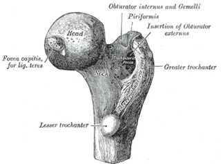
The upper extremity, proximal extremity or superior epiphysis of the femur is the part of the femur closest to the pelvic bone and the trunk. It contains the following structures:

The anterior inferior iliac spine is a bony eminence on the anterior border of the hip bone, or, more precisely, the wing of the ilium.

The iliofemoral ligament is a ligament of the hip joint which extends from the ilium to the femur in front of the joint. It is also referred to as the Y-ligament or the ligament of Bigelow, and any combinations of these names.
A femoral head ostectomy is a surgical operation to remove the head and neck from the femur. It is performed to alleviate pain, and is a salvage procedure, reserved for condition where pain can not be alleviated in any other way. It is common in veterinary surgery. Other names are excision arthroplasty of the femoral head and neck, Girdlestone's operation, Girdlestone procedure, and femoral head and neck ostectomy.

The femur neck is a flattened pyramidal process of bone, connecting the femoral head with the femoral shaft, and forming with the latter a wide angle opening medialward.
The femoral head is the highest part of the thigh bone (femur). It is supported by the femoral neck.
Protrusio acetabuli is an uncommon defect of the acetabulum. The acetabulum is the socket that receives the femoral head to make the hip joint. The hip bone of the pelvic bone/girdle is composed of three bones, the ilium, the ischium and the pubis. In protrusio deformity, there is medial displacement of the femoral head in that the medial aspect of the femoral cortex is medial to the ilioischial line. The socket is too deep and may protrude into the pelvis.

The hip bone is a large irregular bone, constricted in the center and expanded above and below. In some vertebrates it is composed of three parts: the ilium, ischium, and the pubis.
Projectional radiography ("X-ray") is one of the two main modalities of medical imaging to diagnose hip dysplasia, the other one being medical ultrasonography.. Ultrasound imaging yields better results defining the anatomy until the cartilage is ossified. When the infant is around 3 months old a clear roentgenographic image can be achieved. Unfortunately the time the joint gives a good x-ray image is also the point at which nonsurgical treatment methods cease to give good results.
The public domain consists of all the creative works to which no exclusive intellectual property rights apply. Those rights may have expired, been forfeited, expressly waived, or may be inapplicable.

Gray's Anatomy is an English language textbook of human anatomy originally written by Henry Gray and illustrated by Henry Vandyke Carter. Earlier editions were called Anatomy: Descriptive and Surgical and Gray's Anatomy: Descriptive and Applied, but the book's name is commonly shortened to, and later editions are titled, Gray's Anatomy. The book is widely regarded as an extremely influential work on the subject, and has continued to be revised and republished from its initial publication in 1858 to the present day. The latest edition of the book, the 41st, was published in September 2015.