The skull is a bone protective cavity for the brain. The skull is composed of three types of bone: cranial bones, facial bones, and ear ossicles. Two parts are more prominent: the cranium and the mandible. In humans, these two parts are the neurocranium (braincase) and the viscerocranium that includes the mandible as its largest bone. The skull forms the anterior-most portion of the skeleton and is a product of cephalisation—housing the brain, and several sensory structures such as the eyes, ears, nose, and mouth. In humans, these sensory structures are part of the facial skeleton.

A fontanelle is an anatomical feature of the infant human skull comprising soft membranous gaps (sutures) between the cranial bones that make up the calvaria of a fetus or an infant. Fontanelles allow for stretching and deformation of the neurocranium both during birth and later as the brain expands faster than the surrounding bone can grow. Premature complete ossification of the sutures is called craniosynostosis.

The occipital bone is a cranial dermal bone and the main bone of the occiput. It is trapezoidal in shape and curved on itself like a shallow dish. The occipital bone overlies the occipital lobes of the cerebrum. At the base of the skull in the occipital bone, there is a large oval opening called the foramen magnum, which allows the passage of the spinal cord.
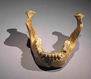
Homo heidelbergensis is an extinct species or subspecies of archaic human which existed during the Middle Pleistocene. It was subsumed as a subspecies of H. erectus in 1950 as H. e. heidelbergensis, but towards the end of the century, it was more widely classified as its own species. It is debated whether or not to constrain H. heidelbergensis to only Europe or to also include African and Asian specimens, and this is further confounded by the type specimen being a jawbone, because jawbones feature few diagnostic traits and are generally missing among Middle Pleistocene specimens. Thus, it is debated if some of these specimens could be split off into their own species or a subspecies of H. erectus. Because the classification is so disputed, the Middle Pleistocene is often called the "muddle in the middle".

Homo antecessor is an extinct species of archaic human recorded in the Spanish Sierra de Atapuerca, a productive archaeological site, from 1.2 to 0.8 million years ago during the Early Pleistocene. Populations of this species may have been present elsewhere in Western Europe, and were among the first to settle that region of the world, hence the name. The first fossils were found in the Gran Dolina cave in 1994, and the species was formally described in 1997 as the last common ancestor of modern humans and Neanderthals, supplanting the more conventional H. heidelbergensis in this position. H. antecessor has since been reinterpreted as an offshoot from the modern human line, although probably one branching off just before the modern human/Neanderthal split.
In the human skull, a sagittal keel, or sagittal torus, is a thickening of part or all of the midline of the frontal bone, or parietal bones where they meet along the sagittal suture, or on both bones. Sagittal keels differ from sagittal crests, which are found in some earlier hominins and in a range of other mammals. While a proper crest functions in anchoring the muscles of mastication to the cranium, the keel is lower and rounded in cross-section, and the jaw muscles do not attach to it.

Craniosynostosis is a condition in which one or more of the fibrous sutures in a young infant's skull prematurely fuses by turning into bone (ossification), thereby changing the growth pattern of the skull. Because the skull cannot expand perpendicular to the fused suture, it compensates by growing more in the direction parallel to the closed sutures. Sometimes the resulting growth pattern provides the necessary space for the growing brain, but results in an abnormal head shape and abnormal facial features. In cases in which the compensation does not effectively provide enough space for the growing brain, craniosynostosis results in increased intracranial pressure leading possibly to visual impairment, sleeping impairment, eating difficulties, or an impairment of mental development combined with a significant reduction in IQ.

Homo rhodesiensis is the species name proposed by Arthur Smith Woodward (1921) to classify Kabwe 1, a Middle Stone Age fossil recovered from Broken Hill mine in Kabwe, Northern Rhodesia. In 2020, the skull was dated to 324,000 to 274,000 years ago. Other similar older specimens also exist.

Ceprano Man, Argil, and Ceprano Calvarium, is a Middle Pleistocene archaic human fossil, a single skull cap (calvarium), accidentally unearthed in a highway construction project in 1994 near Ceprano in the Province of Frosinone, Italy. It was initially considered Homo cepranensis, Homo erectus, or possibly Homo antecessor; but in recent studies, most regard it either as a form of Homo heidelbergensis sharing affinities with African forms, or an early morph of Neanderthal.
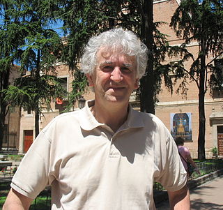
Juan Luis Arsuaga Ferreras is a Spanish paleoanthropologist and author known for his work in the Atapuerca Archaeological Site.

The sagittal suture, also known as the interparietal suture and the sutura interparietalis, is a dense, fibrous connective tissue joint between the two parietal bones of the skull. The term is derived from the Latin word sagitta, meaning arrow.
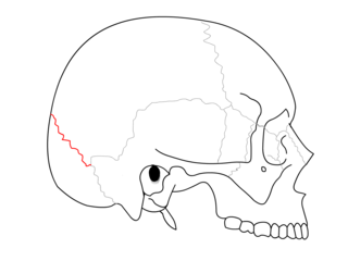
The lambdoid suture is a dense, fibrous connective tissue joint on the posterior aspect of the skull that connects the parietal bones with the occipital bone. It is continuous with the occipitomastoid suture.
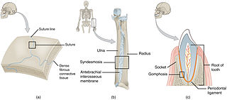
In anatomy, fibrous joints are joints connected by fibrous tissue, consisting mainly of collagen. These are fixed joints where bones are united by a layer of white fibrous tissue of varying thickness. In the skull, the joints between the bones are called sutures. Such immovable joints are also referred to as synarthroses.

The calvaria is the top part of the skull. It is the superior part of the neurocranium and covers the cranial cavity containing the brain. It forms the main component of the skull roof.

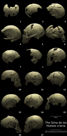
Miguelón is the popular nickname for a human skull, classified as either late Homo heidelbergensis or as early Homo neanderthalensis. It has been estimated to date to 430,000 years ago. It is one of more than 5,500 fossils belonging to early human populations which have been found in the Sima de los Huesos site in the Sierra de Atapuerca in northern Spain.
The fetal head, from an obstetrical viewpoint, and in particular its size, is important because an essential feature of labor is the adaptation between the fetal head and the maternal bony pelvis. Only a comparatively small part of the head at term is represented by the face. The rest of the head is composed of the firm skull, which is made up of two frontal, two parietal, and two temporal bones, along with the upper portion of the occipital bone and the wings of the sphenoid.

The Ndutu skull is the partial cranium of a hominin that has been assigned variously to late Homo erectus, Homo rhodesiensis, and early Homo sapiens, from the Middle Pleistocene, found at Lake Ndutu in northern Tanzania.

The archaeological site of Atapuerca is located in the province of Burgos in the north of Spain and is notable for its evidence of early human occupation. Bone fragments from around 800,000 years ago, found in its Gran Dolina cavern, provide the oldest known evidence of hominid settlement in Western Europe and of hominid cannibalism anywhere in the world.
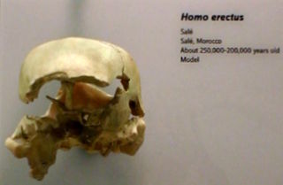
The Salé cranium is a pathological specimen of enigmatic Middle Pleistocene hominin discovered from Salé, Morocco by quarrymen in 1971. Since its discovery, the specimen has variously been classified as Homo sapiens, Homo erectus, Homo rhodesiensis/bodoensis, or Homo heidelbergensis. Its pathological condition and mosaic anatomy has proved difficult to classify. It was discovered with few faunal fossils and no lithics, tentatively dated to 400 ka by some sources.

Agamenón, also known as Agamemnon, is a fossil calvarium belonging to an early Neanderthal that lived at the site of Atapuerca around 430,000 years ago. The crania recovered from Sima de los Huesos have multiple specimen catalogues including Sima de los Huesos 4, SH 4, Cranium 4, Cr4, and Skull 4. Original analyses of the specimen concluded that the individual was deaf, although further study has proven that this is not the case. It is now held at the Museum of Human Evolution.