Bernheim syndrome

Last updated
Hippolyte Bernheim (1840-1919) Bernheim.jpg
Hippolyte Bernheim (1840-1919)

Bernheim syndrome is a presumed disorder wherein the right ventricle is severely compressed due to a shift in the ventricular septal wall of the heart, leading to heart failure. It was first described by Hippolyte Bernheim in 1910. Today, it is argued whether or not Bernheim syndrome is indeed a syndrome or a side effect of other cardiac conditions, such as left ventrical heart failure where the left ventricle is substantially enlarged, encroaching on the space of the right ventricle. [1]

Contents

Signs and symptoms

Signs and symptoms of Bernheim syndrome are ill-defined and typically follow those of heart failure. Bernheim distinguished Bernheim syndrome from the typical heart failure manifestations via the engorgement of veins due to congestion without evidence of pulmonary congestion. [2] There is also evidence of venous blockage going into pulmonary circulation and is therefore isolated to the right side of the heart. Manifestations of Bernheim syndrome include symptoms of hypertension, ronchi in the lungs, edema, vein distention, and signs of poor perfusion. [2] It is important to note that there are no manifestations of dyspnea nor pulmonary congestion until it is presumed to be a terminal stage of Bernheim syndrome. [2]

Etiology

Bernheim syndrome is believed to be the rightward shift of the ventricular septum compressing the right ventricle without causing pulmonary congestion. [3] This was first described by Hippolyte Bernheim in which he presented 10 patients with signs and symptoms of right sided heart failure whose postmortem autospy reveals a ventricular septum that invaded the right ventricle space. [1] This opposed the traditional view of right sided heart failure, right ventricular hypertrophy, where the right ventricle is enlarged. Bernheim describes right ventricles the size of a slit,which was due to the bulging ventricular septum wall.[ citation needed ]

Bernheim syndrome is believed to occur in two stages: the anatomical and clinical stages. During the anatomical stage, there are no clinical signs of the syndrome, but the stenosis of the right ventricle makes it difficult to fill to its normal capacity. This is a offset by the dilation of the right atrium as it takes in different volume. In the clinical period, there are signs and symptoms present. In the clinical period, there are two stages. In the first stage of the clinical signs of venous obstruction due to the right ventricular stenosis become apparent while pulmonary blood flow continues normally. In the second stage of the symptoms of poor circulation becomes apparent such as systemic venous engorgement. [2] It is at this point where the patient appears to have heart failure.[ citation needed ]

A normal view of right ventricle flow via cardiac MRI. In Bernheim syndrome, the movement in the right ventricle is severely impaired.

Diagnosis

Most cases of Bernheim syndrome have been identified postmortem in necropsy. A cross-sectional view of the heart muscle will show a greatly reduced right ventricle size. In necropsy, it is typical for the heart and lungs to be weighed with a higher weight indicating a build up of blood in the lungs: pulmonary congestion. The weight of the lungs is therefore expected to be within normal limits to rule out pulmonary congestion (900-1,280g). [4] The weight of the liver was also part of diagnosis with a significantly greater weight than what is in normal limits (1,440-1,680g) indicative of vein distention. [4]

In a clinical setting, Bernheim claims that the presence of isolated right ventricular failure clearly came from the presence of left ventricular hypertrophy which came secondary resulting to the presence of Bernheims symptom. [2] This is especially considered when the heart failure is not due to a weakness in the myocardium but in the stenosis of the myocardial wall. Fluoroscopy is used to view the blood flow in the heart has also been deemed as a reliable tool. It would be expected for the left ventricle and right atrium to be enlarged with the other two chambers appearing "normal". [2] However, it was typical example to only confirm the presence of Bernheim syndrome in the setting of autopsy.[ citation needed ]

History

In the medical community, however, it is believed that Bernheim syndrome does not actually exist and is only an observed side effect of another condition such as left ventricular hypertrophy. This is due to the lack of finding regarding sole right ventricle compression, without accompaniment of left ventricular hypertrophy, which is expected to encroach into the right ventricular space. It is claimed that there is no observation of a rightward shift of the ventricular septum as is described by Bernheim. Furthermore, using evidence from right and left peak systolic pressures, they determined there was no evidence of right ventricular stenosis to begin with. [1] When right ventricular heart failure is found without left ventricular heart failure, it was accompanied by pulmonary embolism and/or mitral valve stenosis. [4] It is because of these findings that there has been a movement to remove Bernheim syndrome from medical terminology.[ citation needed ]

Related Research Articles

<span class="mw-page-title-main">Tetralogy of Fallot</span> Type of congenital heart defect

Tetralogy of Fallot (TOF), formerly known as Steno-Fallot tetralogy, is a congenital heart defect characterized by four specific cardiac defects. Classically, the four defects are:

<span class="mw-page-title-main">Pulmonary heart disease</span> Medical condition

Pulmonary heart disease, also known as cor pulmonale, is the enlargement and failure of the right ventricle of the heart as a response to increased vascular resistance or high blood pressure in the lungs.

<span class="mw-page-title-main">Mitral stenosis</span> Heart disease with narrowing of valve

Mitral stenosis is a valvular heart disease characterized by the narrowing of the opening of the mitral valve of the heart. It is almost always caused by rheumatic valvular heart disease. Normally, the mitral valve is about 5 cm2 during diastole. Any decrease in area below 2 cm2 causes mitral stenosis. Early diagnosis of mitral stenosis in pregnancy is very important as the heart cannot tolerate increased cardiac output demand as in the case of exercise and pregnancy. Atrial fibrillation is a common complication of resulting left atrial enlargement, which can lead to systemic thromboembolic complications such as stroke.

<span class="mw-page-title-main">Atrial septal defect</span> Human heart defect present at birth

Atrial septal defect (ASD) is a congenital heart defect in which blood flows between the atria of the heart. Some flow is a normal condition both pre-birth and immediately post-birth via the foramen ovale; however, when this does not naturally close after birth it is referred to as a patent (open) foramen ovale (PFO). It is common in patients with a congenital atrial septal aneurysm (ASA).

Kussmaul's sign is a paradoxical rise in jugular venous pressure (JVP) on inspiration, or a failure in the appropriate fall of the JVP with inspiration. It can be seen in some forms of heart disease and is usually indicative of limited right ventricular filling due to right heart dysfunction.

<span class="mw-page-title-main">Jugular venous pressure</span> Blood pressure in a vein of the neck

The jugular venous pressure is the indirectly observed pressure over the venous system via visualization of the internal jugular vein. It can be useful in the differentiation of different forms of heart and lung disease. Classically three upward deflections and two downward deflections have been described.

<span class="mw-page-title-main">Congenital heart defect</span> Defect in the structure of the heart that is present at birth

A congenital heart defect (CHD), also known as a congenital heart anomaly, congenital cardiovascular malformation, and congenital heart disease, is a defect in the structure of the heart or great vessels that is present at birth. A congenital heart defect is classed as a cardiovascular disease. Signs and symptoms depend on the specific type of defect. Symptoms can vary from none to life-threatening. When present, symptoms are variable and may include rapid breathing, bluish skin (cyanosis), poor weight gain, and feeling tired. CHD does not cause chest pain. Most congenital heart defects are not associated with other diseases. A complication of CHD is heart failure.

<span class="mw-page-title-main">Atrium (heart)</span> Part of the human heart

The atrium is one of the two upper chambers in the heart that receives blood from the circulatory system. The blood in the atria is pumped into the heart ventricles through the atrioventricular mitral and tricuspid heart valves.

<span class="mw-page-title-main">Hypoplastic left heart syndrome</span> Type of congenital heart defect

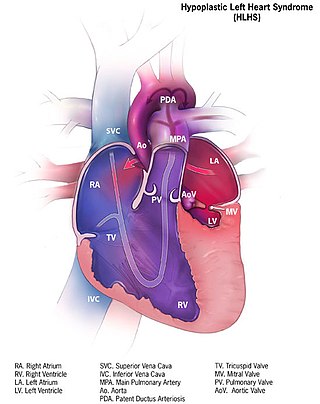
Hypoplastic left heart syndrome (HLHS) is a rare congenital heart defect in which the left side of the heart is severely underdeveloped and incapable of supporting the systemic circulation. It is estimated to account for 2-3% of all congenital heart disease. Early signs and symptoms include poor feeding, cyanosis, and diminished pulse in the extremities. The etiology is believed to be multifactorial resulting from a combination of genetic mutations and defects resulting in altered blood flow in the heart. Several structures can be affected including the left ventricle, aorta, aortic valve, or mitral valve all resulting in decreased systemic blood flow.

<span class="mw-page-title-main">Valvular heart disease</span> Disease in the valves of the heart

Valvular heart disease is any cardiovascular disease process involving one or more of the four valves of the heart. These conditions occur largely as a consequence of aging, but may also be the result of congenital (inborn) abnormalities or specific disease or physiologic processes including rheumatic heart disease and pregnancy.

<span class="mw-page-title-main">Atrioventricular septal defect</span> Medical condition

Atrioventricular septal defect (AVSD) or atrioventricular canal defect (AVCD), also known as "common atrioventricular canal" or "endocardial cushion defect" (ECD), is characterized by a deficiency of the atrioventricular septum of the heart that creates connections between all four of its chambers. It is a very specific combination of 3 defects:

Pulsus paradoxus, also paradoxic pulse or paradoxical pulse, is an abnormally large decrease in stroke volume, systolic blood pressure and pulse wave amplitude during inspiration. Pulsus paradoxus is not related to pulse rate or heart rate, and it is not a paradoxical rise in systolic pressure. Normally, blood pressure drops less precipitously than 10 mmHg during inhalation. Pulsus paradoxus is a sign that is indicative of several conditions most commonly pericardial effusion.

<span class="mw-page-title-main">Pulmonary valve stenosis</span> Medical condition

Pulmonary valve stenosis (PVS) is a heart valve disorder. Blood going from the heart to the lungs goes through the pulmonary valve, whose purpose is to prevent blood from flowing back to the heart. In pulmonary valve stenosis this opening is too narrow, leading to a reduction of flow of blood to the lungs.

Pulmonic stenosis, is a dynamic or fixed obstruction of flow from the right ventricle of the heart to the pulmonary artery. It is usually first diagnosed in childhood.

A right-to-left shunt is a cardiac shunt which allows blood to flow from the right heart to the left heart. This terminology is used both for the abnormal state in humans and for normal physiological shunts in reptiles.

<span class="mw-page-title-main">Right ventricular hypertrophy</span> Medical condition

Right ventricular hypertrophy (RVH) is a condition defined by an abnormal enlargement of the cardiac muscle surrounding the right ventricle. The right ventricle is one of the four chambers of the heart. It is located towards the lower-end of the heart and it receives blood from the right atrium and pumps blood into the lungs.

<span class="mw-page-title-main">Lutembacher's syndrome</span> Medical condition

Lutembacher's syndrome is a very rare form of congenital heart disease that affects one of the chambers of the heart as well as a valve. It is commonly known as both congenital atrial septal defect (ASD) and acquired mitral stenosis (MS). Congenital atrial septal defect refers to a hole being in the septum or wall that separates the two atria; this condition is usually seen in fetuses and infants. Mitral stenosis refers to mitral valve leaflets sticking to each other making the opening for blood to pass from the atrium to the ventricles very small. With the valve being so small, blood has difficulty passing from the left atrium into the left ventricle. Septal defects that may occur with Lutembacher's syndrome include: Ostium primum atrial septal defect or ostium secundum which is more prevalent.

<span class="mw-page-title-main">Right axis deviation</span> Medical condition

The electrical axis of the heart is the net direction in which the wave of depolarization travels. It is measured using an electrocardiogram (ECG). Normally, this begins at the sinoatrial node ; from here the wave of depolarisation travels down to the apex of the heart. The hexaxial reference system can be used to visualise the directions in which the depolarisation wave may travel.

The following outline is provided as an overview of and topical guide to cardiology, the branch of medicine dealing with disorders of the human heart. The field includes medical diagnosis and treatment of congenital heart defects, coronary artery disease, heart failure, valvular heart disease and electrophysiology. Physicians who specialize in cardiology are called cardiologists.

<span class="mw-page-title-main">Ventricular outflow tract obstruction</span> Medical condition

A ventricular outflow tract obstruction is a heart condition in which either the right or left ventricular outflow tract is blocked or obstructed. These obstructions represent a spectrum of disorders. Majority of these cases are congenital, but some are acquired throughout life.

References

  1. 1 2 3 Chung, Monica S.; Ko, Jo Mi; Chamogeorgakis, Themistokles; Hall, Shelley A.; Roberts, William C. (October 2013). "The Myth of the Bernheim Syndrome". Baylor University Medical Center Proceedings. 26 (4): 401–404. doi:10.1080/08998280.2013.11929018. ISSN   0899-8280. PMC   3777094 . PMID   24082420.
  2. 1 2 3 4 5 6 Russek, Henry I.; Zohman, Burton L. (1945-11-01). "The syndrome of Bernheim". American Heart Journal. 30 (5): 427–441. doi:10.1016/0002-8703(45)90039-1. ISSN   0002-8703. PMID   21003402.
  3. Atlas, Donald H.; Eisenberg, Herman L.; Gaberman, Peter (April 1950). "Bernheim's Syndrome: Report of a Case". Circulation. 1 (4): 753–758. doi:10.1161/01.CIR.1.4.753. ISSN   0009-7322.
  4. 1 2 3 Drago, Eugene E.; Aquilina, Joseph T. (1964-10-01). "Bernheim's syndrome". The American Journal of Cardiology. American College of Cardiology Annual Meeting. 14 (4): 568–572. doi:10.1016/0002-9149(64)90044-X. ISSN   0002-9149. PMID   14215071.