
Magnetic resonance imaging (MRI) is a medical imaging technique used in radiology to form pictures of the anatomy and the physiological processes of the body. MRI scanners use strong magnetic fields, magnetic field gradients, and radio waves to generate images of the organs in the body. MRI does not involve X-rays or the use of ionizing radiation, which distinguishes it from computed tomography (CT) and positron emission tomography (PET) scans. MRI is a medical application of nuclear magnetic resonance (NMR) which can also be used for imaging in other NMR applications, such as NMR spectroscopy.

Molecular nanotechnology (MNT) is a technology based on the ability to build structures to complex, atomic specifications by means of mechanosynthesis. This is distinct from nanoscale materials. Based on Richard Feynman's vision of miniature factories using nanomachines to build complex products, this advanced form of nanotechnology would make use of positionally-controlled mechanosynthesis guided by molecular machine systems. MNT would involve combining physical principles demonstrated by biophysics, chemistry, other nanotechnologies, and the molecular machinery of life with the systems engineering principles found in modern macroscale factories.
Nanomedicine is the medical application of nanotechnology. Nanomedicine ranges from the medical applications of nanomaterials and biological devices, to nanoelectronic biosensors, and even possible future applications of molecular nanotechnology such as biological machines. Current problems for nanomedicine involve understanding the issues related to toxicity and environmental impact of nanoscale materials.
Medical physics deals with the application of the concepts and methods of physics to the prevention, diagnosis and treatment of human diseases with a specific goal of improving human health and well-being. Since 2008, medical physics has been included as a health profession according to International Standard Classification of Occupation of the International Labour Organization.

Medical imaging is the technique and process of imaging the interior of a body for clinical analysis and medical intervention, as well as visual representation of the function of some organs or tissues (physiology). Medical imaging seeks to reveal internal structures hidden by the skin and bones, as well as to diagnose and treat disease. Medical imaging also establishes a database of normal anatomy and physiology to make it possible to identify abnormalities. Although imaging of removed organs and tissues can be performed for medical reasons, such procedures are usually considered part of pathology instead of medical imaging.

Molecular imaging is a field of medical imaging that focuses on imaging molecules of medical interest within living patients. This is in contrast to conventional methods for obtaining molecular information from preserved tissue samples, such as histology. Molecules of interest may be either ones produced naturally by the body, or synthetic molecules produced in a laboratory and injected into a patient by a doctor. The most common example of molecular imaging used clinically today is to inject a contrast agent into a patient's bloodstream and to use an imaging modality to track its movement in the body. Molecular imaging originated from the field of radiology from a need to better understand fundamental molecular processes inside organisms in a noninvasive manner.
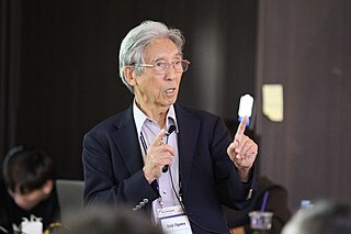
Seiji Ogawa is a Japanese biophysicist and neuroscientist known for discovering the technique that underlies Functional Magnetic Resonance Imaging (fMRI). He is regarded as the father of modern functional brain imaging. He determined that the changes in blood oxygen levels cause its magnetic resonance imaging properties to change, allowing a map of blood, and hence, functional, activity in the brain to be created. This map reflected which neurons of the brain responded with electrochemical signals to mental processes. He was the first scientist who demonstrated that the functional brain imaging is dependent on the oxygenation status of the blood, the BOLD effect. The technique was therefore called blood oxygenation level-dependent or BOLD contrast. Functional MRI (fMRI) has been used to map the visual, auditory, and sensory regions and moving toward higher brain functions such as cognitive functions in the brain.
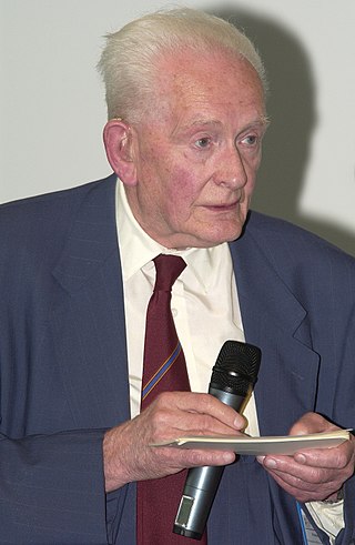
John Rowland Mallard OBE FRSE FREng was an English physicist and professor of Medical Physics at the University of Aberdeen from 1965 until his retirement in 1992. He was known for setting up and leading the team that developed the first magnetic resonance imaging (MRI) full body scanner and, in particular, positron emission tomography (PET). He was born in Kingsthorpe, Northampton, England.

Saint Boniface Hospital is Manitoba's second-largest hospital, located in the Saint Boniface neighbourhood of Winnipeg. Founded by the Sisters of Charity of Montreal in 1871, it was the first hospital in Western Canada. The hospital was incorporated in 1960, and as of 2020 has 436 beds and 30 bassinets.
Preclinical imaging is the visualization of living animals for research purposes, such as drug development. Imaging modalities have long been crucial to the researcher in observing changes, either at the organ, tissue, cell, or molecular level, in animals responding to physiological or environmental changes. Imaging modalities that are non-invasive and in vivo have become especially important to study animal models longitudinally. Broadly speaking, these imaging systems can be categorized into primarily morphological/anatomical and primarily molecular imaging techniques. Techniques such as high-frequency micro-ultrasound, magnetic resonance imaging (MRI) and computed tomography (CT) are usually used for anatomical imaging, while optical imaging, positron emission tomography (PET), and single photon emission computed tomography (SPECT) are usually used for molecular visualizations.
The Athinoula A. Martinos Center for Biomedical Imaging, usually referred to as just the "Martinos Center," is a major hub of biomedical imaging technology development and translational research. Bruce Rosen is the Director of the Center and Monica Langone is the Administrative Director.

Positron emission tomography–magnetic resonance imaging (PET–MRI) is a hybrid imaging technology that incorporates magnetic resonance imaging (MRI) soft tissue morphological imaging and positron emission tomography (PET) functional imaging.

Huntington Medical Research Institutes (HMRI) is an independent, nonprofit, applied medical research organization in Pasadena, California. The Institutes conduct laboratory and clinical work for the development of technology used in the diagnosis and treatment of disease. The Molecular Medicine programs, such as cancer genetics, molecular neurology, molecular pathology and tissue engineering, were conducted at the 99 North El Molino Avenue facility until April 2018. The Neural Engineering program is conducted at the 734 Fairmount Avenue building directly adjacent to Huntington Hospital. The Advanced Imaging Laboratory is located nearby at 10 Pico Street, as is the Liver Center at 660 South Fair Oaks Avenue. A new 35,000 square foot laboratory building for HMRI opened at 686 South Fair Oaks Avenue, Pasadena in April 2018. Programs in the new facility include neurolological and cardiovascular studies, as well as preeclampsia research.

Jin-Suck Suh is a South Korean medical professor. He graduated with MD from Yonsei University in 1979. He received PhD in 1999 from Ajou University. He is the Avison Distinguished Professor, 2011 at Yonsei University. Suh is a director as well as a principal investigator in imaging development projects of medical convergence research institute at Yonsei University.
Bruce Rosen is an American physicist and radiologist and a leading expert in the area of functional neuroimaging. His research for the past 30 years has focused on the development and application of physiological and functional nuclear magnetic resonance techniques, as well as new approaches to combine functional magnetic resonance imaging (fMRI) data with information from other modalities such as positron emission tomography (PET), magnetoencephalography (MEG) and noninvasive optical imaging. The techniques his group has developed to measure physiological and metabolic changes associated with brain activation and cerebrovascular insult are used by research centers and hospitals throughout the world.
James S. Hyde was an American biophysicist. He held the James S. Hyde chair in Biophysics at the Medical College of Wisconsin (MCW) where he specialized in magnetic resonance instrumentation and methodology development in two distinct areas: electron paramagnetic resonance (EPR) spectroscopy and magnetic resonance imaging (MRI). He is senior author of the widely cited 1995 paper by B.B. Biswal et al. reporting the discovery of resting state functional connectivity (fcMRI) in the human brain. He also served as Director of the National Biomedical EPR Center, a Research Resource supported by the National Institutes of Health. He was author of more than 400 peer-reviewed papers and review articles and held 35 U.S. Patents. He was recognized by Festschrifts in both EPR and fcMRI.
The history of magnetic resonance imaging (MRI) includes the work of many researchers who contributed to the discovery of nuclear magnetic resonance (NMR) and described the underlying physics of magnetic resonance imaging, starting early in the twentieth century. MR imaging was invented by Paul C. Lauterbur who developed a mechanism to encode spatial information into an NMR signal using magnetic field gradients in September 1971; he published the theory behind it in March 1973. The factors leading to image contrast had been described nearly 20 years earlier by physician and scientist Erik Odeblad and Gunnar Lindström. Among many other researchers in the late 1970s and 1980s, Peter Mansfield further refined the techniques used in MR image acquisition and processing, and in 2003 he and Lauterbur were awarded the Nobel Prize in Physiology or Medicine for their contributions to the development of MRI. The first clinical MRI scanners were installed in the early 1980s and significant development of the technology followed in the decades since, leading to its widespread use in medicine today.
Denis Le Bihan is a medical doctor, physicist, member of the Institut de France, member of the French Academy of Technologies and director since 2007 of NeuroSpin, an institution of the Atomic Energy and Alternative Energy Commission (CEA) in Saclay, dedicated to the study of the brain by magnetic resonance imaging (MRI) with a very high magnetic field. Denis Le Bihan has received international recognition for his outstanding work, introducing new imaging methods, particularly for the study of the human brain, as evidenced by the many international awards he has received, such as the Gold Medal of the International Society of Magnetic Resonance in Medicine (2001), the coveted Lounsbery Prize, the Louis D. Prize from the Institut de France, the prestigious Honda Prize (2012), the Louis-Jeantet Prize (2014), the Rhein Foundation Award (2021). His work has focused on the introduction, development and application of highly innovative methods, notably diffusion MRI.
R. Mark Henkelman is a Canadian biophysics researcher in the field of medical imaging, now retired, who was appointed as a Fellow of the Royal Society of Canada (2005) and the Order of Canada (2019) in recognition of his pioneering contributions to the field of magnetic resonance imaging.

Katherine Whittaker Ferrara is an American engineer who is a professor of radiology at Stanford University. Ferrara has been elected a Fellow of the American Association for the Advancement of Science, Institute of Electrical and Electronics Engineers and American Institute for Medical and Biological Engineering.