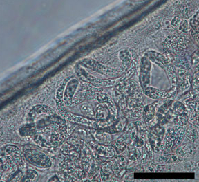
Loa loa filariasis is a skin and eye disease caused by the nematode worm Loa loa. Humans contract this disease through the bite of a deer fly or mango fly, the vectors for Loa loa. The adult Loa loa filarial worm migrates throughout the subcutaneous tissues of humans, occasionally crossing into subconjunctival tissues of the eye where it can be easily observed. Loa loa does not normally affect one's vision but can be painful when moving about the eyeball or across the bridge of the nose. The disease can cause red itchy swellings below the skin called "Calabar swellings". The disease is treated with the drug diethylcarbamazine (DEC), and when appropriate, surgical methods may be employed to remove adult worms from the conjunctiva. Loiasis belongs to the so-called neglected diseases.

Loa loa is a filarial (arthropod-borne) nematode (roundworm) that causes Loa loa filariasis. Loa loa actually means "worm worm", but is commonly known as the "eye worm", as it localizes to the conjunctiva of the eye. Loa loa is commonly found in Africa. It mainly inhabits rain forests in West Africa and has native origins in Ethiopia. The disease caused by Loa loa is called loiasis and is one of the neglected tropical diseases.

Filariasis is a parasitic disease caused by an infection with roundworms of the Filarioidea type. These are spread by blood-feeding insects such as black flies and mosquitoes. They belong to the group of diseases called helminthiases.

Wuchereria bancrofti is a filarial (arthropod-borne) nematode (roundworm) that is the major cause of lymphatic filariasis. It is one of the three parasitic worms, together with Brugia malayi and B. timori, that infect the lymphatic system to cause lymphatic filariasis. These filarial worms are spread by a variety of mosquito vector species. W. bancrofti is the most prevalent of the three and affects over 120 million people, primarily in Central Africa and the Nile delta, South and Central America, the tropical regions of Asia including southern China, and the Pacific islands. If left untreated, the infection can develop into lymphatic filariasis. In rare conditions, it also causes tropical pulmonary eosinophilia. No vaccine is commercially available, but high rates of cure have been achieved with various antifilarial regimens, and lymphatic filariasis is the target of the World Health Organization Global Program to Eliminate Lymphatic Filariasis with the aim to eradicate the disease as a public-health problem by 2020. However, this goal was not met by 2020.

Wolbachia is a genus of gram-negative bacteria that can either infect many species of arthropod as an intracellular parasite, or act as a mutualistic microbe in filarial nematodes. It is one of the most common parasitic microbes of arthropods, and is possibly the most common reproductive parasite in the biosphere. Its interactions with its hosts are often complex. Some host species cannot reproduce, or even survive, without Wolbachia colonisation. One study concluded that more than 16% of neotropical insect species carry bacteria of this genus, and as many as 25 to 70% of all insect species are estimated to be potential hosts.

Brugia malayi is a filarial (arthropod-borne) nematode (roundworm), one of the three causative agents of lymphatic filariasis in humans. Lymphatic filariasis, also known as elephantiasis, is a condition characterized by swelling of the lower limbs. The two other filarial causes of lymphatic filariasis are Wuchereria bancrofti and Brugia timori, which both differ from B. malayi morphologically, symptomatically, and in geographical extent.

Dirofilaria immitis, also known as heartworm or dog heartworm, is a parasitic roundworm that is a type of filarial worm, a small thread-like worm, and which causes dirofilariasis. It is spread from host to host through the bites of mosquitoes. Four genera of mosquitoes transmit dirofilariasis, Aedes, Culex, Anopheles, and Mansonia. The definitive host is the dog, but it can also infect cats, wolves, coyotes, jackals, foxes, ferrets, bears, seals, sea lions and, under rare circumstances, humans.

Onchocerca volvulus is a filarial (arthropod-borne) nematode (roundworm) that causes onchocerciasis, and is the second-leading cause of blindness due to infection worldwide after trachoma. It is one of the 20 neglected tropical diseases listed by the World Health Organization, with elimination from certain countries expected by 2025.
Acanthocheilonema is a genus within the family Onchocercidae which comprises mainly tropical parasitic worms. Cobbold created the genus Acanthocheilonema with only one species, Acanthocheilonema dracunculoides, which was collected from aardwolf in the region of South Africa in the nineteenth century. These parasites have a wide range of mammalian species as hosts, including members of Carnivora, Macroscelidea, Rodentia, Pholidota, Edentata, and Marsupialia. Many species among several genera of filarioids exhibit a high degree of endemicity in studies done on mammalian species in Japan. However, no concrete evidence has confirmed any endemic species in the genus Acanthocheilonema.

Lymphatic filariasis is a human disease caused by parasitic worms known as filarial worms. Usually acquired in childhood, it is a leading cause of permanent disability worldwide, impacting over a hundred million people and manifesting itself in a variety of severe clinical pathologies While most cases have no symptoms, some people develop a syndrome called elephantiasis, which is marked by severe swelling in the arms, legs, breasts, or genitals. The skin may become thicker as well, and the condition may become painful. Affected people are often unable to work and are often shunned or rejected by others because of their disfigurement and disability.
In population ecology, density-dependent processes occur when population growth rates are regulated by the density of a population. This article will focus on density dependence in the context of macroparasite life cycles.

Mansonella perstans is a filarial (arthropod-borne) nematode (roundworm), transmitted by tiny blood-sucking flies called midges. Mansonella perstans is one of two filarial nematodes that causes serous cavity filariasis in humans. The other filarial nematode is Mansonella ozzardi. M. perstans is widespread in many parts of sub-Saharan Africa, parts of Central and South America, and the Caribbean.
Mansonelliasis is the condition of infection by the nematode Mansonella. The disease exists in Africa and tropical Americas, spread by biting midges or blackflies. It is usually asymptomatic.
Brugia timori is a filarial (arthropod-borne) nematode (roundworm) which causes the disease "Timor filariasis", or "Timorian filariasis". While this disease was first described in 1965, the identity of Brugia timori as the causative agent was not known until 1977. In that same year, Anopheles barbirostris was shown to be its primary vector. There is no known animal reservoir host.

The microfilaria is an early stage in the life cycle of certain parasitic nematodes in the family Onchocercidae. In these species, the adults live in a tissue or the circulatory system of vertebrates. They release microfilariae into the bloodstream of the vertebrate host. The microfilariae are taken up by blood-feeding arthropod vectors. In the intermediate host the microfilariae develop into infective larvae that can be transmitted to a new vertebrate host.
Mansonella ozzardi is a filarial (arthropod-borne) nematode (roundworm). This filarial nematode is one of two that causes serous cavity filariasis in humans. The other filarial nematode that causes it in humans is Mansonella perstans. M. ozzardi is an endoparasite that inhabits the serous cavity of the abdomen in the human host. It lives within the mesenteries, peritoneum, and in the subcutaneous tissue.

Dirofilaria repens is a filarial nematode that affects dogs and other carnivores such as cats, wolves, coyotes, foxes, and sea lions, as well as muskrats. It is transmitted by mosquitoes. Although humans may become infected as aberrant hosts, the worms fail to reach adulthood while infecting a human body.

The Filarioidea are a superfamily of highly specialised parasitic nematodes. Species within this superfamily are known as filarial worms or filariae. Infections with parasitic filarial worms cause disease conditions generically known as filariasis. Drugs against these worms are known as filaricides.
Dirofilaria tenuis is a species of nematode, a parasitic roundworm that infects the subcutaneous tissue of vertebrates. D. tenuis most commonly infects raccoons, but some human cases have been reported. They are vectored by mosquitoes and follow similar development and transmission patterns as other Dirofilaria.

Brugia is a genus for a group of small roundworms. They are among roundworms that cause the parasitic disease filariasis. Specifically, of the three species known, Brugia malayi and Brugia timori cause lymphatic filariasis in humans; and Brugia pahangi and Brugia patei infect domestic cats, dogs and other animals. They are transmitted by the bite of mosquitos.


