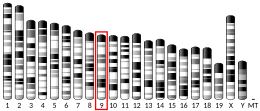
CDON

Last updated

CDON
Protein CDON PDB 3D1M.png
Available structures
PDB Ortholog search: PDBe RCSB
Identifiers
Aliases CDON , CDO, CDON1, HPE11, ORCAM, cell adhesion associated, oncogene regulated, Ihog
External IDs OMIM: 608707; MGI: 1926387; HomoloGene: 22996; GeneCards: CDON; OMA:CDON - orthologs
Orthologs
SpeciesHumanMouse
Entrez
Ensembl
UniProt
RefSeq (mRNA)

NM_001243597
NM_016952
NM_001378964

NM_021339

RefSeq (protein)

NP_001230526
NP_058648
NP_001365893

Location (UCSC) Chr 11: 125.96 – 126.06 Mb Chr 9: 35.42 – 35.51 Mb
PubMed search [3] [4]
Wikidata
View/Edit Human View/Edit Mouse

Cell adhesion molecule-related/down-regulated by oncogenes is a protein that in humans is encoded by the CDON gene. [5] [6]

Contents

CDON and BOC are cell surface receptors of the immunoglobulin (Ig)/fibronectin type III (FNIII) repeat family involved in myogenic differentiation. CDON and BOC are coexpressed during development, form complexes with each other in a cis fashion, and are related to each other in their ectodomains, but each has a unique long cytoplasmic tail. [6]

Structure and function

Cell adhesion molecule-related/down-regulated by oncogenes (CDON) is a conserved transmembrane glycoprotein belonging to a subgroup of the immunoglobulin superfamily of cell adhesion molecules. [7] It is highly expressed in both the somites and dorsal lips of the neural tube of embryonic day 8.5 mice. It is expressed in proliferating and differentiating myoblast cell lines, there is evidence showing its role in mediating the effects of cell–cell interactions between muscle precursors that are critical in myogenesis. [8] It is also expressed in neural crest precursor cells, it regulates the localization of N-cadherin providing a mechanism for directed neural crest migration. [9] CDON protein was shown to bind to all three mammalian isoforms of hedgehog proteins: Sonic Hh, Indian Hh, and Desert Hh. [10]

Clinical significance

CDON mutations are thought to diminish sonic hedgehog (SHH)-pathway activity which is important in stimulating cell proliferation, differentiation, and tissue patterning at multiple points in animal development. CDON was shown to play a role in differentiation of midbrain dopaminergic neurons through the interference with of Shh signaling pathway. [11] Mutations in CDON gene has been associated with Holoprosencephaly which is structural anomaly of the brain, in which the developing forebrain fails to correctly separate into right and left hemispheres. [12] CDON mutations synergistically interact with prenatal alcohol exposure to increase susceptibility to Holoprosencephaly. [13]

Gene knockdown studies

CDON knockdown using morpholinos in zebra fish altered the eye development, CDON was shown important in restraining the size of the optic stalk and ventral retina in chick embryos. [13] Additionally, double CDON knock out mice display optic nerve hypoplasia (ONH), a prominent feature of septo-optic dysplasia (SOD), the same phenotype shown by treating mice prenatally with ethanol. [14] CDON−/− animals also show cardiac dysfunction with increased fibrosis, those cardiac effects are mediated through hyperactivation of WNT/β-catenin signaling. [15]

Interactions

CDON has been shown to interact with CDH1 [16] and BOC. [17]

Related Research Articles

<span class="mw-page-title-main">Holoprosencephaly</span> Failure of the forebrain to develop into two hemispheres during embryonic growth

Holoprosencephaly (HPE) is a cephalic disorder in which the prosencephalon fails to develop into two hemispheres, typically occurring between the 18th and 28th day of gestation. Normally, the forebrain is formed and the face begins to develop in the fifth and sixth weeks of human pregnancy. The condition also occurs in other species.

<span class="mw-page-title-main">Sonic hedgehog protein</span> Signaling molecule in animals

Sonic hedgehog protein (SHH) is encoded for by the SHH gene. The protein is named after the video game character Sonic the Hedgehog.

<span class="mw-page-title-main">Retinal ganglion cell</span> Type of cell within the eye

A retinal ganglion cell (RGC) is a type of neuron located near the inner surface of the retina of the eye. It receives visual information from photoreceptors via two intermediate neuron types: bipolar cells and retina amacrine cells. Retina amacrine cells, particularly narrow field cells, are important for creating functional subunits within the ganglion cell layer and making it so that ganglion cells can observe a small dot moving a small distance. Retinal ganglion cells collectively transmit image-forming and non-image forming visual information from the retina in the form of action potential to several regions in the thalamus, hypothalamus, and mesencephalon, or midbrain.

Cell adhesion molecules (CAMs) are a subset of cell surface proteins that are involved in the binding of cells with other cells or with the extracellular matrix (ECM), in a process called cell adhesion. In essence, CAMs help cells stick to each other and to their surroundings. CAMs are crucial components in maintaining tissue structure and function. In fully developed animals, these molecules play an integral role in generating force and movement and consequently ensuring that organs are able to execute their functions normally. In addition to serving as "molecular glue", CAMs play important roles in the cellular mechanisms of growth, contact inhibition, and apoptosis. Aberrant expression of CAMs may result in a wide range of pathologies, ranging from frostbite to cancer.

<span class="mw-page-title-main">L1 (protein)</span> Mammalian protein found in Homo sapiens

L1, also known as L1CAM, is a transmembrane protein member of the L1 protein family, encoded by the L1CAM gene. This protein, of 200 to 220 kDa, is a neuronal cell adhesion molecule with a strong implication in cell migration, adhesion, neurite outgrowth, myelination and neuronal differentiation. It also plays a key role in treatment-resistant cancers due to its function. It was first identified in 1984 by M. Schachner who found the protein in post-mitotic mice neurons.
<span class="mw-page-title-main">GLI1</span> Protein-coding gene in humans

Zinc finger protein GLI1 also known as glioma-associated oncogene is a protein that in humans is encoded by the GLI1 gene. It was originally isolated from human glioblastoma cells.

<span class="mw-page-title-main">GLI2</span> Protein-coding gene in the species Homo sapiens

Zinc finger protein GLI2 also known as GLI family zinc finger 2 is a protein that in humans is encoded by the GLI2 gene. The protein encoded by this gene is a transcription factor.

<span class="mw-page-title-main">GLI3</span> Protein-coding gene in the species Homo sapiens

Zinc finger protein GLI3 is a protein that in humans is encoded by the GLI3 gene.

<span class="mw-page-title-main">Cyclopamine</span> Chemical compound

Cyclopamine (11-deoxojervine) is a naturally occurring steroidal alkaloid. It is a teratogenic component of corn lily, which when consumed during gestation has been demonstrated to induce birth defects, including the development of a single eye (cyclopia) in offspring. The molecule was named after this effect, which was originally observed by Idaho lamb farmers in 1957 after their herds gave birth to cycloptic lambs. It then took more than a decade to identify corn lily as the culprit. Later work suggested that differing rain patterns had changed grazing behaviours, which led to a greater quantity of corn lily to be ingested by pregnant sheep. Cyclopamine interrupts the sonic hedgehog signalling pathway, instrumental in early development, ultimately causing birth defects.

The Hedgehog signaling pathway is a signaling pathway that transmits information to embryonic cells required for proper cell differentiation. Different parts of the embryo have different concentrations of hedgehog signaling proteins. The pathway also has roles in the adult. Diseases associated with the malfunction of this pathway include cancer.

<span class="mw-page-title-main">Myogenesis</span> Formation of muscular tissue, particularly during embryonic development

Myogenesis is the formation of skeletal muscular tissue, particularly during embryonic development.

<span class="mw-page-title-main">CXCL3</span> Mammalian protein found in Homo sapiens

Chemokine ligand 3 (CXCL3) is a small cytokine belonging to the CXC chemokine family that is also known as GRO3 oncogene (GRO3), GRO protein gamma (GROg) and macrophage inflammatory protein-2-beta (MIP2b). CXCL3 controls migration and adhesion of monocytes and mediates its effects on its target cell by interacting with a cell surface chemokine receptor called CXCR2. More recently, it has been shown that Cxcl3 regulates cell autonomously the migration of the precursors of cerebellar granule neurons toward the internal layers of cerebellum, during the morphogenesis of cerebellum. Moreover, if the expression of Cxcl3 is reduced in cerebellar granule neuron precursors, this highly enhances the frequency of the medulloblastoma, the tumor of cerebellum. In fact, the reduced expression of Cxcl3 forces the cerebellar granule neuron precursors to remain at the surface of the cerebellum, where they highly proliferate under the stimulus of Sonic hedgehog, becoming target of transforming insults. Remarkably, the treatment with CXCL3 completely prevents the growth of medulloblastoma lesions in a Shh-type mouse model of medulloblastoma. Thus, CXCL3 is a target for medulloblastoma therapy. Cxcl3 is directly regulated transcriptionally by BTG2

<span class="mw-page-title-main">GPR56</span> Protein-coding gene in the species Homo sapiens

G protein-coupled receptor 56 also known as TM7XN1 is a protein encoded by the ADGRG1 gene. GPR56 is a member of the adhesion GPCR family. Adhesion GPCRs are characterized by an extended extracellular region often possessing N-terminal protein modules that is linked to a TM7 region via a domain known as the GPCR-Autoproteolysis INducing (GAIN) domain.
<span class="mw-page-title-main">SNAI1</span> Protein

Zinc finger protein SNAI1 is a protein that in humans is encoded by the SNAI1 gene. Snail is a family of transcription factors that promote the repression of the adhesion molecule E-cadherin to regulate epithelial to mesenchymal transition (EMT) during embryonic development.

<span class="mw-page-title-main">Indian hedgehog (protein)</span> Protein-coding gene in the species Homo sapiens

Indian hedgehog homolog (Drosophila), also known as IHH, is a protein which in humans is encoded by the IHH gene. This cell signaling protein is in the hedgehog signaling pathway. The several mammalian variants of the Drosophila hedgehog gene have been named after the various species of hedgehog; the Indian hedgehog is honored by this one. The gene is not specific to Indian hedgehogs.

<span class="mw-page-title-main">SUFU</span> Protein-coding gene in the species Homo sapiens

Suppressor of fused homolog is a protein that in humans is encoded by the SUFU gene. In molecular biology, the protein domain suppressor of fused protein (Sufu) has an important role in the cell. The Sufu is important in negatively regulating an important signalling pathway in the cell, the Hedgehog signalling pathway (HH). This particular pathway is crucial in embryonic development. There are several homologues of Sufu, found in a wide variety of organisms.

<span class="mw-page-title-main">CDH15</span> Protein-coding gene in humans

Cadherin-15 is a protein that in humans is encoded by the CDH15 gene.

<span class="mw-page-title-main">BOC (gene)</span> Protein-coding gene in the species Homo sapiens

Brother of CDO is a protein that in humans is encoded by the BOC gene.

<span class="mw-page-title-main">MYF5</span> Protein-coding gene in the species Homo sapiens

Myogenic factor 5 is a protein that in humans is encoded by the MYF5 gene. It is a protein with a key role in regulating muscle differentiation or myogenesis, specifically the development of skeletal muscle. Myf5 belongs to a family of proteins known as myogenic regulatory factors (MRFs). These basic helix loop helix transcription factors act sequentially in myogenic differentiation. MRF family members include Myf5, MyoD (Myf3), myogenin, and MRF4 (Myf6). This transcription factor is the earliest of all MRFs to be expressed in the embryo, where it is only markedly expressed for a few days. It functions during that time to commit myogenic precursor cells to become skeletal muscle. In fact, its expression in proliferating myoblasts has led to its classification as a determination factor. Furthermore, Myf5 is a master regulator of muscle development, possessing the ability to induce a muscle phenotype upon its forced expression in fibroblastic cells.

<span class="mw-page-title-main">MYF6</span> Protein-coding gene in the species Homo sapiens

Myogenic factor 6 is a protein that in humans is encoded by the MYF6 gene. This gene is also known in the biomedical literature as MRF4 and herculin. MYF6 is a myogenic regulatory factor (MRF) involved in the process known as myogenesis.

References

  1. 1 2 3 GRCh38: Ensembl release 89: ENSG00000064309 Ensembl, May 2017
  2. 1 2 3 GRCm38: Ensembl release 89: ENSMUSG00000038119 Ensembl, May 2017
  3. "Human PubMed Reference:". National Center for Biotechnology Information, U.S. National Library of Medicine.
  4. "Mouse PubMed Reference:". National Center for Biotechnology Information, U.S. National Library of Medicine.
  5. Kang JS, Gao M, Feinleib JL, Cotter PD, Guadagno SN, Krauss RS (July 1997). "CDO: an oncogene-, serum-, and anchorage-regulated member of the Ig/fibronectin type III repeat family". The Journal of Cell Biology. 138 (1): 203–13. doi:10.1083/jcb.138.1.203. PMC   2139939 . PMID   9214393.
  6. 1 2 "Entrez Gene: CDON Cdon homolog (mouse)".
  7. Sanchez-Arrones L, Cardozo M, Nieto-Lopez F, Bovolenta P (May 2012). "Cdon and Boc: Two transmembrane proteins implicated in cell-cell communication". The International Journal of Biochemistry & Cell Biology. 44 (5): 698–702. doi:10.1016/j.biocel.2012.01.019. PMID   22326621.
  8. Kang JS, Mulieri PJ, Miller C, Sassoon DA, Krauss RS (October 1998). "CDO, a robo-related cell surface protein that mediates myogenic differentiation". The Journal of Cell Biology. 143 (2): 403–13. doi:10.1083/jcb.143.2.403. PMC   2132836 . PMID   9786951.
  9. Powell DR, Williams JS, Hernandez-Lagunas L, Salcedo E, O'Brien JH, Artinger KB (November 2015). "Cdon promotes neural crest migration by regulating N-cadherin localization". Developmental Biology. 407 (2): 289–99. doi:10.1016/j.ydbio.2015.07.025. PMC   4663112 . PMID   26256768.
  10. Kavran JM, Ward MD, Oladosu OO, Mulepati S, Leahy DJ (August 2010). "All mammalian Hedgehog proteins interact with cell adhesion molecule, down-regulated by oncogenes (CDO) and brother of CDO (BOC) in a conserved manner". The Journal of Biological Chemistry. 285 (32): 24584–90. doi: 10.1074/jbc.M110.131680 . PMC   2915694 . PMID   20519495.
  11. Kwon YR, Jeong MH, Leem YE, Lee SJ, Kim HJ, Bae GU, Kang JS (September 2014). "The Shh coreceptor Cdo is required for differentiation of midbrain dopaminergic neurons". Stem Cell Research. 13 (2): 262–74. doi: 10.1016/j.scr.2014.07.004 . PMID   25117422.
  12. Bae GU, Domené S, Roessler E, Schachter K, Kang JS, Muenke M, Krauss RS (August 2011). "Mutations in CDON, encoding a hedgehog receptor, result in holoprosencephaly and defective interactions with other hedgehog receptors". American Journal of Human Genetics. 89 (2): 231–40. doi:10.1016/j.ajhg.2011.07.001. PMC   3155179 . PMID   21802063.
  13. 1 2 Hong M, Krauss RS (October 2012). "Cdon mutation and fetal ethanol exposure synergize to produce midline signaling defects and holoprosencephaly spectrum disorders in mice". PLOS Genetics. 8 (10): e1002999. doi: 10.1371/journal.pgen.1002999 . PMC   3469434 . PMID   23071453.
  14. Kahn BM, Corman TS, Lovelace K, Hong M, Krauss RS, Epstein DJ (January 2017). "Prenatal ethanol exposure in mice phenocopies Cdon mutation by impeding Shh function in the etiology of optic nerve hypoplasia". Disease Models & Mechanisms. 10 (1): 29–37. doi:10.1242/dmm.026195. PMC   5278523 . PMID   27935818.
  15. Jeong MH, Kim HJ, Pyun JH, Choi KS, Lee DI, Solhjoo S, O'Rourke B, Tomaselli GF, Jeong DS, Cho H, Kang JS (February 2017). "Cdon deficiency causes cardiac remodeling through hyperactivation of WNT/β-catenin signaling". Proceedings of the National Academy of Sciences of the United States of America. 114 (8): E1345–E1354. Bibcode:2017PNAS..114E1345J. doi: 10.1073/pnas.1615105114 . PMC   5338397 . PMID   28154134.
  16. Kang JS, Feinleib JL, Knox S, Ketteringham MA, Krauss RS (April 2003). "Promyogenic members of the Ig and cadherin families associate to positively regulate differentiation". Proceedings of the National Academy of Sciences of the United States of America. 100 (7): 3989–94. Bibcode:2003PNAS..100.3989K. doi: 10.1073/pnas.0736565100 . PMC   153035 . PMID   12634428.
  17. Kang JS, Mulieri PJ, Hu Y, Taliana L, Krauss RS (January 2002). "BOC, an Ig superfamily member, associates with CDO to positively regulate myogenic differentiation". The EMBO Journal. 21 (1–2): 114–24. doi:10.1093/emboj/21.1.114. PMC   125805 . PMID   11782431.

Further reading