Cardiac allograft vasculopathy

Last updated
Cardiac allograft vasculopathy
Coronary arteries.svg
Coronary arteries
Specialty Cardiology, angiology
Symptoms None, breathlessness, tiredness [1]
Usual onsetAfter heart transplantation [1]
Diagnostic method Coronary angiography, intravascular ultrasound, dobutamine stress echocardiography, positron emission tomography, CT angiography, biomarkers, endomyocardial biopsy [2]
TreatmentControl cardiovascular risk factors, mTOR inhibitors, re-transplantation [1]
Medication Sirolimus, everolimus [1]
Prognosis Progressive [2]
FrequencyAround 50% (in 10 years) [2]
Deaths11-13% of heart transplants one year from surgery [1]

Cardiac allograft vasculopathy (CAV) is a progressive type of coronary artery disease in people who have had a heart transplant. [1] As the donor heart has lost its nerve supply there is typically no chest pain, and CAV is usually detected on routine testing. [2] It may present with symptoms such as tiredness and breathlessness. [2]

Contents

It arises when the blood vessels supplying the transplanted heart change in structure. [3] They gradually narrow and restrict its blood flow, subsequently leading to impairment of the heart muscle or sudden death. [4] In addition to the same risk factors for coronary artery disease due to the build up of plaque, CAV is more likely to occur if the donor was older or died from explosive brain death, and if there is cytomegalovirus infection. [2] Its mechanism involves immunological (innate and adaptive) and nonimmunological factors, with distinct features on histological samples of coronary arteries. [2] Other major causes of death following heart transplantation include graft failure, organ rejection and infection. [5]

Diagnosis is by regular follow-up and monitoring of the transplanted heart for early signs of disease. [2] Tests include coronary angiography, intravascular ultrasound, dobutamine stress echocardiography, positron emission tomography, computed tomographic angiography (CT angiography) and several biomarkers. [2]

Statins and aspirin are commenced early after transplantation and on detection of CAV. [2] Medications including sirolimus and everolimus can slow disease progression. [2] A repeat heart transplantation may be required. [6]

CAV affects around half of heart transplant recipients within 10 years. [2] It contributes to the death of 11-13% one year from heart transplantation. [1]

Definition

Cardiac allograft vasculopathy is an accelerated type of coronary artery disease in people who have had a heart transplantation. [7]

Signs and symptoms

Unlike the chest tightness of angina in those who have not had a heart transplant, people with CAV typically do not experience chest pain because the donor heart has lost its nerve supply. [2] A few regain nerves some years later and may develop unusual chest pain. [8] People with CAV may present with a broad spectrum of symptoms including tiredness, nausea, or abdominal discomfort or may have no symptoms at all. [2] Shortness of breath and arrythmias may also occur. [8]

Risk factors

Similar to coronary artery disease in those who have not had a heart transplant, risk factors to CAV include high blood pressure, high cholesterol, and diabetes mellitus. [2] Other risk factors exclusive to CAV include older donors, cytomegalovirus infection and circulating antibodies after heart transplantation. [2] The mechanism of donor brain death, [8] particularly explosive brain death in the donor has been shown to be a significant factor. It is probably the combination of injuries to the allograft that determine the risk of developing CAV. [2]

Mechanism

Artery wall structure, showing intimal layer Blausen 0055 ArteryWallStructure.png
Artery wall structure, showing intimal layer

Immunological (innate and adaptive) and nonimmunological factors contribute to the complex pathogenesis of CAV. [2]

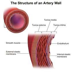
In those nontransplanted people who develop coronary artery disease due to atherosclerosis, progression of disease is slow, histological changes are confined mainly to the main coronary arteries and arterial dilatation is observed as a form of compensatory remodelling. [8] However, in CAV, histology specimens typically show concentric thickening of the intimal layer of the main coronary arteries on the surface of the heart and in intramyocardial arteries which can become obliterated within a few years. [2] [8] There is smooth muscle cell migration, foamy macrophages and lymphocytic infiltrates. This can be seen to affect the whole length of the coronary arteries and often the smaller arteries. [2] Calcification does not always occur in CAV and if it does appear, it happens late. [8] The compensatory arterial dilation does not occur in CAV. [8] Unlike in nontransplanted people with coronary artery disease due to atherosclerosis, in CAV occlusion with thrombus of the vessel lumen is rare. [2]

Inflammation and endothelial injury can be triggered by the donor arrest, organ procurement, and allograft ischaemia and reperfusion. [2]

Diagnosis

As symptoms are so variable and often absent, diagnosis has been a challenge. Hence, regular follow-up and monitoring of the allograft for early signs of disease is advocated. [2]

Coronary angiography

Surveillance is performed by regularly repeating coronary angiography in the cardiac catheterization laboratory, the diagnostic test of choice. [2] This is typically performed annually for the first five years after transplantation. [8] Angiography in CAV characteristically demonstrates diffuse stenoses in large coronary arteries and a reduced number of smaller coronary arteries, also known as "peripheral pruning". [2] [6] However, because CAV frequently affects the entire length of the coronary artery, CAV may not be apparent by angiography alone. [2]

Intravascular ultrasound (IVUS)

Intravascular ultrasound (IVUS) is more sensitive at reliably detecting subtle changes in the thickness of the intimal layer of the artery walls and provide measurements of artery lumen. Following transplantation, serial measurements are compared to the baseline. A greater than 0.5 mm increase in intimal thickness one year after transplantation is predictive of CAV changes on angiography within five years. The paradoxical reduction in the number of blood vessels, can also be detected by intravascular ultrasound. [2] [8]

IVUS, however, tends to be used for research due to its drawbacks of being invasive, requiring the use of contrast material and cost. [8]

Dobutamine stress-echocardiography (DSE)

Alternatively, dobutamine stress echocardiography (DSE) is commonly performed and has an 85% sensitivity for the presence of CAV. A negative DSE correlates with a good prognosis. [8]

Other noninvasive diagnostics include positron emission tomography and computed tomographic angiography (CT angiography). [2] In addition, ECGs may show atypical features of ischaemia. [8]

Biomarkers

Biomarkers for increased risk of CAV include C-reactive protein, serum brain natriuretic peptide and troponin I have been suggested. [2]

Classification

The degree of CAV after heart transplantation has been obtained from a variety of sources including The Cardiac Transplant Research Database, the ISHLT registry and The United Network for Organ Sharing registry. [8]

The International Society for Heart and Lung Transplantation (ISHLT) have formulated and standardized a terminology, based on diagnostic findings, to define the presence and severity of CAV, which in turn reflects prognosis. [8] [10] The severity of CAV is defined by the degree of narrowing of the coronary arteries and the presence of restrictive heart disease. [8]

CodeSeverityDiagnostic findings [10]
ISHLT CAV0Not significantNo detectable lesions on angiography
ISHLT CAV1MildAngiographic left main (LM) 􏰁50%, or primary vessel with maximum lesion of 􏰁70%, or any branch stenosis 􏰁70% (including diffuse narrowing) without allograft dysfunction
ISHLT CAV2ModerateAngiographic LM 􏰁50%; a single primary vessel 􏰄70%, or isolated branch stenosis 􏰄70% in branches of 2 systems, without allograft dysfunction
ISHLT CAV3SevereAngiographic LM 􏰄50%, or two or more primary vessels 􏰄70% stenosis, or isolated branch stenosis 􏰄70% in all 3 systems; or ISHLT CAV1 or CAV2 with allograft dysfunction (defined as LVEF 􏰅45% usually in the presence of regional wall motion abnormalities) or evidence of significant restrictive physiology

Treatment

Primary angioplasty procedure in a person who had CAV after heart transplantation. [11]

Statins

Prevention of CAV progression is important as once developed, CAV existing treatments are often ineffective. [2] Commencing the statins pravastatin and simvastatin early after transplantation reduces the incidence and severity of CAV. [2] [8]

Vitamins

When combined with immunosuppressants, the progression of CAV could possibly be slowed by vitamins C and E. [8]

Aspirin

Since the role of aspirin is already established in coronary artery disease in those who have not had a heart transplant, it is usually given after heart transplantation too. [2]

Antiproliferative agents

On detection of CAV, medications including mTOR inhibitors sirolimus and everolimus have been shown to slow disease progression. [2]

Other treatment options

Clinically significant CAV may require percutaneous coronary interventions for focal disease, but the likelihood of restenosis is high. [2] A repeat heart transplantation may be considered. [2]

Outcome

Once there is reduced left ventricular ejection fraction and symptoms of heart failure, the outcome is typically poor. [2] The risk of major adverse cardiovascular events is increased by 3.4 fold if CAV is present on angiography. [8]

Epidemiology

The frequency of CAV after heart transplantation has been obtained from a variety of sources including The Cardiac Transplant Research Database, the ISHLT registry and The United Network for Organ Sharing registry. [8] In comparison to between 1994 and 2001, there has been a decline in incidence of CAV between 2001 and 2007. [8] ISHLT figures show an incidence of CAV of around 50% at 10 years after heart transplantation. [8]

CAV is a leading cause of late mortality following heart transplantation. [2] Most are not severe but it contributes to the death of 11-13% one year from heart transplantation. [1]

History

Unlike rejection and infection, CAV in the transplanted heart was not initially a predicted outcome. [12] Early survivors of heart transplants soon developed this form of vasculopathy of their coronary arteries, initially identified at post-mortems. [12] There were early suggestions that preventing cytomegalovirus (CMV) infection could decrease the prevalence of CAV. [12] The impact of CAV has changed over time, with early recipients being younger, having more rejection and cardiovascular risk factors and less use of statins. [12] Later recipients used statins routinely and were introduced to the immunosuppressive agent mycophenolate mofetil (MMF) and CMV prophylaxis. [12] In addition, the later recipients were monitored for antibody-mediated cardiac allograft rejection (AMR). [12]

Before 2010 there was no uniform international standards for the nomenclature of CAV. [4] A consensus statement on a standard language for CAV was first published in 2010 by the ISHLT. [4] This was devised in a similar way to the earlier acute rejection grading system by endomyocardial biopsy. [10] [13]

Research directions

Antibody-mediated cardiac allograft rejection (AMR) is a significant factor leading to the rapid progression of CAV. [12] Future research directions in this area may include prospective databases that correlate clinical factors with surveillance of the incidence and severity of AMR, the frequency of CMV infection, and the use of immunosuppressants. The role of inducing immune tolerance has yet to be established. [12]

Related Research Articles

<span class="mw-page-title-main">Coronary artery disease</span> Reduction of blood flow to the heart muscle due to plaque buildup in the hearts arteries

Coronary artery disease (CAD), also called coronary heart disease (CHD), ischemic heart disease (IHD), myocardial ischemia, or simply heart disease, involves the reduction of blood flow to the heart muscle due to build-up of atherosclerotic plaque in the arteries of the heart. It is the most common of the cardiovascular diseases. Types include stable angina, unstable angina, myocardial infarction, and sudden cardiac death. A common symptom is chest pain or discomfort which may travel into the shoulder, arm, back, neck, or jaw. Occasionally it may feel like heartburn. Usually symptoms occur with exercise or emotional stress, last less than a few minutes, and improve with rest. Shortness of breath may also occur and sometimes no symptoms are present. In many cases, the first sign is a heart attack. Other complications include heart failure or an abnormal heartbeat.

<span class="mw-page-title-main">Angina</span> Chest discomfort due to not enough blood flow to heart muscle

Angina, also known as angina pectoris, is chest pain or pressure, usually caused by insufficient blood flow to the heart muscle (myocardium). It is most commonly a symptom of coronary artery disease.

<span class="mw-page-title-main">Atherosclerosis</span> Form of arteriosclerosis

Atherosclerosis is a pattern of the disease arteriosclerosis in which the wall of the artery develops abnormalities, called lesions. These lesions may lead to narrowing due to the buildup of atheromatous plaque. At onset there are usually no symptoms, but if they develop, symptoms generally begin around middle age. When severe, it can result in coronary artery disease, stroke, peripheral artery disease, or kidney problems, depending on which arteries are affected.

<span class="mw-page-title-main">Peripheral artery disease</span> Medical condition

Peripheral artery disease (PAD) is an abnormal narrowing of arteries other than those that supply the heart or brain. PAD can happen in any blood vessel, but it is more common in the legs than the arms.

<span class="mw-page-title-main">Everolimus</span> Chemical compound

Everolimus, sold under the brand name Afinitor among others, is a medication used as an immunosuppressant to prevent rejection of organ transplants and as a targeted therapy in the treatment of renal cell cancer and other tumours.

<span class="mw-page-title-main">Atheroma</span> Accumulation of degenerative material in the inner layer of artery walls

An atheroma, or atheromatous plaque, is an abnormal accumulation of material in the inner layer of an arterial wall.

Intravascular ultrasound (IVUS) or intravascular echocardiography is a medical imaging methodology using a specially designed catheter with a miniaturized ultrasound probe attached to the distal end of the catheter. The proximal end of the catheter is attached to computerized ultrasound equipment. It allows the application of ultrasound technology, such as piezoelectric transducer or CMUT, to see from inside blood vessels out through the surrounding blood column, visualizing the endothelium of blood vessels.

A vulnerable plaque is a kind of atheromatous plaque – a collection of white blood cells and lipids in the wall of an artery – that is particularly unstable and prone to produce sudden major problems such as a heart attack or stroke.

<span class="mw-page-title-main">Cardiomegaly</span> Medical condition

Cardiomegaly is a medical condition in which the heart becomes enlarged. It is more commonly referred to simply as "having an enlarged heart". It is usually the result of underlying conditions that make the heart work harder, such as obesity, heart valve disease, high blood pressure (hypertension), and coronary artery disease. Cardiomyopathy is also associated with cardiomegaly.

A vascular bypass is a surgical procedure performed to redirect blood flow from one area to another by reconnecting blood vessels. Often, this is done to bypass around a diseased artery, from an area of normal blood flow to another relatively normal area. It is commonly performed due to inadequate blood flow (ischemia) caused by atherosclerosis, as a part of organ transplantation, or for vascular access in hemodialysis. In general, someone's own vein (autograft) is the preferred graft material for a vascular bypass, but other types of grafts such as polytetrafluoroethylene (Teflon), polyethylene terephthalate (Dacron), or a different person's vein (allograft) are also commonly used. Arteries can also serve as vascular grafts. A surgeon sews the graft to the source and target vessels by hand using surgical suture, creating a surgical anastomosis.

Sean Patrick Pinney is an American cardiologist and the Director of both the Advanced Heart Failure and Cardiac Transplant Program and the Pulmonary Hypertension Program at Mount Sinai Medical Center in New York City.

<span class="mw-page-title-main">Coronary ischemia</span> Medical condition

Coronary ischemia, myocardial ischemia, or cardiac ischemia, is a medical term for a reduced blood flow in the coronary circulation through the coronary arteries. Coronary ischemia is linked to heart disease, and heart attacks. Coronary arteries deliver oxygen-rich blood to the heart muscle. Reduced blood flow to the heart associated with coronary ischemia can result in inadequate oxygen supply to the heart muscle. When oxygen supply to the heart is unable to keep up with oxygen demand from the muscle, the result is the characteristic symptoms of coronary ischemia, the most common of which is chest pain. Chest pain due to coronary ischemia commonly radiates to the arm or neck. Certain individuals such as women, diabetics, and the elderly may present with more varied symptoms. If blood flow through the coronary arteries is stopped completely, cardiac muscle cells may die, known as a myocardial infarction, or heart attack.

<span class="mw-page-title-main">Myocardial infarction</span> Interruption of blood supply to a part of the heart

A myocardial infarction (MI), commonly known as a heart attack, occurs when blood flow decreases or stops in one of the coronary arteries of the heart, causing damage to the heart muscle. The most common symptom is chest pain or discomfort which may travel into the shoulder, arm, back, neck or jaw. Often it occurs in the center or left side of the chest and lasts for more than a few minutes. The discomfort may occasionally feel like heartburn. Other symptoms may include shortness of breath, nausea, feeling faint, a cold sweat or feeling tired. About 30% of people have atypical symptoms. Women more often present without chest pain and instead have neck pain, arm pain or feel tired. Among those over 75 years old, about 5% have had an MI with little or no history of symptoms. An MI may cause heart failure, an irregular heartbeat, cardiogenic shock or cardiac arrest.

A Cytomegalovirus vaccine is a vaccine to prevent cytomegalovirus (CMV) infection or curb virus re-activation in persons already infected. Challenges in developing a vaccine include adeptness of CMV in evading the immune system and limited animal models. As of 2018 no such vaccine exists, although a number of vaccine candidates are under investigation. They include recombinant protein, live attenuated, DNA and other vaccines.

<span class="mw-page-title-main">Spontaneous coronary artery dissection</span> Uncommon cause of heart attacks mostly affecting younger, healthy women

Spontaneous coronary artery dissection (SCAD) is an uncommon but potentially lethal condition in which one of the coronary arteries that supply the heart, spontaneously develops a blood collection, or hematoma, within the artery wall due to a tear in the wall. SCAD is one of the arterial dissections that can occur.

<span class="mw-page-title-main">Heart transplantation</span> Surgical transplant procedure

A heart transplant, or a cardiac transplant, is a surgical transplant procedure performed on patients with end-stage heart failure or severe coronary artery disease when other medical or surgical treatments have failed. As of 2018, the most common procedure is to take a functioning heart, with or without both lungs, from a recently deceased organ donor and implant it into the patient. The patient's own heart is either removed and replaced with the donor heart or, much less commonly, the recipient's diseased heart is left in place to support the donor heart.

<span class="mw-page-title-main">Coronary CT angiography</span> Use of computed tomography angiography to assess the coronary arteries of the heart

Coronary CT angiography is the use of computed tomography (CT) angiography to assess the coronary arteries of the heart. The patient receives an intravenous injection of radiocontrast and then the heart is scanned using a high speed CT scanner, allowing physicians to assess the extent of occlusion in the coronary arteries, usually in order to diagnose coronary artery disease.

Mandeep R. Mehra is The William Harvey Distinguished Chair in Advanced Cardiovascular Medicine and a professor of medicine at Harvard Medical School. He is the medical director of the Brigham Heart and Vascular Center in Boston, Massachusetts, and specializes in advanced heart failure, mechanical circulatory support and cardiac transplantation.

Sharon Ann Hunt is a cardiology professor and Director of the Post Heart Transplant Programme in Palo Alto, California and is affiliated with Stanford University Medical Center, professionally known for her work in the care of patients after heart transplantation.

<span class="mw-page-title-main">Endomyocardial biopsy</span> Invasive medical procedure to obtain small samples of heart muscle

Endomyocardial biopsy (EMB) is an invasive procedure used routinely to obtain small samples of heart muscle, primarily for detecting rejection of a donor heart following heart transplantation. It is also used as a diagnostic tool in some heart diseases.

References

  1. 1 2 3 4 5 6 7 8 Shanmuganathan, Mayooran; Dar, Owais (2020). "73. Complications of heart transplantation". In Raja, Shahzad G. (ed.). Cardiac Surgery: A Complete Guide. Switzerland: Springer. p. 669. ISBN   978-3-030-24176-6.
  2. 1 2 3 4 5 6 7 8 9 10 11 12 13 14 15 16 17 18 19 20 21 22 23 24 25 26 27 28 29 30 31 32 33 34 35 36 37 Stehlik, Josef; Kobashigawa, Jon; Hunt, Sharon A.; Reichenspurner, Hermann; Kirklin, James K. (2 January 2018). "Honoring 50 Years of Clinical Heart Transplantation in Circulation". Circulation . 137 (1): 71–87. doi: 10.1161/CIRCULATIONAHA.117.029753 . PMID   29279339.
  3. Pober, Jordan S; Chih, Sharon; Kobashigawa, Jon; Madsen, Joren C; Tellides, George (3 August 2021). "Cardiac allograft vasculopathy: current review and future research directions". Cardiovascular Research. 117 (13): 2624–2638. doi:10.1093/cvr/cvab259. PMC   8783389 . PMID   34343276.
  4. 1 2 3 Elsevier (2010). "New ISHLT Cardiac Allograft Vasculopathy Standardized Nomenclature: A Common International Definition Will Benefit Heart Transplant Patients". www.elsevier.com. Elsevier. Retrieved 27 January 2019.
  5. Dipchand, Anne I. (2018-01-02). "Current state of pediatric cardiac transplantation". Annals of Cardiothoracic Surgery. 7 (1): 31–55–55. doi: 10.21037/acs.2018.01.07 . ISSN   2225-319X. PMC   5827130 . PMID   29492382.
  6. 1 2 Lee, Michael S.; Tadwalkar, Rigved V.; Fearon, William F.; Kirtane, Ajay J.; Patel, Amisha J.; Patel, Chetan B.; Ali, Ziad; Rao, Sunil V. (2018-12-01). "Cardiac allograft vasculopathy: A review". Catheterization and Cardiovascular Interventions. 92 (7): E527–E536. doi:10.1002/ccd.27893. ISSN   1522-726X. PMID   30265435. S2CID   52880607.(subscription required)
  7. Lee, Felicity; Nair, Vidhya; Chih, Sharon (2020). "Cardiac allograft vasculopathy: Insights on pathogenesis and therapy" . Clinical Transplantation. 34 (3): e13794. doi:10.1111/ctr.13794. ISSN   1399-0012. PMID   31991002. S2CID   210948957.
  8. 1 2 3 4 5 6 7 8 9 10 11 12 13 14 15 16 17 18 19 20 21 Eisen, Howard J. (2016). "19. Complications after cardiac transplantation". In Domanski, Michael J.; Mehra, Mandeep R.; Pfeffer, Marc A. (eds.). Oxford Textbook of Advanced Heart Failure and Cardiac Transplantation. Oxford University Press. p. 323. ISBN   9780198734871.
  9. Blausen.com staff; staff, Blausen com (2014). "Medical gallery of Blausen Medical 2014". WikiJournal of Medicine . 1 (2): 10. doi: 10.15347/wjm/2014.010 . ISSN   2002-4436.
  10. 1 2 3 Mehra, Mandeep R.; Crespo-Leiro, Maria G.; Dipchand, Anne; Ensminger, Stephan M.; Hiemann, Nicola E.; Kobashigawa, Jon A.; Madsen, Joren; Parameshwar, Jayan; Starling, Randall C. (July 2010). Patricia A. Uber. "International Society for Heart and Lung Transplantation working formulation of a standardized nomenclature for cardiac allograft vasculopathy—2010". The Journal of Heart and Lung Transplantation. 29 (7): 717–27. doi: 10.1016/j.healun.2010.05.017 . PMID   20620917 . Retrieved 27 January 2019.
  11. Nascimento, Bruno Ramos; Gomes, Thalles Oliveira; Borges, Júlio César; Athayde, Guilherme Rafael Sant'Anna; de Andrade, Sílvio Amadeu; Moreira, Maria da Consolação Vieira (2013). "Primary Angioplasty for Cardiac Allograft Vasculopathy Presenting as ST-Elevation Acute Myocardial Infarction during Endomyocardial Biopsy". Case Reports in Transplantation. 2013: 606481. doi: 10.1155/2013/606481 . ISSN   2090-6943. PMC   3771468 . PMID   24066253.
  12. 1 2 3 4 5 6 7 8 Hunt, Sharon A. (2017-11-27). "Cardiac Allograft Vasculopathy: It Really Has Changed Over Time". JACC: Heart Failure. 5 (12): 902–903. doi: 10.1016/j.jchf.2017.09.013 . ISSN   2213-1779. PMID   29191296.
  13. Mehra, M. R. (2006). "Contemporary Concepts in Prevention and Treatment of Cardiac Allograft Vasculopathy". American Journal of Transplantation . 6 (6): 1248–1256. doi:10.1111/j.1600-6143.2006.01314.x. ISSN   1600-6143. PMID   16686747. S2CID   22840034.