This article needs additional citations for verification .(November 2008) |

Cell disruption, sometimes referred to as digestion, is a method or process for releasing biological molecules from inside a cell.
This article needs additional citations for verification .(November 2008) |
Cell disruption, sometimes referred to as digestion, is a method or process for releasing biological molecules from inside a cell.
The production of biologically interesting molecules using cloning and culturing methods allows the study and manufacture of relevant molecules. Except for excreted molecules, cells producing molecules of interest must be disrupted. This page discusses various methods. Another method of disruption is called cell unroofing.
A common laboratory-scale mechanical method for cell disruption uses glass, ceramic, or steel beads, 0.1–2 mm (0.004–0.08 in) in diameter, mixed with a sample suspended in an aqueous solution. First developed by Tim Hopkins in the late 1970s, the sample and bead mix is subjected to high level agitation by stirring or shaking. Beads collide with the cellular sample, cracking open the cell to release the intracellular components. Unlike some other methods, mechanical shear is moderate during homogenization resulting in excellent membrane or subcellular preparations. The method, often called "bead beating", works well for all types of cellular material - from spores to animal and plant tissues. It is the most widely used method of yeast lysis, and can yield breakage of well over 50% (up to 95%). [1] It has the advantage over other mechanical cell disruption methods of being able to disrupt very small sample sizes, process many samples at a time with no cross-contamination concerns, and does not release potentially harmful aerosols in the process.
In the simplest example of the method, an equal volume of beads are added to a cell or tissue suspension in a test tube and the sample is vigorously mixed on a common laboratory vortex mixer. While processing times are slow, taking 3–10 times longer than that in specialty shaking machines, it works well for easily disrupted cells and is inexpensive.
Successful bead beating is dependent not only on design features of the shaking machine (which take into consideration shaking oscillations frequency, shaking throw or distance, shaking orientation and vial orientation), but also the selection of correct bead size (0.1–6 mm (0.004–0.2 in) diameter), bead composition (glass, ceramic, steel) and bead load in the vial.
In most laboratories, bead beating is done in batch sizes of one to twenty-four sealed, plastic vials or centrifuge tubes. The sample and tiny beads are agitated at about 2000 oscillations per minute in specially designed reciprocating shakers driven by high power electric motors. Cell disruption is complete in 1–3 minutes of shaking. Significantly faster rates of cell disruption are achieved with a bead beater variation called SoniBeast. Differing from conventional machines, it agitates the beads using a vortex motion at 20,000 oscillations per minute. Larger bead beater machines that hold deep-well microtiter plates also shorten process times, as do Bead Dispensers designed to quickly load beads into multiple vials or microplates. [2] [3] Pre-loaded vials and microplates are also available.
All high energy bead beating machines warm the sample about 10 degrees per minute. This is due to frictional collisions of the beads during homogenization. Cooling of the sample during or after bead beating may be necessary to prevent damage to heat-sensitive proteins such as enzymes. Sample warming can be controlled by bead beating for short time intervals with cooling on ice between each interval, by processing vials in pre-chilled aluminum vial holders or by circulating gaseous coolant through the machine during bead beating.
A different bead beater configuration, suitable for larger sample volumes, uses a rotating fluorocarbon rotor inside a 15, 50 or 200 ml chamber to agitate the beads. In this configuration, the chamber can be surrounded by a static cooling jacket. Using this same rotor/chamber configuration, large commercial machines are available to process many liters of cell suspension. Currently, these machines are limited to processing unicellular organisms such as yeast, algae and bacteria.
Samples with a tough extracellular matrix, such as animal connective tissue, some tumor biopsy samples, venous tissue, cartilage, seeds, etc., are reduced to a fine powder by impact pulverization at liquid nitrogen temperatures. This technique, known as cryopulverization, is based on the fact that biological samples containing a significant fraction of water become brittle at extremely cold temperatures. This technique was first described by Smucker and Pfister in 1975, who referred to the technique as cryo-impacting. The authors demonstrated cells are effectively broken by this method, confirming by phase and electron microscopy that breakage planes cross cell walls and cytoplasmic membranes. [4]
The technique can be done by using a mortar and pestle cooled to liquid nitrogen temperatures, but use of this classic apparatus is laborious and sample loss is often a concern. Specialised stainless steel pulverizers generically known as Tissue Pulverizers are also available for this purpose. They require less manual effort, give good sample recovery and are easy to clean between samples. Advantages of this technique are higher yields of proteins and nucleic acids from small, hard tissue samples - especially when used as a preliminary step to mechanical or chemical/solvent cell disruption methods mentioned above.
Since the 1940s high pressure has been used as a method of cell disruption, most notably by the French Pressure Cell Press, or French Press for short. This method was developed by Charles Stacy French and utilises high pressure to force cells through a narrow orifice, causing the cells to lyse due to the shear forces experienced across the pressure differential. [5] [6] While French Presses have become a staple item in many microbiology laboratories, their production has been largely discontinued, leading to a resurgence in alternate applications of similar technology.
Modern physical cell disruptors typically operate via either pneumatic or hydraulic pressure. Although pneumatic machines are typically lower cost, their performance can be unreliable due to variations in the processing pressure throughout the stroke of the air pump. It is generally considered that hydraulic machines offer superior lysing ability, especially when processing harder to break samples such as yeast or Gram-positive bacteria, due to their ability to maintain constant pressure throughout the piston stroke. As the French Press, which is operated by hydraulic pressure, is capable of over 90% lysis of most commonly used cell types it is often taken as the gold standard in lysis performance and modern machines are often compared against it not only in terms of lysis efficiency but also in terms of safety and ease of use. Some manufacturers are also trying to improve on the traditional design by altering properties within these machines other than the pressure driving the sample through the orifice. One such example is Constant Systems, who have recently shown that their Cell Disruptors not only match the performance of a traditional French Press, but also that they are striving towards attaining the same results at a much lower power. [7]
Pressure Cycling Technology ("PCT"). PCT is a patented, enabling technology platform that uses alternating cycles of hydrostatic pressure between ambient and ultra-high levels (up to 90,000 psi) to safely, conveniently and reproducibly control the actions of molecules in biological samples, e.g., the rupture (lysis) of cells and tissues from human, animal, plant, and microbial sources, and the inactivation of pathogens. PCT-enhanced systems (instruments and consumables) address some challenging problems inherent in biological sample preparation. PCT advantages include: (a) extraction and recovery of more membrane proteins, (b) enhanced protein digestion, (c) differential lysis in a mixed sample base, (d) pathogen inactivation, (e) increased DNA detection, and (f) exquisite sample preparation process control. [8]
The Microfluidizer method used for cell disruption strongly influences the physicochemical properties of the lysed cell suspension, such as particle size, viscosity, protein yield and enzyme activity. In recent years the Microfluidizer method has gained popularity in cell disruption due to its ease of use and efficiency at disrupting many different kinds of cells. The Microfluidizer technology was licensed from a company called Arthur D. Little and was first developed and utilized in the 1980s, initially starting as a tool for liposome creation. It has since been used in other applications such as cell disruption nanoemulsions, and solid particle size reduction, among others.
By using microchannels with fixed geometry, and an intensifier pump, high shear rates are generated that rupture the cells. This method of cell lysis can yield breakage of over 90% of E. coli cells. [9]
Many proteins are extremely temperature-sensitive, and in many cases can start to denature at temperatures of only 4 degrees Celsius. Within the microchannels, temperatures exceed 4 degrees Celsius, but the machine is designed to cool quickly so that the time the cells are exposed to elevated temperatures is extremely short (residence time 25 ms-40 ms). Because of this effective temperature control, the Microfluidizer yields higher levels of active proteins and enzymes than other mechanical methods when the proteins are temperature-sensitive. [10]
Viscosity changes are also often observed when disrupting cells. If the cell suspension viscosity is high, it can make downstream handling—such as filtration and accurate pipetting—quite difficult. The viscosity changes observed with a Microfluidizer are relatively low, and decreases with further additional passes through the machine. [11]
In contrast to other mechanical disruption methods the Microfluidizer breaks the cell membranes efficiently but gently, resulting in relatively large cell wall fragments (450 nm), and thus making it easier to separate the cell contents. This can lead to shorter filtration times and better centrifugation separation. [12]
Microfluidizer technology scales from one milliliter to thousands of liters.
For nitrogen decompression, large quantities of nitrogen are first dissolved in the cell under high pressure within a suitable pressure vessel. Then, when the gas pressure is suddenly released, the nitrogen comes out of the solution as expanding bubbles that stretch the membranes of each cell until they rupture and release the contents of the cell.
Nitrogen decompression is more protective of enzymes and organelles than ultrasonic and mechanical homogenizing methods and compares favorably to the controlled disruptive action obtained in a PTFE and glass mortar and pestle homogenizer. [13] While other disruptive methods depend upon friction or a mechanical shearing action that generate heat, the nitrogen decompression procedure is accompanied by an adiabatic expansion that cools the sample instead of heating it.
The blanket of inert nitrogen gas that saturates the cell suspension and the homogenate offers protection against oxidation of cell components. Although other gases: carbon dioxide, nitrous oxide, carbon monoxide and compressed air have been used in this technique, nitrogen is preferred because of its non-reactive nature and because it does not alter the pH of the suspending medium. In addition, nitrogen is preferred because it is generally available at low cost and at pressures suitable for this procedure.
Once released, subcellular substances are not exposed to continued attrition that might denature the sample or produce unwanted damage. There is no need to watch for a peak between enzyme activity and percent disruption. Since nitrogen bubbles are generated within each cell, the same disruptive force is applied uniformly throughout the sample, thus ensuring unusual uniformity in the product. Cell-free homogenates can be produced.
The technique is used to homogenize cells and tissues, release intact organelles, prepare cell membranes, release labile biochemicals, and produce uniform and repeatable homogenates without subjecting the sample to extreme chemical or physical stress.
The method is particularly well suited for treating mammalian and other membrane-bound cells. [14] It has also been used successfully for treating plant cells, for releasing virus from fertilized eggs and for treating fragile bacteria. It is not recommended for untreated bacterial cells. Yeast, fungus, spores and other materials with tough cell walls do not respond well to this method.

Proteins are large biomolecules and macromolecules that comprise one or more long chains of amino acid residues. Proteins perform a vast array of functions within organisms, including catalysing metabolic reactions, DNA replication, responding to stimuli, providing structure to cells and organisms, and transporting molecules from one location to another. Proteins differ from one another primarily in their sequence of amino acids, which is dictated by the nucleotide sequence of their genes, and which usually results in protein folding into a specific 3D structure that determines its activity.

The lipid bilayer is a thin polar membrane made of two layers of lipid molecules. These membranes are flat sheets that form a continuous barrier around all cells. The cell membranes of almost all organisms and many viruses are made of a lipid bilayer, as are the nuclear membrane surrounding the cell nucleus, and membranes of the membrane-bound organelles in the cell. The lipid bilayer is the barrier that keeps ions, proteins and other molecules where they are needed and prevents them from diffusing into areas where they should not be. Lipid bilayers are ideally suited to this role, even though they are only a few nanometers in width, because they are impermeable to most water-soluble (hydrophilic) molecules. Bilayers are particularly impermeable to ions, which allows cells to regulate salt concentrations and pH by transporting ions across their membranes using proteins called ion pumps.
Lysis is the breaking down of the membrane of a cell, often by viral, enzymic, or osmotic mechanisms that compromise its integrity. A fluid containing the contents of lysed cells is called a lysate. In molecular biology, biochemistry, and cell biology laboratories, cell cultures may be subjected to lysis in the process of purifying their components, as in protein purification, DNA extraction, RNA extraction, or in purifying organelles.
A lysis buffer is a buffer solution used for the purpose of breaking open cells for use in molecular biology experiments that analyze the labile macromolecules of the cells. Most lysis buffers contain buffering salts and ionic salts to regulate the pH and osmolarity of the lysate. Sometimes detergents are added to break up membrane structures. For lysis buffers targeted at protein extraction, protease inhibitors are often included, and in difficult cases may be almost required. Lysis buffers can be used on both animal and plant tissue cells.
In cell biology, cell fractionation is the process used to separate cellular components while preserving individual functions of each component. This is a method that was originally used to demonstrate the cellular location of various biochemical processes. Other uses of subcellular fractionation is to provide an enriched source of a protein for further purification, and facilitate the diagnosis of various disease states.
Protein purification is a series of processes intended to isolate one or a few proteins from a complex mixture, usually cells, tissues or whole organisms. Protein purification is vital for the specification of the function, structure and interactions of the protein of interest. The purification process may separate the protein and non-protein parts of the mixture, and finally separate the desired protein from all other proteins. Ideally, to study a protein of interest, it must be separated from other components of the cell so that contaminants will not interfere in the examination of the protein of interest's structure and function. Separation of one protein from all others is typically the most laborious aspect of protein purification. Separation steps usually exploit differences in protein size, physico-chemical properties, binding affinity and biological activity. The pure result may be termed protein isolate.

The lytic cycle is one of the two cycles of viral reproduction, the other being the lysogenic cycle. The lytic cycle results in the destruction of the infected cell and its membrane. Bacteriophages that can only go through the lytic cycle are called virulent phages.
The first isolation of deoxyribonucleic acid (DNA) was done in 1869 by Friedrich Miescher. DNA extraction is the process of isolating DNA from the cells of an organism isolated from a sample, typically a biological sample such as blood, saliva, or tissue. It involves breaking open the cells, removing proteins and other contaminants, and purifying the DNA so that it is free of other cellular components. The purified DNA can then be used for downstream applications such as PCR, sequencing, or cloning. Currently, it is a routine procedure in molecular biology or forensic analyses.
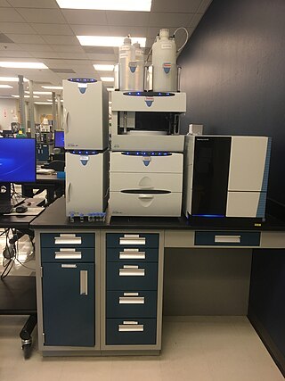
Ion chromatography is a form of chromatography that separates ions and ionizable polar molecules based on their affinity to the ion exchanger. It works on almost any kind of charged molecule—including small inorganic anions, large proteins, small nucleotides, and amino acids. However, ion chromatography must be done in conditions that are one pH unit away from the isoelectric point of a protein.

The French pressure cell press, or French press, is an apparatus used in biological experimentation to disrupt the plasma membrane of cells by passing them through a narrow valve under high pressure. The French press can also be used for disintegration of chloroplasts, homogenates of animal tissue, and other biological particles. It is capable of disrupting cell walls while leaving the cell nucleus undisturbed. The French press was invented by Charles Stacy French of the Carnegie Institution of Washington. The press uses an external hydraulic pump to drive a piston within a larger cylinder that contains the liquid sample. The highly pressurized sample is then squeezed past a needle valve. As the sample passes through the valve, the fluid experiences shear stress and decompression, causing cellular disruption. The major components of a French press are made of stainless steel to prevent sample contamination.

A plasmid preparation is a method of DNA extraction and purification for plasmid DNA. It is an important step in many molecular biology experiments and is essential for the successful use of plasmids in research and biotechnology. Many methods have been developed to purify plasmid DNA from bacteria. During the purification procedure, the plasmid DNA is often separated from contaminating proteins and genomic DNA.

Immunocytochemistry (ICC) is a common laboratory technique that is used to anatomically visualize the localization of a specific protein or antigen in cells by use of a specific primary antibody that binds to it. The primary antibody allows visualization of the protein under a fluorescence microscope when it is bound by a secondary antibody that has a conjugated fluorophore. ICC allows researchers to evaluate whether or not cells in a particular sample express the antigen in question. In cases where an immunopositive signal is found, ICC also allows researchers to determine which sub-cellular compartments are expressing the antigen.

Cryogenic grinding, also known as freezer milling, freezer grinding, and cryomilling, is the act of cooling or chilling a material and then reducing it into a small particle size. For example, thermoplastics are difficult to grind to small particle sizes at ambient temperatures because they soften, adhere in lumpy masses and clog screens. When chilled by dry ice, liquid carbon dioxide or liquid nitrogen, the thermoplastics can be finely ground to powders suitable for electrostatic spraying and other powder processes. Cryogenic grinding of plant and animal tissue is a technique used by microbiologists. Samples that require extraction of nucleic acids must be kept at −80 °C or lower during the entire extraction process. For samples that are soft or flexible at room temperature, cryogenic grinding may be the only viable technique for processing samples. A number of recent studies report on the processing and behavior of nanostructured materials via cryomilling.
Electromanipulation is a micro-material analyzing method mostly used for manipulations of biological cells that uses properties of diverse electric fields. In nanotechnology, nanomaterials are so small that they can hardly be directly mechanically manipulated. Hence, electric fields are applied to them to make field-induced movements or deformations. It is a recently developed technology and is still in progress of widening applications. Types of Electronmanipulation includes dielectrophoresis, electro-rotation, electro-deformation, electro-disruption, electro-destruction, electroporation, and electro-fusion. Diverse electromanipulations are achieved using various electric fields including AC(alternating current), DC(direct current), and pulsed(deliver high-energy discharges at very short periods) electrical fields. Electromanipulation of cells permits diverse cell manipulations with minimal mechanical contact between cells and device structures. Although predominantly used in cells, elctromanipulation also contributes to other scientific fields such as Hybridoma technology and nanoelectronic devices development.

A homogenizer is a piece of laboratory or industrial equipment used for the homogenization of various types of material, such as tissue, plant, food, soil, and many others. Many different models have been developed using various physical technologies for disruption. The mortar and pestle, already used for thousands of years, is a standard tool even in modern laboratories. More modern solutions are based on blender type instruments, bead mills, ultrasonic treatment, rotor-stator mechanical, high pressure, and many other physical forces. While there are many application overlaps between methods, each homogenization method has distinct advantages and disadvantages.

Spin column-based nucleic acid purification is a solid phase extraction method to quickly purify nucleic acids. This method relies on the fact that nucleic acid will bind to the solid phase of silica under certain conditions.
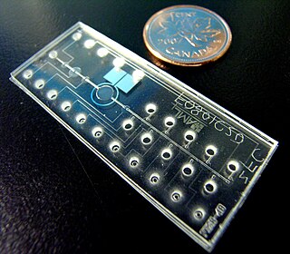
Bio-MEMS is an abbreviation for biomedical microelectromechanical systems. Bio-MEMS have considerable overlap, and is sometimes considered synonymous, with lab-on-a-chip (LOC) and micro total analysis systems (μTAS). Bio-MEMS is typically more focused on mechanical parts and microfabrication technologies made suitable for biological applications. On the other hand, lab-on-a-chip is concerned with miniaturization and integration of laboratory processes and experiments into single chips. In this definition, lab-on-a-chip devices do not strictly have biological applications, although most do or are amenable to be adapted for biological purposes. Similarly, micro total analysis systems may not have biological applications in mind, and are usually dedicated to chemical analysis. A broad definition for bio-MEMS can be used to refer to the science and technology of operating at the microscale for biological and biomedical applications, which may or may not include any electronic or mechanical functions. The interdisciplinary nature of bio-MEMS combines material sciences, clinical sciences, medicine, surgery, electrical engineering, mechanical engineering, optical engineering, chemical engineering, and biomedical engineering. Some of its major applications include genomics, proteomics, molecular diagnostics, point-of-care diagnostics, tissue engineering, single cell analysis and implantable microdevices.
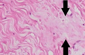
Homogenization, in cell biology or molecular biology, is a process whereby different fractions of a biological sample become equal in composition. It can be a disease sign in histopathology, or an intentional process in research: A homogenized sample is equal in composition throughout, so that removing a fraction does not alter the overall molecular make-up of the sample remaining, and is identical to the fraction removed. Induced homogenization in biology is often followed by molecular extraction and various analytical techniques, including ELISA and western blot.
Single cell oil, also known as Microbial oil consists of the intracellular storage lipids, triacyglycerols. It is similar to vegetable oil, another biologically produced oil. They are produced by oleaginous microorganisms, which is the term for those bacteria, molds, algae and yeast, which can accumulate 20% to 80% lipids of their biomass. The accumulation of lipids take place by the end of logarithmic phase and continues during station phase until carbon source begins to reduce with nutrition limitation.
This glossary of cellular and molecular biology is a list of definitions of terms and concepts commonly used in the study of cell biology, molecular biology, and related disciplines, including genetics, biochemistry, and microbiology. It is split across two articles: