Related Research Articles

An eyelid is a thin fold of skin that covers and protects an eye. The levator palpebrae superioris muscle retracts the eyelid, exposing the cornea to the outside, giving vision. This can be either voluntarily or involuntarily. The human eyelid features a row of eyelashes along the eyelid margin, which serve to heighten the protection of the eye from dust and foreign debris, as well as from perspiration. "Palpebral" means relating to the eyelids. Its key function is to regularly spread the tears and other secretions on the eye surface to keep it moist, since the cornea must be continuously moist. They keep the eyes from drying out when asleep. Moreover, the blink reflex protects the eye from foreign bodies.

Dry eye syndrome (DES), also known as keratoconjunctivitis sicca (KCS), is the condition of having dry eyes. Other associated symptoms include irritation, redness, discharge, and easily fatigued eyes. Blurred vision may also occur. Symptoms range from mild and occasional to severe and continuous. Scarring of the cornea may occur in untreated cases.

A chalazion or meibomian cyst is a cyst in the eyelid usually due to a blocked meibomian gland, typically in the middle of the eyelid, red, and not painful. They tend to come on gradually over a few weeks.

The lacrimal glands are paired exocrine glands, one for each eye, found in most terrestrial vertebrates and some marine mammals, that secrete the aqueous layer of the tear film. In humans, they are situated in the upper lateral region of each orbit, in the lacrimal fossa of the orbit formed by the frontal bone. Inflammation of the lacrimal glands is called dacryoadenitis. The lacrimal gland produces tears which are secreted by the lacrimal ducts, and flow over the ocular surface, and then into canals that connect to the lacrimal sac. From that sac, the tears drain through the lacrimal duct into the nose.
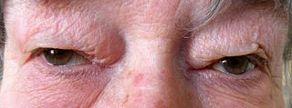
Dermatochalasis is a medical condition, defined as an excess of skin in the upper or lower eyelid, also known as "baggy eyes." It may be either an acquired or a congenital condition. It is generally treated with blepharoplasty.

Exophthalmos is a bulging of the eye anteriorly out of the orbit. Exophthalmos can be either bilateral or unilateral. Complete or partial dislocation from the orbit is also possible from trauma or swelling of surrounding tissue resulting from trauma.

The ophthalmic nerve (V1) is a sensory nerve of the face. It is one of three divisions of the trigeminal nerve (CN V). It has three branches that provide sensory innervation to the eye, the skin of the upper face, and the skin of the anterior scalp.

The tarsi are two comparatively thick, elongated plates of dense connective tissue, about 10 mm (0.39 in) in length for the upper eyelid and 5 mm for the lower eyelid; one is found in each eyelid, and contributes to its form and support. They are located directly above the lid margins. The tarsus has a lower and upper part making up the palpebrae.

The lacrimal nerve is the smallest branch of the ophthalmic nerve (V1), itself a branch of the trigeminal nerve (CN V). The other branches of the ophthalmic nerve are the frontal nerve and nasociliary nerve.

The lacrimal artery is an artery of the orbit around the eye. It arises close to the optic foramen, and is a branch of the ophthalmic artery. It accompanies the lacrimal nerve along the upper border of the lateral rectus muscle. It supplies the lacrimal gland, two rectus muscles of the eye, the eyelids, and the conjunctiva.
The lacrimal apparatus is the physiological system containing the orbital structures for tear production and drainage.
It consists of:
The lacrimal canaliculi,, are the small channels in each eyelid that drain lacrimal fluid, from the lacrimal puncta to the lacrimal sac. This forms part of the lacrimal apparatus that drains lacrimal fluid from the surface of the eye to the nasal cavity.
The superior transverse ligament of the eye is a transverse ligament surrounding the levator palpebrae superioris muscle close to its partial implantation into the skin of the upper eyelid. The muscle also attaches to the superior tarsal plate and into orbital bone.
The medial palpebral ligament is a ligament of the face. It attaches to the frontal process of the maxilla, the lacrimal groove, and the tarsus of each eyelid. It has a superficial (anterior) and a deep (posterior) layer, with many surrounding attachments. It connects the medial canthus of each eyelid to the medial part of the orbit. It is a useful point of fixation during eyelid reconstructive surgery.

Tenon's capsule, also known as the Tenon capsule, fascial sheath of the eyeball or the fascia bulbi, is a thin membrane which envelops the eyeball from the optic nerve to the corneal limbus, separating it from the orbital fat and forming a socket in which it moves.

The lacrimal caruncle, or caruncula lacrimalis, is the small, pink, globular nodule at the inner corner of the eye. It consists of tissue types of neighbouring eye structures. It may suffer from lesions and allergic inflammation.
Krause's glands are small, mucous accessory lacrimal glands that are found underneath the eyelid where the upper and lower conjunctivae meet. Their ducts unite into a rather long sinus which open into the fornix conjunctiva. There are approximately forty Krause glands in the region of the upper eyelid, and around 6 to 8 in the region of the lower lid. The function of these glands are to produce tears which are secreted onto the surface of the conjunctiva.

Hidrocystoma is an adenoma of the sweat glands.
The accessory visual structures are the protecting and supporting structures (adnexa) of the eye, including the eyebrow, eyelids, and lacrimal apparatus. The eyebrows, eyelids, eyelashes, lacrimal gland and drainage apparatus all play a crucial role with regards to globe protection, lubrication, and minimizing the risk of ocular infection. The adnexal structures also help to keep the cornea moist and clean.

Krause's glands, Wolfring's glands and Popov's gland are the accessory lacrimal glands of the lacrimal system of human eye. These glands are structurally and histologically similar to the main lacrimal gland. Glands of Krause are located in the stroma of the conjunctival fornix, and the glands of Wolfring are located along the orbital border of the tarsal plate. These glands are oval and display numerous acini. The acini are surrounded, sometimes incompletely, by a row of myoepithelial cells. Animal studies suggest that the ducts of Wolfring glands have a tortuous course and open onto the palpebral conjunctiva. Like the main lacrimal gland, the accessory lacrimal glands are also densely innervated, but they lack parasympathetic innervation. These glands are exocrine glands, responsible for the basal (unstimulated) secretion of the middle aqueous layer of the tear film. 20 to 40 glands of Krause are found in the upper fornix, and 6-8 glands appear in the lower fornix. There are usually 2 to 5 Ciaccio's glands, and are found along the superior tarsal border of the upper eye lid. Popov’s glands are located within the substance of the caruncle.
References
- ↑ Presutti, Livio; Mattiolu, Francesco (2015). Endoscopic Surgery of the Lacrimal Drainage System. Springer. p. 4. ISBN 978-3319206332.
- ↑ Hall, Nikki; Peden, Robert (2016). FRC Ophth Part 1: 400 SBAs and CRQs. JP Medical Ltd. p. 121. ISBN 9781909836365.