Colloidal probe technique

Last updated
Scheme of the colloidal probe technique for direct force measurements in the sphere-plane and sphere-sphere geometries. Colloidal Probe Geometries.png
Scheme of the colloidal probe technique for direct force measurements in the sphere-plane and sphere-sphere geometries.

The colloidal probe technique is commonly used to measure interaction forces acting between colloidal particles and/or planar surfaces in air or in solution. This technique relies on the use of an atomic force microscope (AFM). However, instead of a cantilever with a sharp AFM tip, one uses the colloidal probe. The colloidal probe consists of a colloidal particle of few micrometers in diameter that is attached to an AFM cantilever. The colloidal probe technique can be used in the sphere-plane or sphere-sphere geometries (see figure). One typically achieves a force resolution between 1 and 100 pN and a distance resolution between 0.5 and 2 nm.

Contents

The colloidal probe technique has been developed in 1991 independently by Ducker [1] and Butt. [2] Since its development this tool has gained wide popularity in numerous research laboratories, and numerous reviews are available in the scientific literature. [3] [4] [5]

Alternative techniques to measure force between surfaces involve the surface forces apparatus, total internal reflection microscopy, and optical tweezers techniques to with video microscopy.

Purpose

The possibility to measure forces involving particles and surfaces directly is essential since such forces are relevant in a variety of processes involving colloidal and polymeric systems. Examples include particle aggregation, suspension rheology, particle deposition, and adhesion processes. One can equally study similar biological phenomena, such as deposition of bacteria or the infection of cells by viruses. Forces are equally most informative to investigate the mechanical properties of interfaces, bubbles, capsules, membranes, or cell walls. Such measurements permit to make conclusions about the elastic or plastic deformation or eventual rupture in such systems.

The colloidal probe technique provides a versatile tool to measure such forces between a colloidal particle and a planar substrate or between two colloidal particles (see figure above). The particles used in such experiments have typically a diameter between 1–10 μm. Typical applications involve measurements of electrical double layer forces and the corresponding surface potentials or surface charge, van der Waals forces, or forces induced by adsorbed polymers. [3] [5] [6]

Principle

The colloidal probe technique uses a standard AFM for the force measurements. But instead the AFM cantilever with an attached sharp tip one uses the colloidal probe. This colloidal probe is normally obtained by attaching a colloidal particle to a cantilever. By recording the deflection of the cantilever as a function of the vertical displacement of the AFM scanner one can extract the force acting between the probe and the surface as a function of the surface separation. This type of AFM operation is referred to as the force mode. With this probe, one can study interactions between various surfaces and probe particles in the sphere-plane geometry. It is also possible to study forces between colloidal particles by attaching another particle to the substrate and perform the measurement in the sphere-sphere geometry, see figure above.

Principle of the force measurements by the colloidal probe technique. Illustration of the scanner displacement D, cantilever deflection x, and lever signal S. Colloidal Probe Force Measurement.png
Principle of the force measurements by the colloidal probe technique. Illustration of the scanner displacement D, cantilever deflection ξ, and lever signal S.

The force mode used in the colloidal probe technique is illustrated in the figure on the left. The scanner is fabricated from piezoelectric crystals, which enable its positioning with a precision better than 0.1 nm. The scanner is lifted towards the probe and thereby one records the scanner displacement D. At the same time, the deflection of the cantilever ξ is monitored as well, typically with a comparable precision. One measures the deflection by focusing a light beam originating from a non-coherent laser diode to the back of the cantilever and detecting the reflected beam with a split photodiode. The lever signal S represents the difference in the photocurrents originating from the two halves of the diode. The lever signal is therefore proportional to the deflection ξ.

During an approach-retraction cycle, one records the lever signal S as a function of the vertical displacement D of the scanner. Suppose for the moment that the probe and the substrate are hard and non-deformable objects and that no forces are acting between them when they are not in contact. In such a situation, one refers to a hard-core repulsion. The cantilever will thus not deform as long not being in contact with the substrate. When the cantilever touches the substrate, its deflection will be the same as the displacement of the substrate. This response is referred to as the constant compliance or contact region. The lever signal S as a function of the scanner displacement D is shown in the figure below. This graph consists of two straight lines resembling a hockey-stick. When the surfaces are not in contact, the lever signal will be denoted as S0. This value corresponds to the non-deformed lever. In the constant compliance region, the lever signal is simply a linear function of the displacement, and can be represented as a straight line

S = aD + b

The parameters a and b can be obtained from a least-squares fit of the constant compliance region. The inverse slope a−1 is also referred to as the optical lever sensitivity. By inverting this relation for the lever signal S0, which corresponds to the non-deformed lever, one can accurately obtain the contact point from D0 = (S0b)/a. Depending on the substrate, the precision in determining this contact point is between 0.5–2 nm. In the constant compliance region, the lever deformation is given by

ξ = (SS0)/a

In this fashion, one can detect deflections of the cantilever with typical resolution of better than 0.1 nm.

Response of the colloidal probe to different force profiles. The forces profiles are shown in top row, and the cantilever response in the bottom. From left to right: Hard-core repulsion, soft repulsion, and attraction. Colloidal Probe Response Forces.png
Response of the colloidal probe to different force profiles. The forces profiles are shown in top row, and the cantilever response in the bottom. From left to right: Hard-core repulsion, soft repulsion, and attraction.

Let us now consider the relevant situation where the probe and the substrate interact. Let us denote by F(h) the force between the probe and the substrate. This force depends on the surface separation h. In equilibrium, this force is compensated by the restoring force of the spring, which is given by the Hooke's law

F = kξ

where k is the spring constant of the cantilever. Typical spring constants of AFM cantilevers are in the range of 0.1−10 N/m. Since the deflection is monitored with a precision better 0.1 nm, one typically obtains a force resolution of 1−100 pN. The separation distance can be obtained from the displacement of the scanner and the cantilever deflection

h = ξ + DD0

Figure below illustrates how the cantilever responds to different force profiles. In the case of a soft repulsive force, the cantilever is repelled from the surface and only slowly approaches the constant compliance region. In such situations, it might be actually difficult to identify this region correctly. When the force is attractive, the cantilever is attracted to the surface and may become unstable. From stability considerations one finds that the cantilever will be unstable provided

dF/dh > k

This instability is illustrated in the right panel of the figure on the right. As the cantilever approaches, the slope of the force curve increases. When the slope becomes larger than the spring constant of the cantilever, the cantilever jumps into contact when the slope of the force curve exceeds the force constant of the cantilever. Upon retraction, the same phenomenon happens, but the point where the cantilever jumps out is reached at a smaller separation. Upon approach and retraction, the system will show a hysteresis. In such situations, a part of the force profile cannot be probed. However, this problem can be avoided by using a stiffer cantilever, albeit at the expense of an inferior force resolution.

Extensions

The colloidal probes are normally fabricated by gluing a colloidal particle to a tip-less cantilever with a micromanipulator in air. The subsequent rewetting of the probe may lead to the formation of nanosized bubbles on the probe surface. This problem can be avoided by attaching the colloidal particles under wet conditions in AFM fluid cell to appropriately functionalized cantilevers. [5] While the colloidal probe technique is mostly used in the sphere-plane geometry, it can be also used in the sphere-sphere geometry. [6] The latter geometry further requires a lateral centering of the two particles, which can be either achieved with an optical microscope or an AFM scan. The results obtained in these two different geometries can be related with the Derjaguin approximation.

The force measurements rely on an accurate value of the spring constant of the cantilever. This spring constant can be measured by different techniques. [3] [4] The thermal noise method is the simplest to use, as it is implemented on most AFMs. This approach relies on the determination of the mean square amplitude of the cantilever displacement due to spontaneous thermal fluctuations. This quantity is related to the spring constant by means of the equipartition theorem. In the added mass method one attaches a series of metal beads to the cantilever and each case one determines the resonance frequency. By exploiting the relation for a harmonic oscillator between the resonance frequency and the mass added one can evaluate the spring constant as well. The frictional force method relies on measurement of the approach and retract curves of the cantilever through a viscous fluid. Since the hydrodynamic drag of a sphere close to a planar substrate is known theoretically, the spring constant of the cantilever can be deduced. The geometrical method exploits relations between the geometry of the cantilever and its elastic properties.

The separation is normally measured from the onset of the constant compliance region. While the relative surface separation can be determined with a resolution of 0.1 nm or better, the absolute surface separation is obtained from the onset of the constant compliance region. While this onset can be determined for solid samples with a precision between 0.5–2 nm, the location of this onset can be problematic for soft repulsive interactions and for deformable surfaces. For this reason, techniques have been developed to measure the surface separation independently (e.g., total internal reflection microscopy, reflection interference contrast microscopy). [7]

By scanning the sample with the colloidal probe laterally permits to exploit friction forces between the probe and the substrate. [4] Since this technique exploits the torsion of the cantilever, to obtain quantitative data the torsional spring constant of the cantilever must be determined.

A related technique involving similar type of force measurements with the AFM is the single molecular force spectroscopy. However, this technique uses a regular AFM tip to which a single polymer molecule is attached. From the retraction part of the force curve, one can obtain information about stretching of the polymer or its peeling from the surface.

See also

Related Research Articles

Force spectroscopy is a set of techniques for the study of the interactions and the binding forces between individual molecules. These methods can be used to measure the mechanical properties of single polymer molecules or proteins, or individual chemical bonds. The name "force spectroscopy", although widely used in the scientific community, is somewhat misleading, because there is no true matter-radiation interaction.

<span class="mw-page-title-main">Atomic force microscopy</span> Type of microscopy

Atomic force microscopy (AFM) or scanning force microscopy (SFM) is a very-high-resolution type of scanning probe microscopy (SPM), with demonstrated resolution on the order of fractions of a nanometer, more than 1000 times better than the optical diffraction limit.

<span class="mw-page-title-main">Surface forces apparatus</span>

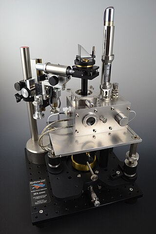
The Surface Force Apparatus (SFA) is a scientific instrument which measures the interaction force of two surfaces as they are brought together and retracted using multiple beam interferometry to monitor surface separation and directly measure contact area and observe any surface deformations occurring in the contact zone. One surface is held by a cantilevered spring, and the deflection of the spring is used to calculate the force being exerted. The technique was pioneered by David Tabor and R.H.S. Winterton in the late 1960s at Cambridge University. By the mid-1970s, J.N. Israelachvili had adapted the original design to operate in liquids, notably aqueous solutions, while at the Australian National University, and further advanced the technique to support friction and electro-chemical surface studies while at the University of California Santa Barbara.

<span class="mw-page-title-main">Magnetic force microscope</span>

Magnetic force microscopy (MFM) is a variety of atomic force microscopy, in which a sharp magnetized tip scans a magnetic sample; the tip-sample magnetic interactions are detected and used to reconstruct the magnetic structure of the sample surface. Many kinds of magnetic interactions are measured by MFM, including magnetic dipole–dipole interaction. MFM scanning often uses non-contact AFM (NC-AFM) mode.

<span class="mw-page-title-main">Kelvin probe force microscope</span> Noncontact variant of atomic force microscopy

Kelvin probe force microscopy (KPFM), also known as surface potential microscopy, is a noncontact variant of atomic force microscopy (AFM). By raster scanning in the x,y plane the work function of the sample can be locally mapped for correlation with sample features. When there is little or no magnification, this approach can be described as using a scanning Kelvin probe (SKP). These techniques are predominantly used to measure corrosion and coatings.

Nanotribology is the branch of tribology that studies friction, wear, adhesion and lubrication phenomena at the nanoscale, where atomic interactions and quantum effects are not negligible. The aim of this discipline is characterizing and modifying surfaces for both scientific and technological purposes.

Electrostatic force microscopy (EFM) is a type of dynamic non-contact atomic force microscopy where the electrostatic force is probed.. This force arises due to the attraction or repulsion of separated charges. It is a long-range force and can be detected 100 nm or more from the sample.

<span class="mw-page-title-main">Nanometrology</span> Metrology of nanomaterials

Nanometrology is a subfield of metrology, concerned with the science of measurement at the nanoscale level. Nanometrology has a crucial role in order to produce nanomaterials and devices with a high degree of accuracy and reliability in nanomanufacturing.

<span class="mw-page-title-main">Chemical force microscopy</span> Method of microscopy which measures chemical bonding between the probe and surface

In materials science, chemical force microscopy (CFM) is a variation of atomic force microscopy (AFM) which has become a versatile tool for characterization of materials surfaces. With AFM, structural morphology is probed using simple tapping or contact modes that utilize van der Waals interactions between tip and sample to maintain a constant probe deflection amplitude or maintain height while measuring tip deflection. CFM, on the other hand, uses chemical interactions between functionalized probe tip and sample. Choice chemistry is typically gold-coated tip and surface with R−SH thiols attached, R being the functional groups of interest. CFM enables the ability to determine the chemical nature of surfaces, irrespective of their specific morphology, and facilitates studies of basic chemical bonding enthalpy and surface energy. Typically, CFM is limited by thermal vibrations within the cantilever holding the probe. This limits force measurement resolution to ~1 pN which is still very suitable considering weak COOH/CH3 interactions are ~20 pN per pair. Hydrophobicity is used as the primary example throughout this consideration of CFM, but certainly any type of bonding can be probed with this method.

<span class="mw-page-title-main">Total internal reflection microscopy</span>

Total internal reflection microscopy is a specialized optical imaging technique for object tracking and detection utilizing the light scattered from an evanescent field in the vicinity of a dielectric interface. Its advantages are a high signal-to-noise ratio and a high spatial resolution in the vertical dimension.

<span class="mw-page-title-main">Piezoresponse force microscopy</span> Microscopy technique for piezoelectric materials

Piezoresponse force microscopy (PFM) is a variant of atomic force microscopy (AFM) that allows imaging and manipulation of piezoelectric/ferroelectric materials domains. This is achieved by bringing a sharp conductive probe into contact with a ferroelectric surface and applying an alternating current (AC) bias to the probe tip in order to excite deformation of the sample through the converse piezoelectric effect (CPE). The resulting deflection of the probe cantilever is detected through standard split photodiode detector methods and then demodulated by use of a lock-in amplifier (LiA). In this way topography and ferroelectric domains can be imaged simultaneously with high resolution.

In microscopy, scanning joule expansion microscopy (SJEM) is a form of scanning probe microscopy heavily based on atomic force microscopy (AFM) that maps the temperature distribution along a surface. Resolutions down to 10 nm have been achieved and 1 nm resolution is theoretically possible. Thermal measurements at the nanometer scale are of both academic and industrial interest, particularly in regards to nanomaterials and modern integrated circuits.

<span class="mw-page-title-main">Photoconductive atomic force microscopy</span> Type of atomic force microscopy

Photoconductive atomic force microscopy (PC-AFM) is a variant of atomic force microscopy that measures photoconductivity in addition to surface forces.

The technique of vibrational analysis with scanning probe microscopy allows probing vibrational properties of materials at the submicrometer scale, and even of individual molecules. This is accomplished by integrating scanning probe microscopy (SPM) and vibrational spectroscopy. This combination allows for much higher spatial resolution than can be achieved with conventional Raman/FTIR instrumentation. The technique is also nondestructive, requires non-extensive sample preparation, and provides more contrast such as intensity contrast, polarization contrast and wavelength contrast, as well as providing specific chemical information and topography images simultaneously.

<span class="mw-page-title-main">Derjaguin approximation</span>

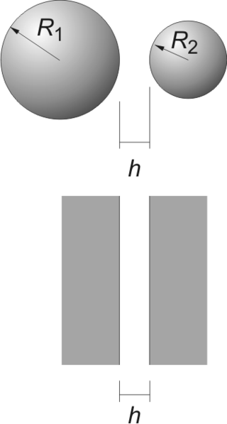
The Derjaguin approximation (or sometimes also referred to as the proximity approximation), named after the Russian scientist Boris Derjaguin, expresses the force profile acting between finite size bodies in terms of the force profile between two planar semi-infinite walls. This approximation is widely used to estimate forces between colloidal particles, as forces between two planar bodies are often much easier to calculate. The Derjaguin approximation expresses the force F(h) between two bodies as a function of the surface separation as

A depletion force is an effective attractive force that arises between large colloidal particles that are suspended in a dilute solution of depletants, which are smaller solutes that are preferentially excluded from the vicinity of the large particles. One of the earliest reports of depletion forces that lead to particle coagulation is that of Bondy, who observed the separation or "creaming" of rubber latex upon addition of polymer depletant molecules to solution. More generally, depletants can include polymers, micelles, osmolytes, ink, mud, or paint dispersed in a continuous phase.

<span class="mw-page-title-main">Non-contact atomic force microscopy</span>

Non-contact atomic force microscopy (nc-AFM), also known as dynamic force microscopy (DFM), is a mode of atomic force microscopy, which itself is a type of scanning probe microscopy. In nc-AFM a sharp probe is moved close to the surface under study, the probe is then raster scanned across the surface, the image is then constructed from the force interactions during the scan. The probe is connected to a resonator, usually a silicon cantilever or a quartz crystal resonator. During measurements the sensor is driven so that it oscillates. The force interactions are measured either by measuring the change in amplitude of the oscillation at a constant frequency just off resonance or by measuring the change in resonant frequency directly using a feedback circuit to always drive the sensor on resonance.

<span class="mw-page-title-main">Infrared Nanospectroscopy (AFM-IR)</span> Infrared microscopy technique

AFM-IR or infrared nanospectroscopy is one of a family of techniques that are derived from a combination of two parent instrumental techniques. AFM-IR combines the chemical analysis power of infrared spectroscopy and the high-spatial resolution of scanning probe microscopy (SPM). The term was first used to denote a method that combined a tuneable free electron laser with an atomic force microscope equipped with a sharp probe that measured the local absorption of infrared light by a sample with nanoscale spatial resolution.

The operation of a photon scanning tunneling microscope (PSTM) is analogous to the operation of an electron scanning tunneling microscope, with the primary distinction being that PSTM involves tunneling of photons instead of electrons from the sample surface to the probe tip. A beam of light is focused on a prism at an angle greater than the critical angle of the refractive medium in order to induce total internal reflection within the prism. Although the beam of light is not propagated through the surface of the refractive prism under total internal reflection, an evanescent field of light is still present at the surface.

A probe tip is an instrument used in scanning probe microscopes (SPMs) to scan the surface of a sample and make nano-scale images of surfaces and structures. The probe tip is mounted on the end of a cantilever and can be as sharp as a single atom. In microscopy, probe tip geometry and the composition of both the tip and the surface being probed directly affect resolution and imaging quality. Tip size and shape are extremely important in monitoring and detecting interactions between surfaces. SPMs can precisely measure electrostatic forces, magnetic forces, chemical bonding, Van der Waals forces, and capillary forces. SPMs can also reveal the morphology and topography of a surface.

References

  1. Ducker, William A.; Senden, Tim J.; Pashley, Richard M. (1991). "Direct measurement of colloidal forces using an atomic force microscope". Nature. 353 (6341): 239–241. Bibcode:1991Natur.353..239D. doi:10.1038/353239a0. ISSN   0028-0836. S2CID   4311419..
  2. Butt, Hans-Jürgen (1991). "Measuring electrostatic, van der Waals, and hydration forces in electrolyte solutions with an atomic force microscope". Biophysical Journal. 60 (6): 1438–1444. Bibcode:1991BpJ....60.1438B. doi:10.1016/S0006-3495(91)82180-4. ISSN   0006-3495. PMC   1260203 . PMID   19431815.
  3. 1 2 3 Butt, Hans-Jürgen; Cappella, Brunero; Kappl, Michael (2005). "Force measurements with the atomic force microscope: Technique, interpretation and applications". Surface Science Reports. 59 (1–6): 1–152. Bibcode:2005SurSR..59....1B. doi:10.1016/j.surfrep.2005.08.003. ISSN   0167-5729.
  4. 1 2 3 Ralston, John; Larson, Ian; Rutland, Mark W.; Feiler, Adam A.; Kleijn, Mieke (2005). "Atomic force microscopy and direct surface force measurements (IUPAC Technical Report)". Pure and Applied Chemistry. 77 (12): 2149–2170. doi: 10.1351/pac200577122149 . ISSN   1365-3075.
  5. 1 2 3 Borkovec, Michal; Szilagyi, Istvan; Popa, Ionel; Finessi, Marco; Sinha, Prashant; Maroni, Plinio; Papastavrou, Georg (2012). "Investigating forces between charged particles in the presence of oppositely charged polyelectrolytes with the multi-particle colloidal probe technique". Advances in Colloid and Interface Science. 179–182: 85–98. doi:10.1016/j.cis.2012.06.005. ISSN   0001-8686. PMID   22795487.
  6. 1 2 I. Larson, Ian; Drummond, Calum J.; Chan, Derek Y. C.; Grieser, Franz (1995). "Direct Force Measurements between Dissimilar Metal Oxides". The Journal of Physical Chemistry. 99 (7): 2114–2118. doi:10.1021/j100007a048. ISSN   0022-3654.; Toikka, Gary; Hayes, Robert A.; Ralston, John (1996). "Surface Forces between Spherical ZnS Particles in Aqueous Electrolyte". Langmuir. 12 (16): 3783–3788. doi:10.1021/la951534u. ISSN   0743-7463..
  7. Clark, Spencer C.; Walz, John Y.; Ducker, William A. (2004). "Atomic Force Microscopy Colloid−Probe Measurements with Explicit Measurement of Particle−Solid Separation". Langmuir. 20 (18): 7616–7622. doi:10.1021/la0497752. ISSN   0743-7463. PMID   15323510.