Emmonsia parva

Last updated

Emmonsia parva
Scientific classification
Kingdom:
Division:
Class:
Order:
Family:
Genus:
Species:
Emmonsia parva
Synonyms
  • Chrysosporium parvum(C.W. Emmons & Ashburn) J.W. Carmich.
  • Haplosporangium parvumC.W. Emmons & Ashburn
  • Blastomyces parvus(C.W. Emmons & Ashburn) Y.P. Jiang, Sigler & de Hoog

Emmonsia parva (formerly Chrysosporium parvum) is a filamentous, saprotrophic fungus and one of three species within the genus Emmonsia. [1] [2] The fungus is most known for its causal association with the lung disease, adiaspiromycosis [3] which occurs most commonly in small mammals but is also seen in humans. [2] The disease was first described from rodents in Arizona, [4] and the first human case was reported in France in 1964. [2] Since then, the disease has been reported from Honduras, Brazil, the Czech Republic, Russia, the United States of America and Guatemala. [2] Infections in general are quite rare, especially in humans. [2]

Contents

Ecology

Emmonsia parva, E. crescens, and E. pasteuriana together comprise the genus Emmonsia, [3] however they exhibit different ecological characteristics. Whereas E. crescens is found worldwide, E. parva is restricted to areas in North and South America, Eastern Europe, Australia and regions in Asia. [5] The fungus is primarily a saprotroph, deriving its nutrition from dead material. [6] It is also soil-dwelling where it release spores into the air. [7] Because of this the main targets of infection are small burrowing mammals such as rodents, although infection of larger mammals such as humans has been documented. Some of the known animal species that it can infect include the beaver, mink, weasel, wood rat, pine marten, pine squirrel, cottontail rabbit, muskrat, skunk, white-tailed mouse and the rock rabbit. [8] The fungus is closely related to the genus Blastomyces. [9]

Growth and morphology

The fungus is dimorphic growing in two distinct forms. [2] It grows as hyphae at room temperature, but when conidia are transferred to 40 °C they convert to larger adiaspores. [8] It has no teleomorphs and no sexual stage. [2] It does not have any particular growth requirements in terms of culture media, but it is known to grow well on pablum cereal agar, potato dextrose agar (PDA) and phytone yeast extract agar. [2] [8] They also grow well on Sabouraud dextrose agar at 25 °C. [7] Growth is slightly inhibited when grown in media containing cycloheximide. [10] E. parva grows at a moderate pace, slower than E. crescens. After 21 days of growth at room temperature colony diameters range from 36 to 85 mm. [8] The colonies are smooth and velvety and are white with tan centers from a top view and cream from the bottom. [2] Hyphae in this form are septate and hyaline. [2] The conidiophores they produce are unicellular, thick-walled (2 μm), [6] and usually simple with a single terminal conidium also called an aleurioconidium. [8] The conidiophore is also known to occasionally branch into 1-3 sections each bearing its own conidium. [6] Before differentiating into adiaspores, the conidia measure 2-4 μm [4] in diameter and are shaped either ovoid, subglobose or pyriform with glabrous walls. [8] After growth at 40 °C the conidia morph into their adiaspore form enlarging to approximately 25 μm in vitro and 40 μm in vivo. [2] These adiaspores are uninucleate and they do not replicate. [2] They are occasionally mistaken for spherules of the organism Coccidioides immitis. [2]

Pathogenicity

Transmission

The main route of infection is inhalation of airborne spores through the respiratory pathway. [8] This can occur in both healthy and immunocompromised individuals, however a disseminated infection is more common in the latter. [8] After inhalation the conidia switch to their adiaspore state, triggered by the temperature increase within the body. These develop without replicating in the alveoli of the lung. [2] Transmission can originate directly from the soil or through an animal reservoir such as mice or bats. [2]

Infection

Adiaspiromycosis, caused by E. parva may lead to pulmonary disease. It is termed an infection, but better described as a bodily reaction to foreign material, [3] invoking various cellular processes within the circulatory and immune systems. Once the adiaspore is formed it finds a place to localize in the alveoli and implants somewhere in the tissue in that section. [2] At this site the spores become calcified which causes a slight localized reaction involving inflammation. [2] Lung function may be obstructed at this stage. The body mounts a multicellular immune response to the presence of these adiaspores leading to the formation of noncaseating granulomas. [2]

The onset of adiaspiromycosis is dependent on the level of exposure to conidia. The disease is self-limiting so the onset of symptoms is determined by the amount of conidia inhaled. [8] Low level exposure induces little to no clinical symptoms, while a greater dosage risks development of pulmonary disease. This is referred to as acute pulmonary adiaspiromycosis, primary progressive pulmonary adiaspiromycosis or disseminated pulmonary adiaspiromycosis. It is characterized by widespread lesions caused by granulomas within both lungs. There are 3 forms of manifestation:

The onset of symptoms is dependent on 2 factors: the dosage of conidia [10] and the immune status of the host. [4] Usually infection is asymptomatic with pulmonary disease developing very rarely. [2] Fatality rates are low. If however the level of spores inhaled is sufficient or the immune system of the host is adequately compromised clinical symptoms may develop which include coughing, dyspnea, low-grade fever, weight loss and conjunctivitis including blurred vision, photophobia and ocular pain. [2] [8] Within the sclera, white, opaque nodules develop in conjunction with local edema or hyperemia. This opacification may extend to other distal parts of the eye such as the limbus and angular corneal opacities. [2] Granulomas also form in the anterior chamber. However purulent conjunctival discharge and hemorrhaging were not observed as opposed to conjunctivitis brought on by viral or bacterial infection. [2]

Diagnosis

Adiaspiromycosis is histopathologically diagnosed. Three criteria must be met for accurate diagnosis:

The adiaspores must be identified as E. parva in culture media or sequencing to separate the infection from E. crescens. Periodic acid–Schiff (PAS) stains can be used to observe the thick-walled adiaspores in tissue regions. [2] Sequencing can be used as a tool to discriminate the fungus from its close relative, the genus Blastomyces. [9] The target is the D2 variable domain found in the large subunit of nuclear rRNA at the 5' end. [2] This region has sufficient variability between the two species. To differentiate between separate Emmonsia species the internal transcribed spacer (ITS) can be targeted. [2] Also the size of the spores varies considerably with E. parva having smaller ones.

Treatment

The onset of adiaspiromycosis is rarely serious and is self-limiting [4] so often no treatment is required and the body clears the spores on its own. Fluconazole (FLC), AmB, and ketoconazole [4] [8] have proven to be therapeutic in progressive or serious infection. Oral intraconazole (ITC) is also an effective aid. Surgical resection may be a last resort if fungal infection persists. [8]

Epidemiology

Exposure to soil and dust is a major route of infection so manual labor in regions with high levels of each can lead to an increased risk of inhalation. As a result, adiaspiromycosis is reported more commonly in men then in women. [5] In most cases exposure is asymptomatic. Cases have been reported in immunocompromised individuals such as those diagnosed with AIDS. [5] There has been only one reported case of E. pasteuriana infection and it occurred in an AIDS patient who had disseminated open wounds. [8] Cleaning, working or playing in areas inhabited by small mammals such as mice or bats may increase risk of infection. [2] Farmers, greenhouse workers or those involved in construction are also at a greater risk of exposure. [8]

Related Research Articles

<span class="mw-page-title-main">Histoplasmosis</span> Human disease

Histoplasmosis is a fungal infection caused by Histoplasma capsulatum. Symptoms of this infection vary greatly, but the disease affects primarily the lungs. Occasionally, other organs are affected; called disseminated histoplasmosis, it can be fatal if left untreated.

<i>Talaromyces marneffei</i> Species of fungus

Talaromyces marneffei, formerly called Penicillium marneffei, was identified in 1956. The organism is endemic to southeast Asia where it is an important cause of opportunistic infections in those with HIV/AIDS-related immunodeficiency. Incidence of T. marneffei infections has increased due to a rise in HIV infection rates in the region.

<span class="mw-page-title-main">Blastomycosis</span> Medical condition

Blastomycosis, also known as Gilchrist's disease, is a fungal infection, typically of the lungs, which can spread to brain, stomach, intestine and skin, where it appears as crusting purplish warty plaques with a roundish bumpy edge and central depression. Only about half of people with the disease have symptoms, which can include fever, cough, night sweats, muscle pains, weight loss, chest pain, and feeling tired. Symptoms usually develop between three weeks and three months after breathing in the spores. In 25% to 40% of cases, the infection also spreads to other parts of the body, such as the skin, bones or central nervous system. Although blastomycosis is especially dangerous for those with weak immune systems, most people diagnosed with blastomycosis have healthy immune systems.

<span class="mw-page-title-main">Conidium</span> Asexual, non-motile spore of a fungus

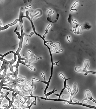
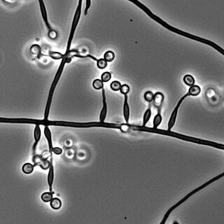
A conidium, sometimes termed an asexual chlamydospore or chlamydoconidium, is an asexual, non-motile spore of a fungus. The word conidium comes from the Ancient Greek word for dust, κόνις (kónis). They are also called mitospores due to the way they are generated through the cellular process of mitosis. They are produced exogenously. The two new haploid cells are genetically identical to the haploid parent, and can develop into new organisms if conditions are favorable, and serve in biological dispersal.

<span class="mw-page-title-main">Hypersensitivity pneumonitis</span> Medical condition

Hypersensitivity pneumonitis (HP) or extrinsic allergic alveolitis (EAA) is a syndrome caused by the repetitive inhalation of antigens from the environment in susceptible or sensitized people. Common antigens include molds, bacteria, bird droppings, bird feathers, agricultural dusts, bioaerosols and chemicals from paints or plastics. People affected by this type of lung inflammation (pneumonitis) are commonly exposed to the antigens by their occupations, hobbies, the environment and animals. The inhaled antigens produce a hypersensitivity immune reaction causing inflammation of the airspaces (alveoli) and small airways (bronchioles) within the lung. Hypersensitivity pneumonitis may eventually lead to interstitial lung disease.

<span class="mw-page-title-main">Pneumonitis</span> General inflammation of lung tissue

Pneumonitis describes general inflammation of lung tissue. Possible causative agents include radiation therapy of the chest, exposure to medications used during chemo-therapy, the inhalation of debris, aspiration, herbicides or fluorocarbons and some systemic diseases. If unresolved, continued inflammation can result in irreparable damage such as pulmonary fibrosis.

<span class="mw-page-title-main">Aspergilloma</span> Medical condition

An aspergilloma is a clump of mold which exists in a body cavity such as a paranasal sinus or an organ such as the lung. By definition, it is caused by fungi of the genus Aspergillus.

<span class="mw-page-title-main">Sporotrichosis</span> Medical condition

Sporotrichosis, also known as rose handler's disease, is a fungal infection that may be localised to skin, lungs, bone and joint, or become systemic. It presents with firm painless nodules that later ulcerate. Following initial exposure to Sporothrix schenckii, the disease typically progresses over a period of a week to several months. Serious complications may develop in people who have a weakened immune system.

<span class="mw-page-title-main">Farmer's lung</span> Hypersensitivity pneumonitis

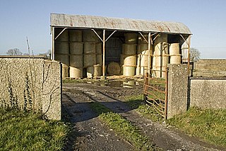
Farmer's lung is a hypersensitivity pneumonitis induced by the inhalation of biologic dusts coming from hay dust or mold spores or any other agricultural products. It results in a type III hypersensitivity inflammatory response and can progress to become a chronic condition which is considered potentially dangerous.

<span class="mw-page-title-main">Aspergillosis</span> Medical condition

Aspergillosis is a fungal infection of usually the lungs, caused by the genus Aspergillus, a common mould that is breathed in frequently from the air around, but does not usually affect most people. It generally occurs in people with lung diseases such as asthma, cystic fibrosis or tuberculosis, or those who have had a stem cell or organ transplant, and those who cannot fight infection because of medications they take such as steroids and some cancer treatments. Rarely, it can affect skin.

<span class="mw-page-title-main">Paracoccidioidomycosis</span> Medical condition

Paracoccidioidomycosis (PCM), also known as South American blastomycosis, is a fungal infection that can occur as a mouth and skin type, lymphangitic type, multi-organ involvement type (particularly lungs), or mixed type. If there are mouth ulcers or skin lesions, the disease is likely to be widespread. There may be no symptoms, or it may present with fever, sepsis, weight loss, large glands, or a large liver and spleen.

<i>Histoplasma capsulatum</i> Species of fungus

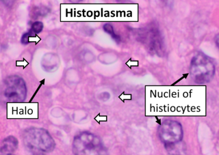
Histoplasma capsulatum is a species of dimorphic fungus. Its sexual form is called Ajellomyces capsulatus. It can cause pulmonary and disseminated histoplasmosis.

<i>Aspergillus terreus</i> Species of fungus

Aspergillus terreus, also known as Aspergillus terrestris, is a fungus (mold) found worldwide in soil. Although thought to be strictly asexual until recently, A. terreus is now known to be capable of sexual reproduction. This saprotrophic fungus is prevalent in warmer climates such as tropical and subtropical regions. Aside from being located in soil, A. terreus has also been found in habitats such as decomposing vegetation and dust. A. terreus is commonly used in industry to produce important organic acids, such as itaconic acid and cis-aconitic acid, as well as enzymes, like xylanase. It was also the initial source for the drug mevinolin (lovastatin), a drug for lowering serum cholesterol.

<i>Blastomyces dermatitidis</i> Species of fungus

Blastomyces dermatitidis is a dimorphic fungus that causes blastomycosis, an invasive and often serious fungal infection found occasionally in humans and other animals. It lives in soil and wet, decaying wood, often in an area close to a waterway such as a lake, river or stream. Indoor growth may also occur, for example, in accumulated debris in damp sheds or shacks. The fungus is endemic to parts of eastern North America, particularly boreal northern Ontario, southeastern Manitoba, Quebec south of the St. Lawrence River, parts of the U.S. Appalachian mountains and interconnected eastern mountain chains, the west bank of Lake Michigan, the state of Wisconsin, and the entire Mississippi Valley including the valleys of some major tributaries such as the Ohio River. In addition, it occurs rarely in Africa both north and south of the Sahara Desert, as well as in the Arabian Peninsula and the Indian subcontinent. Though it has never been directly observed growing in nature, it is thought to grow there as a cottony white mold, similar to the growth seen in artificial culture at 25 °C (77 °F). In an infected human or animal, however, it converts in growth form and becomes a large-celled budding yeast. Blastomycosis is generally readily treatable with systemic antifungal drugs once it is correctly diagnosed; however, delayed diagnosis is very common except in highly endemic areas.

<span class="mw-page-title-main">Chronic pulmonary aspergillosis</span> Fungal infection

Chronic pulmonary aspergillosis is a long-term fungal infection caused by members of the genus Aspergillus—most commonly Aspergillusfumigatus. The term describes several disease presentations with considerable overlap, ranging from an aspergilloma—a clump of Aspergillus mold in the lungs—through to a subacute, invasive form known as chronic necrotizing pulmonary aspergillosis which affects people whose immune system is weakened. Many people affected by chronic pulmonary aspergillosis have an underlying lung disease, most commonly tuberculosis, allergic bronchopulmonary aspergillosis, asthma, or lung cancer.

<i>Ochroconis gallopava</i> Species of fungus

Ochroconis gallopava, also called Dactylaria gallopava or Dactylaria constricta var. gallopava, is a member of genus Dactylaria. Ochroconis gallopava is a thermotolerant, darkly pigmented fungus that causes various infections in fowls, turkeys, poults, and immunocompromised humans first reported in 1986. Since then, the fungus has been increasingly reported as an agent of human disease especially in recipients of solid organ transplants. Ochroconis gallopava infection has a long onset and can involve a variety of body sites. Treatment of infection often involves a combination of antifungal drug therapy and surgical excision.

<i>Penicillium digitatum</i> Species of fungus

Penicillium digitatum is a mesophilic fungus found in the soil of citrus-producing areas. It is a major source of post-harvest decay in fruits and is responsible for the widespread post-harvest disease in Citrus fruit known as green rot or green mould. In nature, this necrotrophic wound pathogen grows in filaments and reproduces asexually through the production of conidiophores and conidia. However, P. digitatum can also be cultivated in the laboratory setting. Alongside its pathogenic life cycle, P. digitatum is also involved in other human, animal and plant interactions and is currently being used in the production of immunologically based mycological detection assays for the food industry.

<i>Aspergillus clavatus</i> Species of fungus

Aspergillus clavatus is a species of fungus in the genus Aspergillus with conidia dimensions 3–4.5 x 2.5–4.5 μm. It is found in soil and animal manure. The fungus was first described scientifically in 1834 by the French mycologist John Baptiste Henri Joseph Desmazières.

<i>Metarhizium granulomatis</i> Species of fungus

Metarhizium granulomatis is a fungus in the family Clavicipitaceae associated with systemic mycosis in veiled chameleons. The genus Metarhizium is known to infect arthropods, and collectively are referred to green-spored asexual pathogenic fungi. This species grows near the roots of plants and has been reported as an agent of disease in captive veiled chameleons. The etymology of the species epithet, "granulomatis" refers to the ability of the fungus to cause granulomatous disease in susceptible reptiles.

Emmonsiosis, also known as emergomycosis, is a systemic fungal infection that can affect the lungs, generally always affects the skin and can become widespread. The lesions in the skin look like small red bumps and patches with a dip, ulcer and dead tissue in the centre.

References

  1. Mcginnis, Michael R. (1980). Laboratory handbook of medical mycology. Elsevier inc. ISBN   9780124828506.
  2. 1 2 3 4 5 6 7 8 9 10 11 12 13 14 15 16 17 18 19 20 21 22 23 24 25 26 27 28 29 Liu, Dongyou (2011). Molecular Detection of Human Fungal Pathogens. Boca Raton, FL: CRC Press. ISBN   9781439812402.
  3. 1 2 3 Berman, Jules (2012). Taxonomic Guide to Infectious Diseases Understanding the Biologic Classes of Pathogenic Organisms (1st ed.). London, UK: Academic Press. ISBN   9780124158955.
  4. 1 2 3 4 5 Stapleton, Philip; Flieder, Douglas (2013). Spencer's Pathology of the Lung (6th ed.). Philadelphia: Cambridge University Press. ISBN   9780521509954.
  5. 1 2 3 Richardson, Malcolm; Warnock, David (2012). Fungal Infection: Diagnosis and Management (4th ed.). New York. NY: Wiley-Blackwell. ISBN   9781405170567.
  6. 1 2 3 Plunkett, Orda; Wilson, Walter (1970). Fungous Diseases of Man (3rd ed.). Los Angeles: University of California Press. ISBN   9780520013445.
  7. 1 2 Carter, G.R.; Carter, M.E.; Markey, Bryan; Quinn, Patrick (1993). Clinical Veterinary Microbiology (1st ed.). Mosby. ISBN   9780723432371.
  8. 1 2 3 4 5 6 7 8 9 10 11 12 13 14 15 16 17 Lyon, G. Marshall; Reiss, Errol; Shadomy, H. Jean (2011). Fundamental Medical Mycology. New York, NY: Wiley-Blackwell. ISBN   9780470177914.
  9. 1 2 Peterson, S. W.; Sigler, L. (October 1998). "Molecular genetic variation in Emmonsia crescens and Emmonsia parva, etiologic agents of adiaspiromycosis, and their phylogenetic relationship to Blastomyces dermatitidis (Ajellomyces dermatitidis) and other systemic fungal pathogens". Journal of Clinical Microbiology. 36 (10): 2918–2925. doi: 10.1128/JCM.36.10.2918-2925.1998 . ISSN   0095-1137. PMC   105088 . PMID   9738044.
  10. 1 2 Howard, Dexter H., ed. (2003). Pathogenic fungi in humans and animals (2nd ed.). New York, NY: Dekker. ISBN   978-0824706838.