FIS1

Last updated
FIS1
Protein FIS1 PDB 1iyg.png
Available structures
PDB Ortholog search: PDBe RCSB
Identifiers
Aliases FIS1 , TTC11, CGI-135, fission, mitochondrial 1
External IDs OMIM: 609003 MGI: 1913687 HomoloGene: 41099 GeneCards: FIS1
Orthologs
SpeciesHumanMouse
Entrez
Ensembl
UniProt
RefSeq (mRNA)

NM_016068

NM_001163243
NM_025562
NM_001347504

RefSeq (protein)

NP_057152

NP_001156715
NP_001334433
NP_079838

Location (UCSC) Chr 7: 101.24 – 101.25 Mb Chr 5: 136.98 – 137 Mb
PubMed search [3] [4]
Wikidata
View/Edit Human View/Edit Mouse

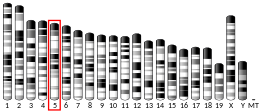
Mitochondrial fission 1 protein (FIS1) is a protein that in humans is encoded by the FIS1 gene on chromosome 7. [5] [6] [7] This protein is a component of a mitochondrial complex, the ARCosome, that promotes mitochondrial fission. [7] [8] Its role in mitochondrial fission thus implicates it in the regulation of mitochondrial morphology, the cell cycle, and apoptosis. [7] [8] [9] [10] By extension, the protein is involved in associated diseases, including neurodegenerative diseases and cancers. [11] [12]

Contents

Structure

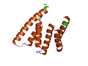
The protein encoded by this gene is a 16 kDa integral protein situated in the outer mitochondrial membrane (OMM). [9] It is composed of a transmembrane domain at the C-terminal and a cytosolic domain at the N-terminal. [9] [13] [14] The transmembrane domain anchors FIS1 in the OMM, though it has been observed to target different cellular compartments, such as the peroxisome, depending on its hydrophobicity, charge, and length. [14] [15] Meanwhile, the cytosolic domain contains a bundle of six helices, four of which contain two tandem tetratricopeptide repeat (TPR)-like motifs. These motifs form a concave surface by their combined superhelical structure and potentially bind another FIS1 protein to form a dimer, or other proteins. [9] [13] Moreover, the N-terminal arm can dock at, and thus obstruct, the TPR motifs, allowing the protein to exist in a dynamic equilibrium between "open" and "closed" states. [13]

Function

FIS1 is indirectly involved in mitochondrial fission via binding dynamin-related protein 1 (DRP1). [12] [15] By extension, FIS1 helps regulate the size and distribution of mitochondria in response to local demand for ATP or calcium ions. [13] In addition, mitochondrial fission may lead to release of cytochrome C, which eventually leads to cell death. [9] In a separate apoptotic signalling pathway, FIS1 interacts with BCAP31 to form a complex, the ARCosome. The ARCosome promotes cell death by bridging the mitochondria and the endoplasmic reticulum (ER), allowing FIS1 to transmit a proapoptotic signal from the mitochondria to the ER and activate procaspase-8. The ARCosome then forms a platform with procaspase-8 to increase calcium load in the mitochondria, resulting in apoptosis. [8] [12] Additionally, FIS1 is involved in other modes of shaping mitochondrial morphology. For example, it interacts with TBC1D15 to regulate mitochondrial morphology, particularly with regard to lysosome and endosome fusion. [14] FIS1 also prevents mitochondria elongation, which would otherwise lead to cell cycle delay or arrest, and ultimately, senescence. Moreover, mitochondrial dysfunction results in elevated reactive oxygen species (ROS) levels, which cause DNA damage and induce transcriptional repression, as well as induce mitophagy. [9] [10]

Clinical Significance

As a fission factor, FIS1 is associated with neurodegenerative diseases. [11] [12] Stress, such as NO, can trigger aberrant mitochondrial fission and fusion, resulting in mitophagy. [9] [11] For example, increased mitochondrial fragmentation and FIS1 levels were observed in Alzheimer's disease (AD) patients. Thus, FIS1 could serve as a biomarker for early detection of AD. [11] FIS1 is also implicated in a variety of cancers, including acute myeloid leukemia, breast cancer, and prostate cancer. [12]

Interactions

FIS1 has been shown to interact with:

Related Research Articles

<span class="mw-page-title-main">Bcl-2</span> Protein found in humans

Bcl-2, encoded in humans by the BCL2 gene, is the founding member of the Bcl-2 family of regulator proteins that regulate cell death (apoptosis), by either inhibiting (anti-apoptotic) or inducing (pro-apoptotic) apoptosis. It was the first apoptosis regulator identified in any organism.

<span class="mw-page-title-main">Apoptosome</span> A protein complex involved in the cellular apoptotic process.

The apoptosome is a large quaternary protein structure formed in the process of apoptosis. Its formation is triggered by the release of cytochrome c from the mitochondria in response to an internal (intrinsic) or external (extrinsic) cell death stimulus. Stimuli can vary from DNA damage and viral infection to developmental cues such as those leading to the degradation of a tadpole's tail.

<span class="mw-page-title-main">FADD</span> Human protein and coding gene

FAS-associated death domain protein, also called MORT1, is encoded by the FADD gene on the 11q13.3 region of chromosome 11 in humans.

<span class="mw-page-title-main">ASK1</span> Protein-coding gene in the species Homo sapiens

Apoptosis signal-regulating kinase 1 (ASK1) also known as mitogen-activated protein kinase 5 (MAP3K5) is a member of MAP kinase family and as such a part of mitogen-activated protein kinase pathway. It activates c-Jun N-terminal kinase (JNK) and p38 mitogen-activated protein kinases in a Raf-independent fashion in response to an array of stresses such as oxidative stress, endoplasmic reticulum stress and calcium influx. ASK1 has been found to be involved in cancer, diabetes, rheumatoid arthritis, cardiovascular and neurodegenerative diseases.

<span class="mw-page-title-main">MFN2</span> Protein-coding gene in the species Homo sapiens

Mitofusin-2 is a protein that in humans is encoded by the MFN2 gene. Mitofusins are GTPases embedded in the outer membrane of the mitochondria. In mammals MFN1 and MFN2 are essential for mitochondrial fusion. In addition to the mitofusins, OPA1 regulates inner mitochondrial membrane fusion, and DRP1 is responsible for mitochondrial fission.

<span class="mw-page-title-main">DNAJA3</span> Protein-coding gene in the species Homo sapiens

DnaJ homolog subfamily A member 3, mitochondrial, also known as Tumorous imaginal disc 1 (TID1), is a protein that in humans is encoded by the DNAJA3 gene on chromosome 16. This protein belongs to the DNAJ/Hsp40 protein family, which is known for binding and activating Hsp70 chaperone proteins to perform protein folding, degradation, and complex assembly. As a mitochondrial protein, it is involved in maintaining membrane potential and mitochondrial DNA (mtDNA) integrity, as well as cellular processes such as cell movement, growth, and death. Furthermore, it is associated with a broad range of diseases, including neurodegenerative diseases, inflammatory diseases, and cancers.
<span class="mw-page-title-main">PRDX5</span> Protein-coding gene in the species Homo sapiens

Peroxiredoxin-5 (PRDX5), mitochondrial is a protein that in humans is encoded by the PRDX5 gene, located on chromosome 11.

<span class="mw-page-title-main">DNM1L</span> Protein-coding gene in humans

Dynamin-1-like protein is a GTPase that regulates mitochondrial fission. In humans, dynamin-1-like protein, which is typically referred to as dynamin-related protein 1 (Drp1), is encoded by the DNM1L gene and is part of the dynamin superfamily (DSP) family of proteins.

<span class="mw-page-title-main">DAP3</span> Protein-coding gene in the species Homo sapiens

28S ribosomal protein S29, mitochondrial, also known as death-associated protein 3 (DAP3), is a protein that in humans is encoded by the DAP3 gene on chromosome 1. This gene encodes a 28S subunit protein of the mitochondrial ribosome (mitoribosome) and plays key roles in translation, cellular respiration, and apoptosis. Moreover, DAP3 is associated with cancer development, but has been observed to aid some cancers while suppressing others.

<span class="mw-page-title-main">DAPK3</span> Protein-coding gene in the species Homo sapiens

Death-associated protein kinase 3 is an enzyme that in humans is encoded by the DAPK3 gene.

<span class="mw-page-title-main">MARCH5</span> Protein-coding gene in the species Homo sapiens

E3 ubiquitin-protein ligase MARCH5, also known as membrane-associated ring finger (C3HC4) 5, is an enzyme that, in humans, is encoded by the MARCH5 gene. It is localized in the mitochondrial outer membrane and has four transmembrane domains.

<span class="mw-page-title-main">VDAC2</span> Protein-coding gene in the species Homo sapiens

Voltage-dependent anion-selective channel protein 2 is a protein that in humans is encoded by the VDAC2 gene on chromosome 10. This protein is a voltage-dependent anion channel and shares high structural homology with the other VDAC isoforms. VDACs are generally involved in the regulation of cell metabolism, mitochondrial apoptosis, and spermatogenesis. Additionally, VDAC2 participates in cardiac contractions and pulmonary circulation, which implicate it in cardiopulmonary diseases. VDAC2 also mediates immune response to infectious bursal disease (IBD).

<span class="mw-page-title-main">VDAC3</span> Protein-coding gene in the species Homo sapiens

Voltage-dependent anion-selective channel protein 3 (VDAC3) is a protein that in humans is encoded by the VDAC3 gene on chromosome 8. The protein encoded by this gene is a voltage-dependent anion channel and shares high structural homology with the other VDAC isoforms. Nonetheless, VDAC3 demonstrates limited pore-forming ability and, instead, interacts with other proteins to perform its biological functions, including sperm flagella assembly and centriole assembly. Mutations in VDAC3 have been linked to male infertility, as well as Parkinson's disease.

Mitochondrial biogenesis is the process by which cells increase mitochondrial numbers. It was first described by John Holloszy in the 1960s, when it was discovered that physical endurance training induced higher mitochondrial content levels, leading to greater glucose uptake by muscles. Mitochondrial biogenesis is activated by numerous different signals during times of cellular stress or in response to environmental stimuli, such as aerobic exercise.
<span class="mw-page-title-main">Mitochondrial fission</span>

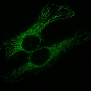
Mitochondrial fission is the process where mitochondria divide or segregate into two separate mitochondrial organelles. Mitochondrial fission is counteracted by the process of mitochondrial fusion, whereby two separate mitochondria can fuse together to form a large one. Mitochondrial fusion in turn can result in elongated mitochondrial networks. Both mitochondrial fission and fusion are balanced in the cell, and mutations interfering with either processes are associated with a variety of diseases. Mitochondria can divide by prokaryotic binary fission and since they require mitochondrial DNA for their function, fission is coordinated with DNA replication. Some of the proteins that are involved in mitochondrial fission have been identified and some of them are associated with mitochondrial diseases. Mitochondrial fission has significant implications in stress response and apoptosis.

<span class="mw-page-title-main">Mitochondrial fission factor</span> Protein-coding gene in the species Homo sapiens

Mitochondrial fission factor (Mff) is a protein that in humans is encoded by the MFF gene. Its primary role is in controlling the division of mitochondria. Mitochondrial morphology changes by continuous fission in order to create interconnected network of mitochondria. This activity is crucial for normal function of mitochondria. Mff is anchored to the mitochondrial outer membrane through the C-terminal transmembrane domain, extruding the bulk of the N-terminal portion containing two short amino acid repeats in the N-terminal half and a coiled-coil domain just upstream of the transmembrane domain into the cytosol. It has also been shown to regulate peroxisome morphology.

<span class="mw-page-title-main">Mitochondrial fusion</span> Merging of two or more mitochondria within a cell to form a single compartment

Mitochondria are dynamic organelles with the ability to fuse and divide (fission), forming constantly changing tubular networks in most eukaryotic cells. These mitochondrial dynamics, first observed over a hundred years ago are important for the health of the cell, and defects in dynamics lead to genetic disorders. Through fusion, mitochondria can overcome the dangerous consequences of genetic malfunction. The process of mitochondrial fusion involves a variety of proteins that assist the cell throughout the series of events that form this process.

<span class="mw-page-title-main">MUL1</span> Protein-coding gene in the species Homo sapiens

Mitochondrial E3 ubiquitin protein ligase 1 (MUL1) is an enzyme that in humans is encoded by the MUL1 gene on chromosome 1. This enzyme localizes to the outer mitochondrial membrane, where it regulates mitochondrial morphology and apoptosis through multiple pathways, including the Akt, JNK, and NF-κB. Its proapoptotic function thus implicates it in cancer and Parkinson's disease.

<span class="mw-page-title-main">Mitochondrial dynamics protein MID49</span> Protein-coding gene in the species Homo sapiens

Mitochondrial elongation factor 2 is a protein that in humans is encoded by the MIEF2 gene.

<span class="mw-page-title-main">David A. Hood</span> Canadian researcher and exercise physiologist

David A. Hood is a Canadian professor, exercise physiologist, and Director of the Muscle Health Research Centre at York University. A holder of an NSERC Tier I Canada Research Chair in Cell Physiology, Hood is credited with making significant research advances in understanding of the biology of exercise, mitochondria and muscle health.

References

  1. 1 2 3 GRCh38: Ensembl release 89: ENSG00000214253 - Ensembl, May 2017
  2. 1 2 3 GRCm38: Ensembl release 89: ENSMUSG00000019054 - Ensembl, May 2017
  3. "Human PubMed Reference:". National Center for Biotechnology Information, U.S. National Library of Medicine.
  4. "Mouse PubMed Reference:". National Center for Biotechnology Information, U.S. National Library of Medicine.
  5. Stojanovski D, Koutsopoulos OS, Okamoto K, Ryan MT (Mar 2004). "Levels of human Fis1 at the mitochondrial outer membrane regulate mitochondrial morphology". Journal of Cell Science. 117 (Pt 7): 1201–10. doi: 10.1242/jcs.01058 . PMID   14996942.
  6. Kong D, Xu L, Yu Y, Zhu W, Andrews DW, Yoon Y, Kuo TH (Apr 2005). "Regulation of Ca2+-induced permeability transition by Bcl-2 is antagonized by Drpl and hFis1". Molecular and Cellular Biochemistry. 272 (1–2): 187–99. doi:10.1007/s11010-005-7323-3. PMID   16010987. S2CID   21452703.
  7. 1 2 3 "Entrez Gene: FIS1 fission 1 (mitochondrial outer membrane) homolog (S. cerevisiae)".
  8. 1 2 3 4 5 Iwasawa R, Mahul-Mellier AL, Datler C, Pazarentzos E, Grimm S (Feb 2011). "Fis1 and Bap31 bridge the mitochondria-ER interface to establish a platform for apoptosis induction". The EMBO Journal. 30 (3): 556–68. doi:10.1038/emboj.2010.346. PMC   3034017 . PMID   21183955.
  9. 1 2 3 4 5 6 7 Gomes LC, Scorrano L (2008). "High levels of Fis1, a pro-fission mitochondrial protein, trigger autophagy". Biochimica et Biophysica Acta (BBA) - Bioenergetics. 1777 (7–8): 860–6. doi: 10.1016/j.bbabio.2008.05.442 . PMID   18515060.
  10. 1 2 Lee S, Park YY, Kim SH, Nguyen OT, Yoo YS, Chan GK, Sun X, Cho H (Feb 2014). "Human mitochondrial Fis1 links to cell cycle regulators at G2/M transition". Cellular and Molecular Life Sciences. 71 (4): 711–25. doi:10.1007/s00018-013-1428-8. PMID   23907611. S2CID   11694077.
  11. 1 2 3 4 Wang S, Song J, Tan M, Albers KM, Jia J (Jul 2012). "Mitochondrial fission proteins in peripheral blood lymphocytes are potential biomarkers for Alzheimer's disease". European Journal of Neurology. 19 (7): 1015–22. doi:10.1111/j.1468-1331.2012.03670.x. PMID   22340708. S2CID   21950507.
  12. 1 2 3 4 5 Tian Y, Huang Z, Wang Z, Yin C, Zhou L, Zhang L, Huang K, Zhou H, Jiang X, Li J, Liao L, Yang M, Meng F (2014). "Identification of novel molecular markers for prognosis estimation of acute myeloid leukemia: over-expression of PDCD7, FIS1 and Ang2 may indicate poor prognosis in pretreatment patients with acute myeloid leukemia". PLOS ONE. 9 (1): e84150. Bibcode:2014PLoSO...984150T. doi: 10.1371/journal.pone.0084150 . PMC   3885535 . PMID   24416201.
  13. 1 2 3 4 Lees JP, Manlandro CM, Picton LK, Tan AZ, Casares S, Flanagan JM, Fleming KG, Hill RB (Oct 2012). "A designed point mutant in Fis1 disrupts dimerization and mitochondrial fission". Journal of Molecular Biology. 423 (2): 143–58. doi:10.1016/j.jmb.2012.06.042. PMC   3456991 . PMID   22789569.
  14. 1 2 3 4 Onoue K, Jofuku A, Ban-Ishihara R, Ishihara T, Maeda M, Koshiba T, Itoh T, Fukuda M, Otera H, Oka T, Takano H, Mizushima N, Mihara K, Ishihara N (Jan 2013). "Fis1 acts as a mitochondrial recruitment factor for TBC1D15 that is involved in regulation of mitochondrial morphology". Journal of Cell Science. 126 (Pt 1): 176–85. doi: 10.1242/jcs.111211 . PMID   23077178.
  15. 1 2 3 Palmer CS, Elgass KD, Parton RG, Osellame LD, Stojanovski D, Ryan MT (Sep 2013). "Adaptor proteins MiD49 and MiD51 can act independently of Mff and Fis1 in Drp1 recruitment and are specific for mitochondrial fission". The Journal of Biological Chemistry. 288 (38): 27584–93. doi: 10.1074/jbc.M113.479873 . PMC   3779755 . PMID   23921378.
  16. Yoon Y, Krueger EW, Oswald BJ, McNiven MA (Aug 2003). "The mitochondrial protein hFis1 regulates mitochondrial fission in mammalian cells through an interaction with the dynamin-like protein DLP1". Molecular and Cellular Biology. 23 (15): 5409–20. doi:10.1128/MCB.23.15.5409-5420.2003. PMC   165727 . PMID   12861026.

Further reading