Liver function tests, also referred to as a hepatic panel, are groups of blood tests that provide information about the state of a patient's liver. These tests include prothrombin time (PT/INR), activated partial thromboplastin time (aPTT), albumin, bilirubin, and others. The liver transaminases aspartate transaminase and alanine transaminase are useful biomarkers of liver injury in a patient with some degree of intact liver function.

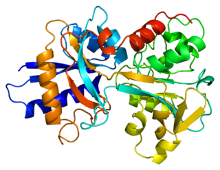
Transferrins are glycoproteins found in vertebrates which bind and consequently mediate the transport of iron (Fe) through blood plasma. They are produced in the liver and contain binding sites for two Fe3+ ions. Human transferrin is encoded by the TF gene and produced as a 76 kDa glycoprotein.

Insulin-like growth factor 1 (IGF-1), also called somatomedin C, is a hormone similar in molecular structure to insulin which plays an important role in childhood growth, and has anabolic effects in adults. In the 1950s IGF-1 was called "sulfation factor" because it stimulated sulfation of cartilage in vitro, and in the 1970s due to its effects it was termed "nonsuppressible insulin-like activity" (NSILA).

Adiponectin is a protein hormone and adipokine, which is involved in regulating glucose levels and fatty acid breakdown. In humans, it is encoded by the ADIPOQ gene and is produced primarily in adipose tissue, but also in muscle and even in the brain.

Liver disease, or hepatic disease, is any of many diseases of the liver. If long-lasting it is termed chronic liver disease. Although the diseases differ in detail, liver diseases often have features in common.

Serum albumin, often referred to simply as blood albumin, is an albumin found in vertebrate blood. Human serum albumin is encoded by the ALB gene. Other mammalian forms, such as bovine serum albumin, are chemically similar.

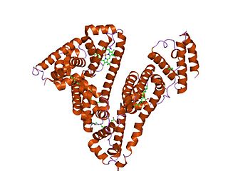
Human serum albumin is the serum albumin found in human blood. It is the most abundant protein in human blood plasma; it constitutes about half of serum protein. It is produced in the liver. It is soluble in water, and it is monomeric.

Calciphylaxis, also known as calcific uremic arteriolopathy (CUA) or “Grey Scale”, is a rare syndrome characterized by painful skin lesions. The pathogenesis of calciphylaxis is unclear but believed to involve calcification of the small blood vessels located within the fatty tissue and deeper layers of the skin, blood clots, and eventual death of skin cells due to lack of blood flow. It is seen mostly in people with end-stage kidney disease but can occur in the earlier stages of chronic kidney disease and rarely in people with normally functioning kidneys. Calciphylaxis is a rare but serious disease, believed to affect 1-4% of all dialysis patients. It results in chronic non-healing wounds and indicates poor prognosis, with typical life expectancy of less than one year.

The liver X receptor (LXR) is a member of the nuclear receptor family of transcription factors and is closely related to nuclear receptors such as the PPARs, FXR and RXR. Liver X receptors (LXRs) are important regulators of cholesterol, fatty acid, and glucose homeostasis. LXRs were earlier classified as orphan nuclear receptors, however, upon discovery of endogenous oxysterols as ligands they were subsequently deorphanized.

Free fatty acid receptor 1 (FFAR1), also known as G-protein coupled receptor 40 (GPR40), is a rhodopsin-like G-protein coupled receptor that is coded by the FFAR1 gene. This gene is located on the short arm of chromosome 19 at position 13.12. G protein-coupled receptors reside on their parent cells' surface membranes, bind any one of the specific set of ligands that they recognize, and thereby are activated to trigger certain responses in their parent cells. FFAR1 is a member of a small family of structurally and functionally related GPRs termed free fatty acid receptors (FFARs). This family includes at least three other FFARs viz., FFAR2, FFAR3, and FFAR4. FFARs bind and thereby are activated by certain fatty acids.

Free fatty acid receptor 3 protein is a G protein coupled receptor that in humans is encoded by the FFAR3 gene. GPRs reside on cell surfaces, bind specific signaling molecules, and thereby are activated to trigger certain functional responses in their parent cells. FFAR3 is a member of the free fatty acid receptor group of GPRs that includes FFAR1, FFAR2, and FFAR4. All of these FFARs are activated by fatty acids. FFAR3 and FFAR2 are activated by certain short-chain fatty acids (SC-FAs), i.e., fatty acids consisting of 2 to 6 carbon atoms whereas FFFAR1 and FFAR4 are activated by certain fatty acids that are 6 to more than 21 carbon atoms long. Hydroxycarboxylic acid receptor 2 is also activated by a SC-FA that activate FFAR3, i.e., butyric acid.

Succinate receptor 1 (SUCNR1), previously named G protein-coupled receptor 91 (GPR91), is a receptor that is activated by succinate, i.e., the anionic form of the dicarboxylic acid, succinic acid. Succinate and succinic acid readily convert into each other by gaining (succinate) or losing (succinic acid) protons, i.e., H+ (see Ions). Succinate is by far the predominant form of this interconversion in living organisms. Succinate is one of the intermediate metabolites in the citric acid cycle (also termed the TCA cycle or tricarboxylic acid cycle). This cycle is a metabolic pathway that operates in the mitochondria of virtually all eucaryotic cells. It consists of a series of biochemical reactions that serve the vital function of releasing the energy stored in nutrient carbohydrates, fats, and proteins. Recent studies have found that some of the metabolites in this cycle are able to regulate various physiological and pathological functions in a wide range of cell types. The succinyl CoA in this cycle may release its bound succinate; succinate is one of these mitochondrial-formed bioactive metabolites.

Free Fatty acid receptor 4 (FFAR4), also termed G-protein coupled receptor 120 (GPR120), is a protein that in humans is encoded by the FFAR4 gene. This gene is located on the long arm of chromosome 10 at position 23.33. G protein-coupled receptors reside on their parent cells' surface membranes, bind any one of the specific set of ligands that they recognize, and thereby are activated to trigger certain responses in their parent cells. FFAR4 is a rhodopsin-like GPR in the broad family of GPRs which in humans are encoded by more than 800 different genes. It is also a member of a small family of structurally and functionally related GPRs that include at least three other free fatty acid receptors (FFARs) viz., FFAR1, FFAR2, and FFAR3. These four FFARs bind and thereby are activated by certain fatty acids.

alpha-2-HS-glycoprotein also known as fetuin-A is a protein that in humans is encoded by the AHSG gene. Fetuin-A belongs to the fetuin class of plasma binding proteins and is more abundant in fetal than adult blood.

Amine oxidase, copper containing 3 (AOC3), also known as vascular adhesion protein (VAP-1) and HPAO is an enzyme that in humans is encoded by the AOC3 gene on chromosome 17. This protein is a member of the semicarbazide-sensitive amine oxidase family of enzymes and is associated with many vascular diseases.

FABP1 is a human gene coding for the protein product FABP1. It is also frequently known as liver-type fatty acid-binding protein (LFABP).

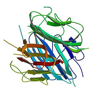
Leukocyte cell-derived chemotaxin-2 (LECT2) is a protein first described in 1996 as a chemotactic factor for neutrophils, i.e. it stimulated human neutrophils to move directionally in an in vitro assay system. The protein was detected in and purified from cultures of Phytohaemagglutinin-activated human T-cell leukemia SKW-3 cells. Subsequent studies have defined LECT2 as a hepatokine, i.e. a substance made and released into the circulation by liver hepatocyte cells that regulates the function of other cells: it is a hepatocyte-derived, hormone-like, signaling protein.

Fetuin-B is a protein that in humans is encoded by the FETUB gene.

Lipotoxicity is a metabolic syndrome that results from the accumulation of lipid intermediates in non-adipose tissue, leading to cellular dysfunction and death. The tissues normally affected include the kidneys, liver, heart and skeletal muscle. Lipotoxicity is believed to have a role in heart failure, obesity, and diabetes, and is estimated to affect approximately 25% of the adult American population.
Hepatokines are proteins produced by liver cells (hepatocytes) that are secreted into the circulation and function as hormones across the organism. Research is mostly focused on hepatokines that play a role in the regulation of metabolic diseases such as diabetes and fatty liver and include: Adropin, ANGPTL4, Fetuin-A, Fetuin-B, FGF-21, Hepassocin, LECT2, RBP4,Selenoprotein P, Sex hormone-binding globulin.


