Gain-field encoding

Last updated

Gain field encoding is a hypothesis about the internal storage and processing of limb motion in the brain. In the motor areas of the brain, there are neurons which collectively have the ability to store information regarding both limb positioning and velocity in relation to both the body (intrinsic) and the individual's external environment (extrinsic). [1] The input from these neurons is taken multiplicatively, forming what is referred to as a gain field. [2] The gain field works as a collection of internal models off of which the body can base its movements. The process of encoding and recalling these models is the basis of muscle memory.

Contents

Physiology

Neuron involved in gain field encoding work multiplicatively, taking the input from several together to form the gain field. [2] It is this process that allows the complexity of motor control. Instead of simply encoding the motion of the limb in which a specific motion is desired, the multiplicative nature of the gain field ensures that the positioning of the rest of the body is taken in to consideration. This process allows for motor coordination of flexible bimanual actions as opposed to restricting the individual to unimanual motion. [3] For example, when considering the movement of both arms, the body calls upon gain field models for each arm in order to compensate for the mechanical interactions created by both.

Location

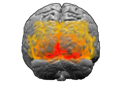
Most gain field activity is based in the premotor cortex found in the frontal lobe anterior to the primary motor cortex, however it receives input from a variety of locations in the brain. [4] These incoming signals provide frame of reference information through the individual's senses. Further evidence suggests that the cerebellum and posterior parietal cortex (PPC) also play major functional roles in gain field encoding. [5] [6] The intrinsic and extrinsic properties of the gain field can be shown as products of the PPC. In Brodmann area 7 of the PPC, the positioning of objects with respect to the eyes is represented completely extrinsically with no input from the positioning of the body involved. [7] [8] This opposes the case of other parts of the PPC such as Brodmann area 5 which represents objects in relation to body defined coordinates. Due to the extrinsic and intrinsic properties of motor functioning, it is speculated that these types of signals are both taken multiplicatively to form the gain field. With input from each area, a three-dimensional representation of the objects in space can be arranged for use by the rest of the motor system.

Unsurprisingly lesions in the parietal cortex lead to deficiencies in an individual's spatial movements and coordination and, in some cases, hemineglect. These effects were widely variable from person to person and depending on the location of the lesion further hinting at the complicated nature of gain modulated neurons. [9]

Gain Modulation

One of the key components of gain-field encoding is the variability in the response amplitude of the action potentials from neurons. This variability, when independent of change in response selectivity, is called gain modulation. Gain Modulation takes place in many cortical areas and is believed to be a common mechanism of neuronal computation. [7] [10] It allows for the combination of different sensory and cognitive information. For example, neurons implicated in processing a part of the visual field see a gain in the response amplitude due to shifting focus to that part of the field of vision. Therefore, neurons that are gain modulated can represent multiple types of information. The multi-modal nature of these neurons makes them ideal for specific types of computations, mainly coordinate transformations. This creates the ability to think spatially, the main contributor to physical coordination.

Encoding Process

The encoding of the neurons involved in the motor gain field follow the same gain modulation principles as most of the neurons within the brain. That is to say, when gain is increased, the connections between the neurons firing increase in strength leading to further gain if the neurons continue to receive stimulation. [11] This observation is why repetition of a particular set of motions is what leads to muscle memory.

Coordinate Manipulation

One of the main results of gain-field encoding is the cognitive ability to manipulate different coordinate planes that are dealt with daily and adjust limb muscle movements accordingly. A good example of this is moving a pointer across a computer screen with a mouse. Depending on the relative location of the user's head to the computer screen as well as the angle at which the screen is being observed, the user's perspective of the screen will be very different. A mentally mapped grid of the screen appears much larger when the user is closer to the screen as opposed to further away, and it is the brain's ability to keep a consistent mental representation that gives people the ability to function under such dynamic conditions. [12]

Mathematical Representation

The equation for the firing rate of a gain modulated neuron is a combination of the two types of information being transmitted to the neuron:

where is the rate of fire, is a function of one type of information input and is another. For example, neural activity for the interaction between gaze direction and retinal image location is almost exactly multiplicative, where represents the location of a stimulus in retinal coordinates and represents gaze angle. The primary process by which this interaction can take place is speculated to be recurrent neural networks where neural connections form a directed cycle. [2] [7] Recurrent circuitry is abundant in cortical networks and reportedly plays a role in sustaining signals, signal amplification, and response selectivity. [13]

Evidence

Early hypotheses of gain field encoding suggested that the gain field works as a model for motion additively. This would mean that if two limbs needed to move, models for each would be called separately but at the same time. However, more recent studies in which more complex motor movements are observed have found that the gain field is created multiplicatively in order to allow the body to adapt to the constantly changing frames of reference experienced in everyday life.

This multiplicative property is an effect of recurrent neural circuitry. A target neuron that takes only two types of direct input can only combine them additively. However mathematical models show that when also receiving recursive input from neighboring neurons, the resulting transformation to the target neurons firing rate is multiplicative. In this model, neurons with overlapping receptive fields excite each other, multiplying the strength. Likewise, neurons with non-overlapping receptive fields are inhibitory. [7] The result is a response curve that is a scaled representation of the simple additive model.

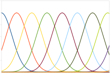
Typical neural population code. Each color represents a separate, adjacent neuron arranged linearly. X axis shows stimulus, such as head position or sound frequency, and Y axis shows neural response. In a gain field these action potentials are taken recurrently with another relevant stimulus. PopulationCode.svg
Typical neural population code. Each color represents a separate, adjacent neuron arranged linearly. X axis shows stimulus, such as head position or sound frequency, and Y axis shows neural response. In a gain field these action potentials are taken recurrently with another relevant stimulus.

Observation of human developmental patterns also lend evidence toward this theory of gain-field encoding and gain modulation. Since arm movements are based on both intrinsic and extrinsic models, in order to build these connections one has to learn by self-generating movements and watching. By moving the arms to different parts of space and following with the eyes, the neurons form connections based on mechanical body movements as well as their positioning in an external space. Ideally this is done from every possible gaze angle and position available. This provides your brain with the proper translations by aligning the retinal (extrinsic) and body-centered (intrinsic) representations of space. It is not surprising that before babies develop motor control of their limbs, they tend to flail and watch their own limbs move. [14]

A similar effect is found when people track moving objects with their eyes. The changing retinal image is referenced with the muscle movements of the eye resulting in the same type of retinal/body-centered alignment. [15] This is one more process that helps the brain properly encode the relationships needed to deal with our changing perception, and also serves as a verification that the proper physical movements are being made.

A contrary hypothesis to gain field encoding involved implicating the neurons of the primary motor cortex (M1) in dynamic muscle movement. An investigation into area M1 shows that when an individual is asked to rotate an object, activation of the neurons in M1 thought to be controlling the motion happened instantaneously with muscle activation. This provides evidence for preliminary steps from higher motor areas communicating with area M1 by means of gain modulation. [16]

See also

Related Research Articles

<span class="mw-page-title-main">Visual cortex</span> Region of the brain that processes visual information

The visual cortex of the brain is the area of the cerebral cortex that processes visual information. It is located in the occipital lobe. Sensory input originating from the eyes travels through the lateral geniculate nucleus in the thalamus and then reaches the visual cortex. The area of the visual cortex that receives the sensory input from the lateral geniculate nucleus is the primary visual cortex, also known as visual area 1 (V1), Brodmann area 17, or the striate cortex. The extrastriate areas consist of visual areas 2, 3, 4, and 5.
<span class="mw-page-title-main">Cerebral cortex</span> Outer layer of the cerebrum of the mammalian brain

The cerebral cortex, also known as the cerebral mantle, is the outer layer of neural tissue of the cerebrum of the brain in humans and other mammals. The cerebral cortex mostly consists of the six-layered neocortex, with just 10% consisting of the allocortex. It is separated into two cortices, by the longitudinal fissure that divides the cerebrum into the left and right cerebral hemispheres. The two hemispheres are joined beneath the cortex by the corpus callosum. The cerebral cortex is the largest site of neural integration in the central nervous system. It plays a key role in attention, perception, awareness, thought, memory, language, and consciousness. The cerebral cortex is part of the brain responsible for cognition.

<span class="mw-page-title-main">Basal ganglia</span> Group of subcortical nuclei involved in the motor and reward systems

The basal ganglia (BG), or basal nuclei, are a group of subcortical nuclei found in the brains of vertebrates. In humans and some primates, differences exist, primarily in the division of the globus pallidus into external and internal regions, and in the division of the striatum. Positioned at the base of the forebrain and the top of the midbrain, they have strong connections with the cerebral cortex, thalamus, brainstem and other brain areas. The basal ganglia are associated with a variety of functions, including regulating voluntary motor movements, procedural learning, habit formation, conditional learning, eye movements, cognition, and emotion.

In biology, a reflex, or reflex action, is an involuntary, unplanned sequence or action and nearly instantaneous response to a stimulus.

<span class="mw-page-title-main">Parietal lobe</span> Part of the brain responsible for sensory input and some language processing

The parietal lobe is one of the four major lobes of the cerebral cortex in the brain of mammals. The parietal lobe is positioned above the temporal lobe and behind the frontal lobe and central sulcus.
<span class="mw-page-title-main">Superior colliculus</span> Structure in the midbrain

In neuroanatomy, the superior colliculus is a structure lying on the roof of the mammalian midbrain. In non-mammalian vertebrates, the homologous structure is known as the optic tectum, or optic lobe. The adjective form tectal is commonly used for both structures.

<span class="mw-page-title-main">Motor cortex</span> Region of the cerebral cortex

The motor cortex is the region of the cerebral cortex involved in the planning, control, and execution of voluntary movements. The motor cortex is an area of the frontal lobe located in the posterior precentral gyrus immediately anterior to the central sulcus.

Central pattern generators (CPGs) are self-organizing biological neural circuits that produce rhythmic outputs in the absence of rhythmic input. They are the source of the tightly-coupled patterns of neural activity that drive rhythmic and stereotyped motor behaviors like walking, swimming, breathing, or chewing. The ability to function without input from higher brain areas still requires modulatory inputs, and their outputs are not fixed. Flexibility in response to sensory input is a fundamental quality of CPG-driven behavior. To be classified as a rhythmic generator, a CPG requires:

  1. "two or more processes that interact such that each process sequentially increases and decreases, and
  2. that, as a result of this interaction, the system repeatedly returns to its starting condition."
<span class="mw-page-title-main">Neural oscillation</span> Brainwaves, repetitive patterns of neural activity in the central nervous system

Neural oscillations, or brainwaves, are rhythmic or repetitive patterns of neural activity in the central nervous system. Neural tissue can generate oscillatory activity in many ways, driven either by mechanisms within individual neurons or by interactions between neurons. In individual neurons, oscillations can appear either as oscillations in membrane potential or as rhythmic patterns of action potentials, which then produce oscillatory activation of post-synaptic neurons. At the level of neural ensembles, synchronized activity of large numbers of neurons can give rise to macroscopic oscillations, which can be observed in an electroencephalogram. Oscillatory activity in groups of neurons generally arises from feedback connections between the neurons that result in the synchronization of their firing patterns. The interaction between neurons can give rise to oscillations at a different frequency than the firing frequency of individual neurons. A well-known example of macroscopic neural oscillations is alpha activity.

A neuronal ensemble is a population of nervous system cells involved in a particular neural computation.

Head direction (HD) cells are neurons found in a number of brain regions that increase their firing rates above baseline levels only when the animal's head points in a specific direction. They have been reported in rats, monkeys, mice, chinchillas and bats, but are thought to be common to all mammals, perhaps all vertebrates and perhaps even some invertebrates, and to underlie the "sense of direction". When the animal's head is facing in the cell's "preferred firing direction" these neurons fire at a steady rate, but firing decreases back to baseline rates as the animal's head turns away from the preferred direction.

<span class="mw-page-title-main">Scratch reflex</span> Response to activation of sensory neurons

The scratch reflex is a response to activation of sensory neurons whose peripheral terminals are located on the surface of the body. Some sensory neurons can be activated by stimulation with an external object such as a parasite on the body surface. Alternatively, some sensory neurons can respond to a chemical stimulus that produces an itch sensation. During a scratch reflex, a nearby limb reaches toward and rubs against the site on the body surface that has been stimulated. The scratch reflex has been extensively studied to understand the functioning of neural networks in vertebrates. Despite decades of research, key aspects of the scratch reflex are still unknown, such as the neural mechanisms by which the reflex is terminated.

The zona incerta (ZI) is a horizontally elongated region of gray matter in the subthalamus below the thalamus. Its connections project extensively over the brain from the cerebral cortex down into the spinal cord.

<span class="mw-page-title-main">Posterior parietal cortex</span>

The posterior parietal cortex plays an important role in planned movements, spatial reasoning, and attention.

Richard Alan Andersen is an American neuroscientist. He is the James G. Boswell Professor of Neuroscience at the California Institute of Technology in Pasadena, California. His research focuses on visual physiology with an emphasis on translational research to humans in the field of neuroprosthetics, brain-computer interfaces, and cortical repair.

The parietal reach region (PRR) is a region within the posterior parietal cortex of the macaque brain that is stimulated through reaching for an object. The PRR was initially proposed by Andersen and Buneo, but they just generally explored the idea as postural modulations. Dr. Steven Chang went further in depth by showing the receptive fields of PRR neurons are multiplicatively stimulated through the combination of initial gaze position and initial hand position. This multiplicative stimulation is known as gain-field encoding. The parietal reach region uses gain-field encoding to calculate the amount of hand displacement needed to reach for an object. A recent study shows that neurons in PRR encodes not only the planned physical movement, but also the anticipated visual sensory consequence of the intended movement once the action is unfolded in time.
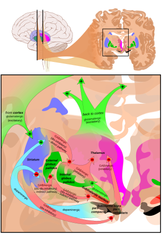
<span class="mw-page-title-main">Cortico-basal ganglia-thalamo-cortical loop</span> System of neural circuits in the brain

The cortico-basal ganglia-thalamo-cortical loop is a system of neural circuits in the brain. The loop involves connections between the cortex, the basal ganglia, the thalamus, and back to the cortex. It is of particular relevance to hyperkinetic and hypokinetic movement disorders, such as Parkinson's disease and Huntington's disease, as well as to mental disorders of control, such as attention deficit hyperactivity disorder (ADHD), obsessive–compulsive disorder (OCD), and Tourette syndrome.

<span class="mw-page-title-main">Laura Busse</span> German neuroscientist

Laura Busse is a German neuroscientist and professor of Systemic Neuroscience within the Division of Neurobiology at the Ludwig Maximilian University of Munich. Busse's lab studies context-dependent visual processing in mouse models by performing large scale in vivo electrophysiological recordings in the thalamic and cortical circuits of awake and behaving mice.

<span class="mw-page-title-main">Eberhard Fetz</span> American neuroscientist, academic and researcher

Eberhard Erich Fetz is an American neuroscientist, academic and researcher. He is a Professor of Physiology and Biophysics and DXARTS at the University of Washington.
<span class="mw-page-title-main">Interlimb coordination</span> Coordination of the left and right limbs

Interlimb coordination is the coordination of the left and right limbs. It could be classified into two types of action: bimanual coordination and hands or feet coordination. Such coordination involves various parts of the nervous system and requires a sensory feedback mechanism for the neural control of the limbs. A model can be used to visualize the basic features, the control centre of locomotor movements, and the neural control of interlimb coordination. This coordination mechanism can be altered and adapted for better performance during locomotion in adults and for the development of motor skills in infants. The adaptive feature of interlimb coordination can also be applied to the treatment for CNS damage from stroke and the Parkinson's disease in the future.

References

  1. Polack, Pierre-Olivier; Friedman, Jonathan; Golshani, Peyman (2013). "Cellular mechanisms of brain state–dependent gain modulation in visual cortex". Nature Neuroscience. 16 (9): 1331–1339. doi:10.1038/nn.3464. ISSN   1097-6256. PMC   3786578 . PMID   23872595.
  2. 1 2 3 De Meyer, K.; Spratling, M. (2011). "Multiplicative Gain Modulation Arises Through Unsupervised Learning in a Predictive Coding Model". Neural Computation. 23 (6): 1536–1567. CiteSeerX   10.1.1.701.2882 . doi:10.1162/neco_a_00130. PMID   21395434. S2CID   11134249.
  3. Yokoi, A.; Hirashima, M.; Nozaki, D. (2011). "Gain Field Encoding of the Kinematics of Both Arms in the Internal Model Enables Flexible Bimanual Action". Journal of Neuroscience. 31 (47): 17058–17068. doi: 10.1523/JNEUROSCI.2982-11.2011 . ISSN   0270-6474. PMC   6623869 . PMID   22114275.
  4. Cherian, A.; Fernandes, H. L.; Miller, L. E. (2013). "Primary motor cortical discharge during force field adaptation reflects muscle-like dynamics". Journal of Neurophysiology. 110 (3): 768–783. doi:10.1152/jn.00109.2012. ISSN   0022-3077. PMC   3742991 . PMID   23657285.
  5. Brayanov, J. B.; Press, D. Z.; Smith, M. A. (2012). "Motor Memory Is Encoded as a Gain-Field Combination of Intrinsic and Extrinsic Action Representations". Journal of Neuroscience. 32 (43): 14951–14965. doi:10.1523/JNEUROSCI.1928-12.2012. ISSN   0270-6474. PMC   3999415 . PMID   23100418.
  6. Chang, S. W. C.; Snyder, L. H. (2010). "Idiosyncratic and systematic aspects of spatial representations in the macaque parietal cortex". Proceedings of the National Academy of Sciences. 107 (17): 7951–7956. Bibcode:2010PNAS..107.7951C. doi: 10.1073/pnas.0913209107 . ISSN   0027-8424. PMC   2867917 . PMID   20375282.
  7. 1 2 3 4 Salinas, E.; Abbott, L.F. (2001). "Chapter 11 Coordinate transformations in the visual system: How to generate gain fields and what to compute with them". Advances in Neural Population Coding. Progress in Brain Research. Vol. 130. pp. 175–190. doi:10.1016/s0079-6123(01)30012-2. ISBN   9780444501103. ISSN   0079-6123. PMID   11480274.
  8. Snyder, L.H.; Grieve, K.L. (1998). "Separate body- and world-referenced representations of visual space in parietalCortex". Nature. 394 (6696): 887–891. Bibcode:1998Natur.394..887S. doi:10.1038/29777. PMID   9732870. S2CID   4319004.
  9. Pouget, A.; Sejnowski, T.J. (1997). "A new view of hemineglect based on the response properties of the parietal neurones". Philosophical Transactions of the Royal Society of London B: Biological Sciences. 352 (1360): 1449–1459. doi:10.1098/rstb.1997.0131. ISSN   0962-8436. PMC   1692050 . PMID   9368933.
  10. Futatsubashi, Genki; Sasada, Shusaku; Tazoe, Toshiki; Komiyama, Tomoyoshi (2013). "Gain modulation of the middle latency cutaneous reflex in patients with chronic joint instability after ankle sprain". Clinical Neurophysiology. 124 (7): 1406–1413. doi:10.1016/j.clinph.2013.01.029. ISSN   1388-2457. PMID   23541471. S2CID   206796851.
  11. Donner, Tobias H; Nieuwenhuis, Sander (2013). "Brain-wide gain modulation: the rich get richer". Nature Neuroscience. 16 (8): 989–990. doi:10.1038/nn.3471. ISSN   1097-6256. PMID   23887133. S2CID   10531112.
  12. Hwang, Eun Jung; Donchin, Opher; Smith, Maurice A.; Shadmehr, Reza (2003). "A Gain-Field Encoding of Limb Position and Velocity in the Internal Model of Arm Dynamics". PLOS Biology. 1 (2): e5. doi: 10.1371/journal.pbio.0000025 . ISSN   1544-9173. PMC   261873 . PMID   14624237. Open Access logo PLoS transparent.svg
  13. Douglas, R. J.; Koch, C.; Mahowald, M.; Martin, K. A. C.; Suarez, H. H. (1995). "Recurrent excitation in neocortical circuits". Science. 269 (5226): 981–985. Bibcode:1995Sci...269..981D. doi:10.1126/science.7638624. PMID   7638624. S2CID   1003907.
  14. van der Meer, A.; van der Weel, F.; Lee, D. (1995). "The functional significance of arm movements in neonates". Science. 267 (5198): 693–695. Bibcode:1995Sci...267..693V. doi:10.1126/science.7839147. ISSN   0036-8075. PMID   7839147. S2CID   32025757.
  15. Lee, J.; Yang, J.; Lisberger, S. G. (2013). "Control of the Gain of Visual-Motor Transmission Occurs in Visual Coordinates for Smooth Pursuit Eye Movements". Journal of Neuroscience. 33 (22): 9420–9430. doi:10.1523/JNEUROSCI.4846-12.2013. ISSN   0270-6474. PMC   3705569 . PMID   23719810.
  16. Kimura, T. (2006). "Transcranial Magnetic Stimulation over Sensorimotor Cortex Disrupts Anticipatory Reflex Gain Modulation for Skilled Action" (PDF). Journal of Neuroscience. 26 (36): 9272–9281. doi: 10.1523/JNEUROSCI.3886-05.2006 . ISSN   0270-6474. PMC   6674505 . PMID   16957083.