Glycoprotein Ib-IX-V complex

Last updated

The GPIb-IX-V complex is a profuse membrane receptor complex originating in megakaryocytes and exclusively functional on the surface of platelets. [1] It primarily functions to mediate the first critical step in platelet adhesion, by facilitating binding to von Willebrand factor (VWF) on damaged sub-endothelium under conditions of high fluid shear stress. [2] [3] Although the primary ligand for the GPIb-V-IX receptor is VWF, it can also bind to a number of other ligands in the circulation such as thrombin, P-selectin, factor XI, factor XII, high molecular weight kininogen as well as bacteria. GPIb-IX-V offers a critical role in thrombosis, metastasis, and the life cycle of platelets, and is implicated in a number of thrombotic pathological processes such as stroke or myocardial infarction. [1] [2]

Contents

Molecular structure

Overview

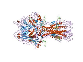
GPIb-IX-V consists of four different subunits namely: GPIbα (molecular weight (MW) 135 kDa), GPIbβ (MW 26 kDa), GPIX (MW 20 kDa) and GPV (MW 82kDa). The complex is assembled such that GPIbα, GPIbβ and GPIX form a highly integrated protein complex in a 1:2:1 stoichiometry; and this associates weakly with GPV resulting in an overall stoichiometric ratio of 1:1. [1] [4] [5] [6]

Each subunit of the complex is a type I transmembrane (TM) protein which consists of a leucine-rich repeat (LRR) ectodomain (extracellular domain), a single transmembrane helix, and a relatively short cytoplasmic tail that lacks enzymatic activity. [1] [7]

The quaternary stabilization of the receptor is facilitated by covalent and non-covalent interactions. The GPIbα subunit is linked to two GPIbβ subunits via membrane-proximal disulfide bonds, while GPIX associates itself tightly through non-covalent interactions with GPIb. [4] [5] [7] The concomitant expression of all three subunits is required to allow the effective expression of GPIb-IX on the platelet cell surface and analysis of receptor expression in transfected Chinese hamster ovary (CHO) cells has further supported that the interaction between these subunits also acts to stabilize them. [1]

View from the top of the membrane proximal portion of GPIb-IX indicating in red the disulfide bonds between GPIba and GPIbb. Disulfide bonds between GPIba and GPIbb.png
View from the top of the membrane proximal portion of GPIb-IX indicating in red the disulfide bonds between GPIbα and GPIbβ.

Each of the four subunits (GPIbα, GPIbβ, GPIX and GPV) is part of the leucine rich repeat motif superfamily. These leucine rich repeat sequences tend to be about 24 amino acids in length either occurring singly or in tandem repeats flanked by conserved N-terminal and C-terminal disulfide loop structures. [3] Nevertheless, even though these structural similarities exist, distinctive genes that exist on different chromosomes of the genome code for the polypeptides that make up the GPIb-V-IX complex.

The four genes that code for the components of the receptor in humans have a simple organization in which the coding sequence is contained within a single exon. This is with the exception of the gene for GPIbβ, which contains an intron 10 bases following the start codon. [3]

Human GPIbα is the product of a gene on chromosome 17 specifically 17p12, GPIbβ is the product of a gene on chromosome 22 specifically 22q11.2, while GPV and GPIX are products of genes found on chromosome 3 specifically 3q21 and 3q29 respectively. [8] Under normal conditions, all four molecules are expressed exclusively in the platelet lineage. GPIbα, GPIbβ and GPIX are necessary for the effective biosynthesis of the receptor and are closely associated at the platelet membrane. Typically, a lack of a single subunit significantly decreases the surface expression of the entire receptor complex. [8] [9]

GPIbα

A ribbon diagram depicting the crystal structure of the GPIba N-terminal domain including the VWF A1 and thrombin binding sites. The crystal structure of the GPIba N-terminal domain depicting important ligand binding sites.png
A ribbon diagram depicting the crystal structure of the GPIbα N-terminal domain including the VWF A1 and thrombin binding sites.

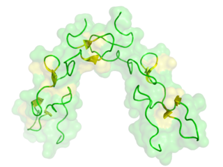
GPIbα (CD42b) consisting of 610 amino acids is the major subunit and contains all known extracellular ligand-binding sites of the complex for example: the A1 domain of von Willebrand factor (VWF) has a binding region as marked in the N-terminal domain of GPIbα; while the thrombin binding site is contained in a conformationally flexible acidic residue-rich sequence containing sulfated tyrosines. [1] [3]

A ribbon diagram depicting the various components of the GPIba subunit. Ribbon diagram depicting the various components of GPIba subunit.png
A ribbon diagram depicting the various components of the GPIbα subunit.

Dissection of the crystal structure of the GPIbα N-terminal leucine rich repeat domain discloses the presence of a single disulfide bond between cysteine (Cys) residues Cys4 and Cys17 in the N-capping region, and two disulfide bonds (Cys209-Cys248 and Cys211-Cys264) in the C-capping region. Furthermore, there are seven tandem leucine rich repeats and their flanking sequences in the central parallel β-coil region. This parallel β-coil region is made up of three sided coils stacked in layers and contains two asparagine residues (Asn21 and Asn159), which serve as N-glycosylation sites. Following the leucine rich repeat domain is the acidic residue-rich sequence containing sulfated tyrosines, the highly O-glycosylated macroglycopeptide, a stalk region of about 40 to 50 residues, a single transmembrane sequence and finally a cytoplasmic tail containing 96 amino acid residues which includes serine residues such as Ser587, Ser590 and Ser609 that can be phosphorylated. [1] [3] [10]

GPIbβ, GPIX, GPV

GPIbβ (CD42c) contains 181 amino acids. In the extracellular domain (ectodomain), both the N-capping and C-capping regions, which flank the leucine rich repeat sequence, contain two interlocking disulfide bonds. Furthermore, there is only a single leucine-rich repeat giving rise to a much less curved parallel β-coil region as compared to that in GPIbα. GPIbβ contains only one N-glycosylation site (Asn41) and is disulfide linked to GPIbα immediately proximal to the plasma membrane of the platelet via Cys122 located at the junction of the extracellular and transmembrane domains. [1] [3]

A ribbon diagram depicting the crystal structure of the GPIbb extracellular domain. The N-terminus is positioned at the top, the concave b-strands sit to the right and the convex loops are positioned to the left. Disulfide bonds present in LRR regions are indicated in yellow. Ribbon diagram depicting the crystal structure of the GPIbb extracellular domain.png
A ribbon diagram depicting the crystal structure of the GPIbβ extracellular domain. The N-terminus is positioned at the top, the concave β-strands sit to the right and the convex loops are positioned to the left. Disulfide bonds present in LRR regions are indicated in yellow.

The GPIbβ cytoplasmic domain has a sequence of 34 amino acids. The region adjacent to the membrane is enriched in basic residues and Ser166 found more distally is phosphorylated and appears to have a role in platelet cytoskeletal rearrangement.

GPIX (CD42a) contains 160 amino acids. The extracellular domain, which also only has a single leucine rich repeat sequence shares more than 45% sequence identity with GPIbβ counterpart. However, the transmembrane and cytoplasmic sequences are considerably different. The GPIX cytoplasmic tail is short consisting of 8 residues and is not known to associate with intracellular proteins. There is also a cysteine residue (Cys154) located at the junction of the transmembrane and cytoplasmic domains. The extracellular domain of GPV contains 13 leucine rich repeats flanked by N- and C- capping regions both containing two interlocking disulfide bonds. This is followed by a stalk region, the transmembrane sequence and a short cytoplasmic tail rich in basic residues. [1] [3]

Interaction of GPV with GPIb-IX via transmembrane (TM) domains. The image demonstrates the accessibility of the GPIba transmembrane helix for direct association with the GPV transmembrane helix as well as the inaccessibility of the GPIX transmembrane helix. Interaction of GPV with GPIb-IX via transmembrane (TM) domains.png
Interaction of GPV with GPIb-IX via transmembrane (TM) domains. The image demonstrates the accessibility of the GPIbα transmembrane helix for direct association with the GPV transmembrane helix as well as the inaccessibility of the GPIX transmembrane helix.

The GPV (CD42d) subunit is only weakly associated with the GPIb-IX part of the receptor complex through interactions between the transmembrane domains and has little impact on the surface expression of GPIb-IX, although GPIb-IX is required for efficient expression of GPV. [1] [6] Furthermore, GPV doesn’t appear to be critical for VWF binding or signal transduction. [7]

Role in disease

Abnormalities of the GPIb-V-IX complex result in abnormal appearance and functioning of platelets resulting in Bernard–Soulier syndrome (BSS), a condition first described by Bernard J and Soulier J.P. [11] It is a rare hereditary bleeding disorder most commonly with an autosomal recessive inheritance and diagnosed based on prolonged skin-bleeding time, a reduced number of very large platelets (macrothrombocytopenia) and defective ristocetin-induced platelet agglutination. [12]

Bernard Soulier Syndrome is characterized by little or no expression of GPIb-IX on the surface of platelets which in turn has the same effect on GPV. There have been a number of mutations associated with BSS patients that have been mapped to GPIbα, GPIbβ and GPIX demonstrating that all three subunits are required for effective surface expression of the complex on platelets. [7]

Related Research Articles

<span class="mw-page-title-main">Integrin</span> Instance of a defined set in Homo sapiens with Reactome ID (R-HSA-374573)

Integrins are transmembrane receptors that help cell-cell and cell-extracellular matrix (ECM) adhesion. Upon ligand binding, integrins activate signal transduction pathways that mediate cellular signals such as regulation of the cell cycle, organization of the intracellular cytoskeleton, and movement of new receptors to the cell membrane. The presence of integrins allows rapid and flexible responses to events at the cell surface.

von Willebrand factor Mammalian protein involved in blood clotting

Von Willebrand factor (VWF) is a blood glycoprotein that promotes hemostasis, specifically, platelet adhesion. It is deficient and/or defective in von Willebrand disease and is involved in many other diseases, including thrombotic thrombocytopenic purpura, Heyde's syndrome, and possibly hemolytic–uremic syndrome. Increased plasma levels in many cardiovascular, neoplastic, metabolic, and connective tissue diseases are presumed to arise from adverse changes to the endothelium, and may predict an increased risk of thrombosis.

Cell adhesion molecules (CAMs) are a subset of cell surface proteins that are involved in the binding of cells with other cells or with the extracellular matrix (ECM), in a process called cell adhesion. In essence, CAMs help cells stick to each other and to their surroundings. CAMs are crucial components in maintaining tissue structure and function. In fully developed animals, these molecules play an integral role in generating force and movement and consequently ensuring that organs are able to execute their functions normally. In addition to serving as "molecular glue", CAMs play important roles in the cellular mechanisms of growth, contact inhibition, and apoptosis. Aberrant expression of CAMs may result in a wide range of pathologies, ranging from frostbite to cancer.

<span class="mw-page-title-main">Hemagglutinin esterase</span> Glycoprotein present in some enveloped viruses

Hemagglutinin esterase (HEs) is a glycoprotein that certain enveloped viruses possess and use as an invading mechanism. HEs helps in the attachment and destruction of certain sialic acid receptors that are found on the host cell surface. Viruses that possess HEs include influenza C virus, toroviruses, and coronaviruses of the subgenus Embecovirus. HEs is a dimer transmembrane protein consisting of two monomers, each monomer is made of three domains. The three domains are: membrane fusion, esterase, and receptor binding domains.

<span class="mw-page-title-main">Disintegrin</span> Proteins from viper venom inhibiting platelets aggregation

Disintegrins are a family of small proteins from viper venoms that function as potent inhibitors of both platelet aggregation and integrin-dependent cell adhesion.

<span class="mw-page-title-main">Bernard–Soulier syndrome</span> Medical condition

Bernard–Soulier syndrome (BSS) is a rare autosomal recessive bleeding disorder that is caused by a deficiency of the glycoprotein Ib-IX-V complex (GPIb-IX-V), the receptor for von Willebrand factor. The incidence of BSS is estimated to be less than 1 case per million persons, based on cases reported from Europe, North America, and Japan. BSS is a giant platelet disorder, meaning that it is characterized by abnormally large platelets.

<span class="mw-page-title-main">Ristocetin</span> Chemical compound

Ristocetin is a glycopeptide antibiotic, obtained from Amycolatopsis lurida, previously used to treat staphylococcal infections. It is no longer used clinically because it caused thrombocytopenia and platelet agglutination. It is now used solely to assay those functions in vitro in the diagnosis of conditions such as von Willebrand disease (vWD) and Bernard–Soulier syndrome. Platelet agglutination caused by ristocetin can occur only in the presence of von Willebrand factor multimers, so if ristocetin is added to blood lacking the factor, the platelets will not clump.

<span class="mw-page-title-main">Convulxin</span>

Convulxin is a snake venom toxin found in a tropical rattlesnake known as Crotalus durissus terrificus. It belongs to the family of hemotoxins, which destroy red blood cells or, as is the case with convulxin, induce blood coagulation.

<span class="mw-page-title-main">Endoglin</span> Protein-coding gene in the species Homo sapiens

Endoglin (ENG) is a type I membrane glycoprotein located on cell surfaces and is part of the TGF beta receptor complex. It is also commonly referred to as CD105, END, FLJ41744, HHT1, ORW and ORW1. It has a crucial role in angiogenesis, therefore, making it an important protein for tumor growth, survival and metastasis of cancer cells to other locations in the body.

Glycoprotein Ib (GPIb), also known as CD42, is a component of the GPIb-V-IX complex on platelets. The GPIb-V-IX complex binds von Willebrand factor, allowing platelet adhesion and platelet plug formation at sites of vascular injury. Glycoprotein Ibα (GPIbα) is the major ligand-binding subunit of the GPIb-V-IX complex. GPIbα is heavily glycosylated.

The mannose receptor is a C-type lectin primarily present on the surface of macrophages, immature dendritic cells and liver sinusoidal endothelial cells, but is also expressed on the surface of skin cells such as human dermal fibroblasts and keratinocytes. It is the first member of a family of endocytic receptors that includes Endo180 (CD280), M-type PLA2R, and DEC-205 (CD205).

<span class="mw-page-title-main">Toll-like receptor 1</span> One of the toll-like receptors and plays a role in the immune system

Toll-like receptor 1 (TLR1) is a member of Toll-like receptors (TLRs), which is a family of pattern recognition receptors (PRRs) that form the cornerstone of the innate immune system. TLR1 recognizes bacterial lipoproteins and glycolipids in complex with TLR2. TLR1 is a cell surface receptor. TLR1 is in humans encoded by the TLR1 gene, which is located on chromosome 4.

<span class="mw-page-title-main">Granulocyte-macrophage colony-stimulating factor receptor</span> Protein-coding gene in humans

The granulocyte-macrophage colony-stimulating factor receptor also known as CD116, is a receptor for granulocyte-macrophage colony-stimulating factor, which stimulates the production of white blood cells. In contrast to M-CSF and G-CSF which are lineage specific, GM-CSF and its receptor play a role in earlier stages of development. The receptor is primarily located on neutrophils, eosinophils and monocytes/macrophages, it is also on CD34+ progenitor cells (myeloblasts) and precursors for erythroid and megakaryocytic lineages, but only in the beginning of their development.

Platelet membrane glycoproteins are surface glycoproteins found on platelets (thrombocytes) which play a key role in hemostasis. When the blood vessel wall is damaged, platelet membrane glycoproteins interact with the extracellular matrix.

<span class="mw-page-title-main">GP1BA</span> Protein-coding gene in the species Homo sapiens

Platelet glycoprotein Ib alpha chain also known as glycoprotein Ib (platelet), alpha polypeptide or CD42b, is a protein that in humans is encoded by the GP1BA gene.

<span class="mw-page-title-main">GP1BB</span> Protein-coding gene in the species Homo sapiens

Glycoprotein Ib (platelet), beta polypeptide (GP1BB) also known as CD42c, is a protein that in humans is encoded by the GP1BB gene.

<span class="mw-page-title-main">Glycoprotein IX</span> Protein-coding gene in the species Homo sapiens

Glycoprotein IX (platelet) (GP9) also known as CD42a (Cluster of Differentiation 42a), is a human gene.

<span class="mw-page-title-main">GP5 (gene)</span> Protein-coding gene in the species Homo sapiens

Glycoprotein V (platelet) (GP5) also known as CD42d (Cluster of Differentiation 42d), is a human gene.

Collagen receptors are membrane proteins that bind the extracellular matrix protein collagen, the most abundant protein in mammals. They control mainly cell proliferation, migration and adhesion, coagulation cascade activation and they affect ECM structure by regulation of MMP.

SNED1 is an extracellular matrix (ECM) protein expressed at low levels in a wide range of tissues. The gene encoding SNED1 is located in the human chromosome 2 at locus q37.3. The corresponding mRNA isolated from the spleen and is 6834bp in length, and the corresponding protein is 1413 amino-acid long. The mouse ortholog of SNED1 was cloned in 2004 from the embryonic kidney by Leimester et al. SNED1 present domains characteristic of ECM proteins, including an amino-terminal NIDO domain, several calcium binding EGF-like domains (EGF_CA), a Sushi domain also known as complement control protein (CCP) domain, and three type III fibronectin (FN3) domains in the carboxy-terminal region.

References

  1. 1 2 3 4 5 6 7 8 9 10 Li R, Emsley J (April 2013). "The organizing principle of the platelet glycoprotein Ib-IX-V complex". J. Thromb. Haemost. 11 (4): 605–14. doi:10.1111/jth.12144. PMC   3696474 . PMID   23336709.
  2. 1 2 McEwan PA, Andrews RK, Emsley J (November 2009). "Glycoprotein Ibalpha inhibitor complex structure reveals a combined steric and allosteric mechanism of von Willebrand factor antagonism". Blood. 114 (23): 4883–5. doi: 10.1182/blood-2009-05-224170 . PMID   19726719.
  3. 1 2 3 4 5 6 7 López JA, Andrews RK, Afshar-Kharghan V, Berndt MC (June 1998). "Bernard-Soulier syndrome". Blood. 91 (12): 4397–418. doi:10.1182/blood.V91.12.4397. PMID   9616133.
  4. 1 2 Du X, Beutler L, Ruan C, Castaldi PA, Berndt MC (May 1987). "Glycoprotein Ib and glycoprotein IX are fully complexed in the intact platelet membrane". Blood. 69 (5): 1524–7. doi: 10.1182/blood.V69.5.1524.1524 . PMID   2436691.
  5. 1 2 Luo SZ, Mo X, Afshar-Kharghan V, Srinivasan S, López JA, Li R (January 2007). "Glycoprotein Ibalpha forms disulfide bonds with 2 glycoprotein Ibbeta subunits in the resting platelet". Blood. 109 (2): 603–9. doi:10.1182/blood-2006-05-024091. PMC   1785083 . PMID   17008541.
  6. 1 2 Mo X, Liu L, López JA, Li R (September 2012). "Transmembrane domains are critical to the interaction between platelet glycoprotein V and glycoprotein Ib-IX complex". J. Thromb. Haemost. 10 (9): 1875–86. doi:10.1111/j.1538-7836.2012.04841.x. PMC   3499136 . PMID   22759073.
  7. 1 2 3 4 McEwan PA, Yang W, Carr KH, et al. (November 2011). "Quaternary organization of GPIb-IX complex and insights into Bernard–Soulier syndrome revealed by the structures of GPIbβ and a GPIbβ/GPIX chimera". Blood. 118 (19): 5292–301. doi:10.1182/blood-2011-05-356253. PMC   3217411 . PMID   21908432.
  8. 1 2 Lanza F (2006). "Bernard-Soulier syndrome (hemorrhagiparous thrombocytic dystrophy)". Orphanet J Rare Dis. 1: 46. doi: 10.1186/1750-1172-1-46 . PMC   1660532 . PMID   17109744.
  9. Nurden AT (August 2005). "Qualitative disorders of platelets and megakaryocytes". J. Thromb. Haemost. 3 (8): 1773–82. doi: 10.1111/j.1538-7836.2005.01428.x . PMID   16102044.
  10. Hollenhorst, Marie A.; Tiemeyer, Katherine H.; Mahoney, Keira E.; Aoki, Kazuhiro; Ishihara, Mayumi; Lowery, Sarah C.; Rangel-Angarita, Valentina; Bertozzi, Carolyn R.; Malaker, Stacy A. (April 2023). "Comprehensive analysis of platelet glycoprotein Ibα ectodomain glycosylation". Journal of Thrombosis and Haemostasis. 21 (4): 995–1009. doi:10.1016/j.jtha.2023.01.009. PMC   10065957 . PMID   36740532.
  11. Bernard J, Soulier JP (1948). "Sur une nouvelle variete de dystrophie thrombocythaire hemorragipare congenitale". Sem Hop Paris. 24: 3217–3223.
  12. Strassel C, David T, Eckly A, et al. (January 2006). "Synthesis of GPIb beta with novel transmembrane and cytoplasmic sequences in a Bernard–Soulier patient resulting in GPIb-defective signaling in CHO cells". J. Thromb. Haemost. 4 (1): 217–28. doi: 10.1111/j.1538-7836.2005.01654.x . PMID   16409472.