
The fornix is a C-shaped bundle of nerve fibers in the brain that acts as the major output tract of the hippocampus. The fornix also carries some afferent fibres to the hippocampus from structures in the diencephalon and basal forebrain. The fornix is part of the limbic system. While its exact function and importance in the physiology of the brain are still not entirely clear, it has been demonstrated in humans that surgical transection – the cutting of the fornix along its body – can cause memory loss. There is some debate over what type of memory is affected by this damage, but it has been found to most closely correlate with recall memory rather than recognition memory. This means that damage to the fornix can cause difficulty in recalling long-term information such as details of past events, but it has little effect on the ability to recognize objects or familiar situations.

A neural pathway is the connection formed by axons that project from neurons to make synapses onto neurons in another location, to enable a signal to be sent from one region of the nervous system to another. Neurons are connected by a single axon, or by a bundle of axons known as a nerve tract, or fasciculus. Shorter neural pathways are found within grey matter in the brain, whereas longer projections, made up of myelinated axons, constitute white matter.

The spinothalamic tract is a sensory pathway from the skin to the thalamus. From the ventral posterolateral nucleus in the thalamus, sensory information is relayed upward to the somatosensory cortex of the postcentral gyrus.
The ventral spinocerebellar tract conveys proprioceptive information from the body to the cerebellum. It is part of the somatosensory system and runs in parallel with the dorsal spinocerebellar tract. Both these tracts involve two neurons.
The ventral spinocerebellar tract will cross to the opposite side of the body first in the spinal cord as part of the anterior white commissure and then cross again to end in the cerebellum, as compared to the dorsal spinocerebellar tract, which does not decussate, or cross sides, at all through its path.

The spinocerebellar tract is a nerve tract originating in the spinal cord and terminating in the same side (ipsilateral) of the cerebellum.

The dorsal root of spinal nerve is one of two "roots" which emerge from the spinal cord. It emerges directly from the spinal cord, and travels to the dorsal root ganglion. Nerve fibres with the ventral root then combine to form a spinal nerve. The dorsal root transmits sensory information, forming the afferent sensory root of a spinal nerve.
The ventral nerve cord (VNC) makes up a part of the central nervous system of some phyla of the bilaterians, particularly within the nematodes, annelids and the arthropods. It usually consists of the segmental ganglia anteriorly with the nerve cords running down the ventral plane of the organism.
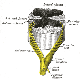
The anterior median fissure of the spinal cord has an average depth of about 3 mm, but this is increased in the lower part of the spinal cord.

The posterior commissure is a rounded band of white fibers crossing the middle line on the dorsal aspect of the rostral end of the cerebral aqueduct. It is important in the bilateral pupillary light reflex.
The lateral grey column is one of the three grey columns of the spinal cord ; the others being the anterior and posterior grey columns. The lateral grey column is primarily involved with activity in the sympathetic division of the autonomic motor system. It projects to the side as a triangular field in the thoracic and upper lumbar regions of the postero-lateral part of the anterior grey column.
The ventral spinothalamic fasciculus situated in the marginal part of the anterior funiculus and intermingled more or less with the vestibulo-spinal fasciculus, is derived from cells in the posterior column or intermediate gray matter of the opposite side. Aβ fibres carry sensory information pertaining to crude touch from the skin. After entering the spinal cord the first order neurons synapse, and the second order neurons decussate via the anterior commissure. These second order neurons ascend synapsing in the VPL of the thalamus. It should also be noted incoming first order neurons can ascend or descend via the Lissauer tract.
The lateral spinothalamic tract, which is a part of the anterolateral system, is a bundle of afferent nerve fibers ascending through the white matter of the spinal cord, carrying sensory information to the brain. It carries pain, crude touch and temperature sensory information to the thalamus. It is composed primarily of fast-conducting, sparsely myelinated A delta fibers and slow-conducting, unmyelinated C fibers. These are secondary sensory neurons which have already synapsed with the primary sensory neurons of the peripheral nervous system in the posterior horn of the spinal cord.

The commissural fibers or transverse fibers are axons that connect the two hemispheres of the brain. In contrast to commissural fibers, association fibers connect regions within the same hemisphere of the brain, and projection fibers connect each region to other parts of the brain or to the spinal cord.
A commissure is the location at which two objects abut or are joined. The term is used especially in the fields of anatomy and biology.

The anterior white commissure is a bundle of nerve fibers which cross the midline of the spinal cord just anterior to the gray commissure. A delta fibers and C fibers carrying pain sensation in the spinothalamic tract contribute to this commissure, as do fibers of the anterior corticospinal tract, which carry motor signals from the primary motor cortex.

A nerve tract is a bundle of nerve fibers (axons) connecting nuclei of the central nervous system. In the peripheral nervous system this is known as a nerve. The main nerve tracts in the central nervous system are of three types: association fibers, commissural fibers, and projection fibers. A tract may also be referred to as a commissure, fasciculus or decussation. A commissure connects the two cerebral hemispheres at the same levels. Examples are the posterior commissure and the corpus callosum. A decussation is a connection made by fibres that cross at different levels (obliquely), such as the sensory decussation. Examples of a fascicle are the subthalamic fasciculus and the lenticular fasciculus.

This article describes anatomical terminology that is used to describe the central and peripheral nervous systems - including the brain, brainstem, spinal cord, and nerves.
The proper fasciculi, or spinospinal fasciculi, or propriospinal tracts, are groups of short fibres, ascending and descending, and crossed and uncrossed, within the spinal cord. These fibres are grouped into anterior, posterior, and lateral regions and make up a spinal pathway. Descending dorsal root collaterals are often included in the pathway.


