Syphilis is a sexually transmitted infection caused by the bacterium Treponema pallidum subspecies pallidum. The signs and symptoms depend on the stage it presents: primary, secondary, latent or tertiary. The primary stage classically presents with a single chancre, though there may be multiple sores. In secondary syphilis, a diffuse rash occurs, which frequently involves the palms of the hands and soles of the feet. There may also be sores in the mouth or vagina. Latent syphilis has no symptoms and can last years. In tertiary syphilis, there are gummas, neurological problems, or heart symptoms. Syphilis has been known as "the great imitator", because it may cause symptoms similar to many other diseases.

Necrosis is a form of cell injury which results in the premature death of cells in living tissue by autolysis. The term "necrosis" came about in the mid-19th century and is commonly attributed to German pathologist Rudolf Virchow, who is often regarded as one of the founders of modern pathology. Necrosis is caused by factors external to the cell or tissue, such as infection, or trauma which result in the unregulated digestion of cell components. In contrast, apoptosis is a naturally occurring programmed and targeted cause of cellular death. While apoptosis often provides beneficial effects to the organism, necrosis is almost always detrimental and can be fatal.

Treponema pallidum, formerly known as Spirochaeta pallida, is a microaerophilic, gram-negative, spirochaete bacterium with subspecies that cause the diseases syphilis, bejel, and yaws. It is known to be transmitted only among humans and baboons. T. pallidum can enter the host through mucosal membranes or open lesions in the skin and is primarily spread through sexual contact. It is a helically coiled microorganism usually 6–15 μm long and 0.1–0.2 μm wide. T. pallidum's lack of both a tricarboxylic acid cycle and processes for oxidative phosphorylation results in minimal metabolic activity. As a chemoorganoheterotroph, Treponema pallidum is an obligate parasite that acquires its glucose carbon source from its host. Glucose can be used not only as a primary carbon source but also in glycolytic mechanisms to generate ATP needed to power the bacterium given its minimal genome. The treponemes have cytoplasmic and outer membranes. Using light microscopy, treponemes are visible only by using dark-field illumination. T. pallidum consists of three subspecies, T. p. pallidum, T. p. endemicum, and T. p. pertenue, each of which has a distinct related disorder. The ability of T. pallidum to avoid host immune defenses has allowed for stealth pathogenicity. The unique outer membrane structure and minimal expression of surface proteins of T. pallidum has made vaccine development difficult. Treponema pallidum can be treated with high efficacy by antibiotics that inhibit bacterial cell wall synthesis such as the beta-lactam antimicrobial penicillin-G.

Yaws is a tropical infection of the skin, bones, and joints caused by the spirochete bacterium Treponema pallidum pertenue. The disease begins with a round, hard swelling of the skin, 2 to 5 cm in diameter. The center may break open and form an ulcer. This initial skin lesion typically heals after 3–6 months. After weeks to years, joints and bones may become painful, fatigue may develop, and new skin lesions may appear. The skin of the palms of the hands and the soles of the feet may become thick and break open. The bones may become misshapen. After 5 years or more, large areas of skin may die, leaving scars.

Granuloma inguinale is a bacterial disease caused by Klebsiella granulomatis characterized by genital ulcers. It is endemic in many less-developed regions. It is also known as donovanosis, granuloma genitoinguinale, granuloma inguinale tropicum, granuloma venereum, granuloma venereum genitoinguinale, lupoid form of groin ulceration, serpiginous ulceration of the groin, ulcerating granuloma of the pudendum, and ulcerating sclerosing granuloma. Oral manifestations are also notably seen. The lesions of oral cavity are usually secondary to active genital lesions.

A granuloma is an aggregation of macrophages that forms in response to chronic inflammation. This occurs when the immune system attempts to isolate foreign substances that it is otherwise unable to eliminate. Such substances include infectious organisms including bacteria and fungi, as well as other materials such as foreign objects, keratin, and suture fragments.

Congenital syphilis is syphilis that occurs when a mother with untreated syphilis passes the infection to her baby during pregnancy or at birth. It may present in the fetus, infant, or later. Clinical features vary and differ between early onset, that is presentation before 2-years of age, and late onset, presentation after age 2-years. Infection in the unborn baby may present as poor growth, non-immune hydrops leading to premature birth or loss of the baby, or no signs. Affected newborns mostly initially have no clinical signs. They may be small and irritable. Characteristic features include a rash, fever, large liver and spleen, a runny and congested nose, and inflammation around bone or cartilage. There may be jaundice, large glands, pneumonia, meningitis, warty bumps on genitals, deafness or blindness. Untreated babies that survive the early phase may develop skeletal deformities including deformity of the nose, lower legs, forehead, collar bone, jaw, and cheek bone. There may be a perforated or high arched palate, and recurrent joint disease. Other late signs include linear perioral tears, intellectual disability, hydrocephalus, and juvenile general paresis. Seizures and cranial nerve palsies may first occur in both early and late phases. Eighth nerve palsy, interstitial keratitis and small notched teeth may appear individually or together; known as Hutchinson's triad.

Peripheral tuberculous lymphadenitis is a form of tuberculosis infection occurring outside of the lungs. In general, it describes tuberculosis infection of the lymph nodes, leading to lymphadenopathy. When cervical lymph nodes are affected, it is commonly referred to as "Scrofula." A majority of tuberculosis infections affect the lungs, and extra-pulmonary tuberculosis infections account for the remainder; these most commonly involve the lymphatic system. Although the cervical region is most commonly affected, tuberculous lymphadenitis can occur all around the body, including the axillary and inguinal regions.

Bejel, or endemic syphilis, is a chronic skin and tissue disease caused by infection by the endemicum subspecies of the spirochete Treponema pallidum. Bejel is one of the "endemic treponematoses", a group that also includes yaws and pinta. Typically, endemic trepanematoses begin with localized lesions on the skin or mucous membranes. Pinta is limited to affecting the skin, whereas bejel and yaws are considered to be invasive because they can also cause disease in bone and other internal tissues.

Gas gangrene is a bacterial infection that produces tissue gas in gangrene. This deadly form of gangrene usually is caused by Clostridium perfringens bacteria. About 1,000 cases of gas gangrene are reported yearly in the United States.
The oral mucosa is the mucous membrane lining the inside of the mouth. It comprises stratified squamous epithelium, termed "oral epithelium", and an underlying connective tissue termed lamina propria. The oral cavity has sometimes been described as a mirror that reflects the health of the individual. Changes indicative of disease are seen as alterations in the oral mucosa lining the mouth, which can reveal systemic conditions, such as diabetes or vitamin deficiency, or the local effects of chronic tobacco or alcohol use. The oral mucosa tends to heal faster and with less scar formation compared to the skin. The underlying mechanism remains unknown, but research suggests that extracellular vesicles might be involved.

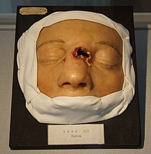
Lupus vulgaris are painful cutaneous tuberculosis skin lesions with nodular appearance, most often on the face around the nose, eyelids, lips, cheeks, ears and neck. It is the most common Mycobacterium tuberculosis skin infection. The lesions may ultimately develop into disfiguring skin ulcers if left untreated.

Neurosyphilis is the infection of the central nervous system by Treponema pallidum, the bacterium that causes the sexually transmitted infection syphilis. In the era of modern antibiotics, the majority of neurosyphilis cases have been reported in HIV-infected patients.
Epulis fissuratum is a benign hyperplasia of fibrous connective tissue which develops as a reactive lesion to chronic mechanical irritation produced by the flange of a poorly fitting denture. More simply, epulis fissuratum is where excess folds of firm tissue form inside the mouth, as a result of rubbing on the edge of dentures that do not fit well. It is a harmless condition and does not represent oral cancer. Treatment is by simple surgical removal of the lesion, and also by adjustment of the denture or provision of a new denture.
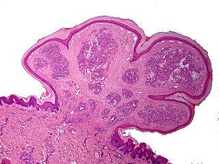
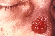
A pyogenic granuloma or lobular capillary hemangioma is a vascular tumor that occurs on both mucosa and skin, and appears as an overgrowth of tissue due to irritation, physical trauma, or hormonal factors. It is often found to involve the gums, skin, or nasal septum, and has also been found far from the head, such as in the thigh.
A peripheral ossifying fibroma, also known as ossifying fibrous epulis, is “a gingival nodule which is composed of a cellular fibroblastic connective tissue stroma which is associated with the formation of randomly dispersed foci of mineralised products, which consists of bone, cementum-like tissue, or a dystrophic calcification. The lesion is considered part of an ossifying fibroma, but that is usually considered to be a jaw tumor. Because of its overwhelming incidence on the gingiva, the condition is associated with two other diseases, though not because they occur together. Instead, the three are associated with each other because they appear frequently on gingiva: pyogenic granuloma and peripheral giant cell granuloma. Some researchers believe peripheral ossifying fibromas to be related to pyogenic fibromas and, in some instances, are the result of a pyogenic granuloma which has undergone fibrosis and calcification.

Commonly known as a dental cyst, the periapical cyst is the most common odontogenic cyst. It may develop rapidly from a periapical granuloma, as a consequence of untreated chronic periapical periodontitis.
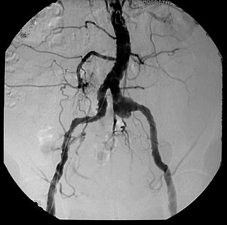
Syphilitic aortitis is inflammation of the aorta associated with the tertiary stage of syphilis infection. SA begins as inflammation of the outermost layer of the blood vessel, including the blood vessels that supply the aorta itself with blood, the vasa vasorum. As SA worsens, the vasa vasorum undergo hyperplastic thickening of their walls thereby restricting blood flow and causing ischemia of the outer two-thirds of the aortic wall. Starved for oxygen and nutrients, elastic fibers become patchy and smooth muscle cells die. If the disease progresses, syphilitic aortitis leads to an aortic aneurysm. Overall, tertiary syphilis is a rare cause of aortic aneurysms. Syphilitic aortitis has become rare in the developed world with the advent of penicillin treatments after World War II.

Touton giant cells are a type of multinucleated giant cell observed in a myriad of pathological disorders and conditions. Specifically, Touton giant cells are found in lipid-rich lesions such as those of fat necrosis, xanthoma, xanthelasma and xanthogranulomas. Touton giant cells are also referred to as xanthelasmatic cells due to the fact they are found in lesions associated with xanthomas which are skin growths with yellow, lipid filled deposits. Touton giant cells are often frequently observed in granulomatous inflammation, which is a type of inflammation caused by the clustering of immune cells, or granulomas. They are also found in dermatofibroma. Touton giant cells are commonly characterized by their very unqiue histological appearance as well as their response to various stimuli associated with the body's immune system.

Meningeal syphilis is a chronic form of syphilis infection that affects the central nervous system. Treponema pallidum, a spirochate bacterium, is the main cause of syphilis, which spreads drastically throughout the body and can infect all its systems if not treated appropriately. Treponema pallidum is the main cause of the onset of meningeal syphilis and other treponemal diseases, and it consists of a cytoplasmic and outer membrane that can cause a diverse array of diseases in the central nervous system and brain.