H2AFX

Last updated • 4 min readFrom Wikipedia, The Free Encyclopedia
H2AX
Protein H2AFX PDB 1aoi.png
Available structures
PDB Ortholog search: PDBe RCSB
Identifiers
Aliases H2AX , H2A.X, H2A/X, H2A histone family member X, H2A.X variant histone, H2AFX
External IDs OMIM: 601772; MGI: 102688; HomoloGene: 134201; GeneCards: H2AX; OMA:H2AX - orthologs
Orthologs
SpeciesHumanMouse
Entrez
Ensembl
UniProt
RefSeq (mRNA)

NM_002105

NM_010436

RefSeq (protein)

NP_002096

NP_034566

Location (UCSC) Chr 11: 119.09 – 119.1 Mb Chr 9: 44.25 – 44.25 Mb
PubMed search [3] [4]
Wikidata
View/Edit Human View/Edit Mouse

H2A histone family member X (usually abbreviated as H2AX) is a type of histone protein from the H2A family encoded by the H2AFX gene. An important phosphorylated form is γH2AX (S139), which forms when double-strand breaks appear.

Contents

In humans and other eukaryotes, the DNA is wrapped around histone octamers, consisting of core histones H2A, H2B, H3 and H4, to form chromatin. H2AX contributes to nucleosome-formation, chromatin-remodeling and DNA repair, and is also used in vitro as an assay for double-strand breaks in dsDNA.

Formation of γH2AX

H2AX becomes phosphorylated on serine 139, then called γH2AX, as a reaction on DNA double-strand breaks (DSB). The kinases of the PI3-family (Ataxia telangiectasia mutated, ATR and DNA-PKcs) are responsible for this phosphorylation, especially ATM. The modification can happen accidentally during replication fork collapse or in the response to ionizing radiation but also during controlled physiological processes such as V(D)J recombination. γH2AX is a sensitive target for looking at DSBs in cells. The presence of γH2AX by itself, however, is not the evidence of the DSBs. [5] The role of the phosphorylated form of the histone in DNA repair is under discussion but it is known that because of the modification the DNA becomes less condensed, potentially allowing space for the recruitment of proteins necessary during repair of DSBs. Mutagenesis experiments have shown that the modification is necessary for the proper formation of ionizing radiation induced foci in response to double strand breaks, but is not required for the recruitment of proteins to the site of DSBs.

Function

DNA damage response

The histone variant H2AX constitutes about 2-25% of the H2A histones in mammalian chromatin. [6] When a double-strand break occurs in DNA, a sequence of events occurs in which H2AX is altered.

Very early after a double-strand break, a specific protein that interacts with and affects the architecture of chromatin is phosphorylated and then released from the chromatin. This protein, heterochromatin protein 1 (HP1)-beta (CBX1), is bound to histone H3 methylated on lysine 9 (H3K9me). Half-maximum release of HP1-beta from damaged DNA occurs within one second. [7] A dynamic alteration in chromatin structure is triggered by HP1-beta release. This alteration in chromatin structure promotes H2AX phosphorylation by ATM, ATR and DNA-PK, [8] allowing formation of γH2AX (H2AX phosphorylated on serine 139). γH2AX can be detected as soon as 20 seconds after irradiation of cells (with DNA double-strand break formation), and half maximum accumulation of γH2AX occurs in one minute. [6] Chromatin with phosphorylated γH2AX extends to about a million base pairs on each side of a DNA double-strand break. [6]

MDC1 (mediator of DNA damage checkpoint protein 1) then binds to γH2AX and the γH2AX/MDC1 complex then orchestrates further interactions in double-strand break repair. [9] The ubiquitin ligases RNF8 and RNF168 bind to the γH2AX/MDC1 complex, ubiquitylating other chromatin components. This allows the recruitment of BRCA1 and 53BP1 to the long, modified γH2AX/MDC1 chromatin. [9] Other proteins that stably assemble on the extensive γH2AX-modified chromatin are the MRN complex (a protein complex consisting of Mre11, Rad50 and Nbs1), RAD51 and the ATM kinase. [10] [11] Further DNA repair components, such as RAD52 and RAD54, rapidly and reversibly interact with the core components stably associated with γH2AX-modified chromatin. [11] The constitutive level of γH2AX expression in live cells, untreated by exogenous agents, likely represents DNA damage by endogenous oxidants generated during cellular respiration. [12]

In chromatin remodeling

The packaging of eukaryotic DNA into chromatin presents a barrier to all DNA-based processes that require recruitment of enzymes to their sites of action. To allow DNA repair, the chromatin must be remodeled.

γH2AX, the phosphorylated form of H2AX, is involved in the steps leading to chromatin decondensation after DNA double-strand breaks. γH2AX does not, itself, cause chromatin decondensation, but within 30 seconds of ionizing radiation, RNF8 protein can be detected in association with γH2AX. [13] RNF8 mediates extensive chromatin decondensation, through its subsequent interaction with CHD4, [14] a component of the nucleosome remodeling and deacetylase complex NuRD.

γH2AX as an assay for double-strand breaks

An assay for γH2AX generally reflects the presence of double-strand breaks in DNA, though the assay may indicate other minor phenomena as well. [15] On the one hand, overwhelming evidence supports a strong, quantitative correlation between γH2AX foci formation and DNA double-strand break induction following ionizing radiation exposure, based on absolute yields and distributions induced per unit dose. [15] On the other hand, not only the formation of distinct γH2AX foci but also the induction of pan-nuclear γH2AX signals have been reported as a cellular reaction to various stressors other than ionizing radiation. [16] The γH2AX signal is always stronger at DNA double-strand breaks than in undamaged chromatin. [16] γH2AX in undamaged chromatin is thought to possibly be generated via direct phosphorylation of H2AX by activated kinases, most likely diffusing from DNA damage sites. In using γH2AX as a marker for double strand breaks, it is important to recognize that it is a down-stream proxy that can be useful for representing DNA damage repair. It does not represent double strand breaks themselves and this needs careful consideration when interpreting data from such assays. [17]

The γH2AX-assay has several disadvantages, therefore new assays have been created. [18]

Interactions

H2AX has been shown to interact with:

Related Research Articles

Chromatin is a complex of DNA and protein found in eukaryotic cells. The primary function is to package long DNA molecules into more compact, denser structures. This prevents the strands from becoming tangled and also plays important roles in reinforcing the DNA during cell division, preventing DNA damage, and regulating gene expression and DNA replication. During mitosis and meiosis, chromatin facilitates proper segregation of the chromosomes in anaphase; the characteristic shapes of chromosomes visible during this stage are the result of DNA being coiled into highly condensed chromatin.

<span class="mw-page-title-main">Histone</span> Protein family around which DNA winds to form nucleosomes

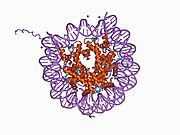
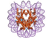
In biology, histones are highly basic proteins abundant in lysine and arginine residues that are found in eukaryotic cell nuclei and in most Archaeal phyla. They act as spools around which DNA winds to create structural units called nucleosomes. Nucleosomes in turn are wrapped into 30-nanometer fibers that form tightly packed chromatin. Histones prevent DNA from becoming tangled and protect it from DNA damage. In addition, histones play important roles in gene regulation and DNA replication. Without histones, unwound DNA in chromosomes would be very long. For example, each human cell has about 1.8 meters of DNA if completely stretched out; however, when wound about histones, this length is reduced to about 90 micrometers (0.09 mm) of 30 nm diameter chromatin fibers.

<span class="mw-page-title-main">DNA repair</span> Cellular mechanism

DNA repair is a collection of processes by which a cell identifies and corrects damage to the DNA molecules that encode its genome. In human cells, both normal metabolic activities and environmental factors such as radiation can cause DNA damage, resulting in tens of thousands of individual molecular lesions per cell per day. Many of these lesions cause structural damage to the DNA molecule and can alter or eliminate the cell's ability to transcribe the gene that the affected DNA encodes. Other lesions induce potentially harmful mutations in the cell's genome, which affect the survival of its daughter cells after it undergoes mitosis. As a consequence, the DNA repair process is constantly active as it responds to damage in the DNA structure. When normal repair processes fail, and when cellular apoptosis does not occur, irreparable DNA damage may occur. This can eventually lead to malignant tumors, or cancer as per the two-hit hypothesis.

<span class="mw-page-title-main">ATM serine/threonine kinase</span> Mammalian protein found in Homo sapiens

ATM serine/threonine kinase or Ataxia-telangiectasia mutated, symbol ATM, is a serine/threonine protein kinase that is recruited and activated by DNA double-strand breaks, oxidative stress, topoisomerase cleavage complexes, splicing intermediates, R-loops and in some cases by single-strand DNA breaks. It phosphorylates several key proteins that initiate activation of the DNA damage checkpoint, leading to cell cycle arrest, DNA repair or apoptosis. Several of these targets, including p53, CHK2, BRCA1, NBS1 and H2AX are tumor suppressors.

<span class="mw-page-title-main">Homologous recombination</span> Genetic recombination between identical or highly similar strands of genetic material

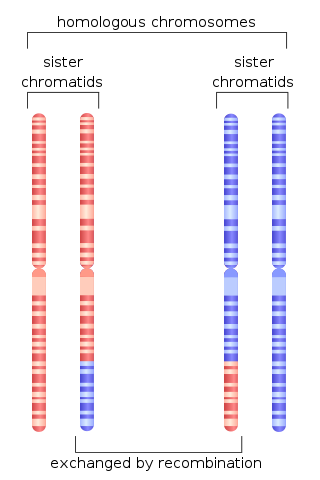
Homologous recombination is a type of genetic recombination in which genetic information is exchanged between two similar or identical molecules of double-stranded or single-stranded nucleic acids.

<span class="mw-page-title-main">Histone H2A</span> One of the five main histone proteins

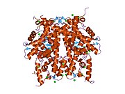
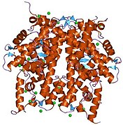
Histone H2A is one of the five main histone proteins involved in the structure of chromatin in eukaryotic cells.

<span class="mw-page-title-main">Nibrin</span> Protein-coding gene in the species Homo sapiens

Nibrin, also known as NBN or NBS1, is a protein which in humans is encoded by the NBN gene.

<span class="mw-page-title-main">Histone-modifying enzymes</span> Type of enzymes

Histone-modifying enzymes are enzymes involved in the modification of histone substrates after protein translation and affect cellular processes including gene expression. To safely store the eukaryotic genome, DNA is wrapped around four core histone proteins, which then join to form nucleosomes. These nucleosomes further fold together into highly condensed chromatin, which renders the organism's genetic material far less accessible to the factors required for gene transcription, DNA replication, recombination and repair. Subsequently, eukaryotic organisms have developed intricate mechanisms to overcome this repressive barrier imposed by the chromatin through histone modification, a type of post-translational modification which typically involves covalently attaching certain groups to histone residues. Once added to the histone, these groups elicit either a loose and open histone conformation, euchromatin, or a tight and closed histone conformation, heterochromatin. Euchromatin marks active transcription and gene expression, as the light packing of histones in this way allows entry for proteins involved in the transcription process. As such, the tightly packed heterochromatin marks the absence of current gene expression.

Chromatin remodeling is the dynamic modification of chromatin architecture to allow access of condensed genomic DNA to the regulatory transcription machinery proteins, and thereby control gene expression. Such remodeling is principally carried out by 1) covalent histone modifications by specific enzymes, e.g., histone acetyltransferases (HATs), deacetylases, methyltransferases, and kinases, and 2) ATP-dependent chromatin remodeling complexes which either move, eject or restructure nucleosomes. Besides actively regulating gene expression, dynamic remodeling of chromatin imparts an epigenetic regulatory role in several key biological processes, egg cells DNA replication and repair; apoptosis; chromosome segregation as well as development and pluripotency. Aberrations in chromatin remodeling proteins are found to be associated with human diseases, including cancer. Targeting chromatin remodeling pathways is currently evolving as a major therapeutic strategy in the treatment of several cancers.

<span class="mw-page-title-main">TP53BP1</span> Protein-coding gene in the species Homo sapiens

Tumor suppressor p53-binding protein 1 also known as p53-binding protein 1 or 53BP1 is a protein that in humans is encoded by the TP53BP1 gene.

<span class="mw-page-title-main">MDC1</span> Protein-coding gene in the species Homo sapiens

Mediator of DNA damage checkpoint protein 1 is a 2080 amino acid long protein that in humans is encoded by the MDC1 gene located on the short arm (p) of chromosome 6. MDC1 protein is a regulator of the Intra-S phase and the G2/M cell cycle checkpoints and recruits repair proteins to the site of DNA damage. It is involved in determining cell survival fate in association with tumor suppressor protein p53. This protein also goes by the name Nuclear Factor with BRCT Domain 1 (NFBD1).

<span class="mw-page-title-main">HIST2H2AC</span> Protein-coding gene in the species Homo sapiens

Histone H2A type 2-C is a protein that in humans is encoded by the HIST2H2AC gene.

<span class="mw-page-title-main">Protein phosphorylation</span> Process of introducing a phosphate group on to a protein

Protein phosphorylation is a reversible post-translational modification of proteins in which an amino acid residue is phosphorylated by a protein kinase by the addition of a covalently bound phosphate group. Phosphorylation alters the structural conformation of a protein, causing it to become activated, deactivated, or otherwise modifying its function. Approximately 13,000 human proteins have sites that are phosphorylated.

<span class="mw-page-title-main">HIST2H2AB</span> Protein-coding gene in the species Homo sapiens

Histone H2A type 2-B is a protein that in humans is encoded by the HIST2H2AB gene.

<span class="mw-page-title-main">RNF8</span> Protein-coding gene in the species Homo sapiens

E3 ubiquitin-protein ligase RNF8 is an enzyme that in humans is encoded by the RNF8 gene. RNF8 has activity both in immune system functions and in DNA repair.

<span class="mw-page-title-main">Homology directed repair</span> Mechanism of DNA repair in cells

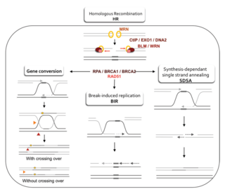
Homology-directed repair (HDR) is a mechanism in cells to repair double-strand DNA lesions. The most common form of HDR is homologous recombination. The HDR mechanism can only be used by the cell when there is a homologous piece of DNA present in the nucleus, mostly in G2 and S phase of the cell cycle. Other examples of homology-directed repair include single-strand annealing and breakage-induced replication. When the homologous DNA is absent, another process called non-homologous end joining (NHEJ) takes place instead.

DNA damage is an alteration in the chemical structure of DNA, such as a break in a strand of DNA, a nucleobase missing from the backbone of DNA, or a chemically changed base such as 8-OHdG. DNA damage can occur naturally or via environmental factors, but is distinctly different from mutation, although both are types of error in DNA. DNA damage is an abnormal chemical structure in DNA, while a mutation is a change in the sequence of base pairs. DNA damages cause changes in the structure of the genetic material and prevents the replication mechanism from functioning and performing properly. The DNA damage response (DDR) is a complex signal transduction pathway which recognizes when DNA is damaged and initiates the cellular response to the damage.

H3S10P is an epigenetic modification to the DNA packaging protein histone H3. It is a mark that indicates the phosphorylation the 10th serine residue of the histone H3 protein.

H3S28P is an epigenetic modification to the DNA packaging protein histone H3. It is a mark that indicates the phosphorylation the 28th serine residue of the histone H3 protein.

H3T6P is an epigenetic modification to the DNA packaging protein histone H3. It is a mark that indicates the phosphorylation of the 6th threonine residue of the histone H3 protein.

References

  1. 1 2 3 GRCh38: Ensembl release 89: ENSG00000188486 Ensembl, May 2017
  2. 1 2 3 GRCm38: Ensembl release 89: ENSMUSG00000049932 Ensembl, May 2017
  3. "Human PubMed Reference:". National Center for Biotechnology Information, U.S. National Library of Medicine.
  4. "Mouse PubMed Reference:". National Center for Biotechnology Information, U.S. National Library of Medicine.
  5. Cleaver JE, Feeney L, Revet I (2011). "Phosphorylated H2Ax is not an unambiguous marker for DNA double strand breaks". Cell Cycle. 10 (19): 3223–4. doi: 10.4161/cc.10.19.17448 . PMID   21921674.
  6. 1 2 3 Rogakou EP, Pilch DR, Orr AH, Ivanova VS, Bonner WM (1998). "DNA double-stranded breaks induce histone H2AX phosphorylation on serine 139". J. Biol. Chem. 273 (10): 5858–68. doi: 10.1074/jbc.273.10.5858 . PMID   9488723.
  7. Ayoub N, Jeyasekharan AD, Bernal JA, Venkitaraman AR (2008). "HP1-beta mobilization promotes chromatin changes that initiate the DNA damage response". Nature. 453 (7195): 682–6. Bibcode:2008Natur.453..682A. doi:10.1038/nature06875. PMID   18438399. S2CID   4348736.
  8. Furuta T, Takemura H, Liao ZY, Aune GJ, Redon C, Sedelnikova OA, Pilch DR, Rogakou EP, Celeste A, Chen HT, Nussenzweig A, Aladjem MI, Bonner WM, Pommier Y (2003). "Phosphorylation of histone H2AX and activation of Mre11, Rad50, and Nbs1 in response to replication-dependent DNA double-strand breaks induced by mammalian DNA topoisomerase I cleavage complexes". J. Biol. Chem. 278 (22): 20303–12. doi: 10.1074/jbc.M300198200 . PMID   12660252.
  9. 1 2 Scully R, Xie A (2013). "Double strand break repair functions of histone H2AX". Mutat. Res. 750 (1–2): 5–14. Bibcode:2013MRFMM.750....5S. doi:10.1016/j.mrfmmm.2013.07.007. PMC   3818383 . PMID   23916969.
  10. Bekker-Jensen S, Lukas C, Kitagawa R, Melander F, Kastan MB, Bartek J, Lukas J (2006). "Spatial organization of the mammalian genome surveillance machinery in response to DNA strand breaks". J. Cell Biol. 173 (2): 195–206. doi:10.1083/jcb.200510130. PMC   2063811 . PMID   16618811.
  11. 1 2 Essers J, Houtsmuller AB, van Veelen L, Paulusma C, Nigg AL, Pastink A, Vermeulen W, Hoeijmakers JH, Kanaar R (2002). "Nuclear dynamics of RAD52 group homologous recombination proteins in response to DNA damage". EMBO J. 21 (8): 2030–7. doi:10.1093/emboj/21.8.2030. PMC   125370 . PMID   11953322.
  12. Tanaka T, Halicka HD, Huang X, Traganos F, Darzynkiewicz Z (2006). "Constitutive histone H2AX phosphorylation and ATM activation, the reporters of DNA damage by endogenous oxidants". Cell Cycle. 5 (17): 1940–5. doi:10.4161/cc.5.17.3191. PMC   3488278 . PMID   16940754.
  13. Mailand N, Bekker-Jensen S, Faustrup H, Melander F, Bartek J, Lukas C, Lukas J (2007). "RNF8 ubiquitylates histones at DNA double-strand breaks and promotes assembly of repair proteins". Cell. 131 (5): 887–900. doi: 10.1016/j.cell.2007.09.040 . PMID   18001824. S2CID   14232192.
  14. Luijsterburg MS, Acs K, Ackermann L, Wiegant WW, Bekker-Jensen S, Larsen DH, Khanna KK, van Attikum H, Mailand N, Dantuma NP (2012). "A new non-catalytic role for ubiquitin ligase RNF8 in unfolding higher-order chromatin structure". EMBO J. 31 (11): 2511–27. doi:10.1038/emboj.2012.104. PMC   3365417 . PMID   22531782.
  15. 1 2 Rothkamm K, Barnard S, Moquet J, Ellender M, Rana Z, Burdak-Rothkamm S (2015). "DNA damage foci: Meaning and significance". Environ. Mol. Mutagen. 56 (6): 491–504. Bibcode:2015EnvMM..56..491R. doi: 10.1002/em.21944 . PMID   25773265. S2CID   32371215.
  16. 1 2 Meyer B, Voss KO, Tobias F, Jakob B, Durante M, Taucher-Scholz G (2013). "Clustered DNA damage induces pan-nuclear H2AX phosphorylation mediated by ATM and DNA-PK". Nucleic Acids Res. 41 (12): 6109–18. doi:10.1093/nar/gkt304. PMC   3695524 . PMID   23620287.
  17. Atkinson, Jake; Bezak, Eva; Kempson, Ivan (4 July 2022). "Imaging DNA double-strand breaks — are we there yet?". Nature Reviews Molecular Cell Biology. 23 (9): 579–580. doi:10.1038/s41580-022-00513-7. PMID   35789205. S2CID   250283224.
  18. Ruprecht N, Hungerbühler MN, Böhm IB, Heverhagen JT (2019). "Improved identification of DNA double strand breaks: γ-H2AX-epitope visualization by confocal microscopy and 3D reconstructed images". Radiat Environ Biophys. 58 (2): 295–302. Bibcode:2019REBio..58..295R. doi: 10.1007/s00411-019-00778-1 . PMID   30799523.
  19. 1 2 Mallery DL, Vandenberg CJ, Hiom K (Dec 2002). "Activation of the E3 ligase function of the BRCA1/BARD1 complex by polyubiquitin chains". The EMBO Journal. 21 (24): 6755–62. doi:10.1093/emboj/cdf691. PMC   139111 . PMID   12485996.
  20. 1 2 Chen A, Kleiman FE, Manley JL, Ouchi T, Pan ZQ (Jun 2002). "Autoubiquitination of the BRCA1*BARD1 RING ubiquitin ligase". The Journal of Biological Chemistry. 277 (24): 22085–92. doi: 10.1074/jbc.M201252200 . PMID   11927591.
  21. Paull TT, Rogakou EP, Yamazaki V, Kirchgessner CU, Gellert M, Bonner WM (2000). "A critical role for histone H2AX in recruitment of repair factors to nuclear foci after DNA damage". Current Biology. 10 (15): 886–95. Bibcode:2000CBio...10..886P. doi: 10.1016/s0960-9822(00)00610-2 . PMID   10959836. S2CID   16108315.
  22. 1 2 Sengupta S, Robles AI, Linke SP, Sinogeeva NI, Zhang R, Pedeux R, Ward IM, Celeste A, Nussenzweig A, Chen J, Halazonetis TD, Harris CC (Sep 2004). "Functional interaction between BLM helicase and 53BP1 in a Chk1-mediated pathway during S-phase arrest". The Journal of Cell Biology. 166 (6): 801–13. doi:10.1083/jcb.200405128. PMC   2172115 . PMID   15364958.
  23. Stewart GS, Wang B, Bignell CR, Taylor AM, Elledge SJ (Feb 2003). "MDC1 is a mediator of the mammalian DNA damage checkpoint". Nature. 421 (6926): 961–6. Bibcode:2003Natur.421..961S. doi:10.1038/nature01446. PMID   12607005. S2CID   4410773.
  24. Xu X, Stern DF (Oct 2003). "NFBD1/MDC1 regulates ionizing radiation-induced focus formation by DNA checkpoint signaling and repair factors". FASEB Journal. 17 (13): 1842–8. doi: 10.1096/fj.03-0310com . PMID   14519663. S2CID   24870579.
  25. Kobayashi J, Tauchi H, Sakamoto S, Nakamura A, Morishima K, Matsuura S, Kobayashi T, Tamai K, Tanimoto K, Komatsu K (Oct 2002). "NBS1 localizes to γH2AX foci through interaction with the FHA/BRCT domain". Current Biology. 12 (21): 1846–51. Bibcode:2002CBio...12.1846K. doi: 10.1016/s0960-9822(02)01259-9 . PMID   12419185. S2CID   10686827.
  26. Fernandez-Capetillo O, Chen HT, Celeste A, Ward I, Romanienko PJ, Morales JC, Naka K, Xia Z, Camerini-Otero RD, Motoyama N, Carpenter PB, Bonner WM, Chen J, Nussenzweig A (Dec 2002). "DNA damage-induced G2-M checkpoint activation by histone H2AX and 53BP1". Nature Cell Biology. 4 (12): 993–7. doi:10.1038/ncb884. PMID   12447390. S2CID   12380387.
  27. Ward IM, Minn K, Jorda KG, Chen J (May 2003). "Accumulation of checkpoint protein 53BP1 at DNA breaks involves its binding to phosphorylated histone H2AX". The Journal of Biological Chemistry. 278 (22): 19579–82. doi: 10.1074/jbc.C300117200 . PMID   12697768.

Further reading