HHV-8-associated MCD

Last updated
HHV-8-associated MCD
Other namesGiant lymph node hyperplasia, lymphoid hamartoma, angiofollicular lymph node hyperplasia
BX2306-20-12043-01-HHV8-06@40x (Enfermedad de Castleman multicentrica asociada a HHV8 y sarcoma de Kaposi).jpg
Micrograph of HHV8-associated Castleman's Disease showing LANA-1 positive lymphoblasts in a regressed germinal center and mantle zone. LANA-1 stain.
Specialty Hematology, immunology, infectious disease, pathology
Diagnostic method Based on patient history, physical exam, laboratory testing, medical imaging, histopathology
FrequencyApproximately 300 new cases per year in the United States

Human herpesvirus 8 associated multicentric Castleman disease (HHV-8-associated MCD) is a subtype of Castleman disease (also known as giant lymph node hyperplasia, lymphoid hamartoma, or angiofollicular lymph node hyperplasia), a group of rare lymphoproliferative disorders characterized by lymph node enlargement, characteristic features on microscopic analysis of enlarged lymph node tissue, and a range of symptoms and clinical findings.

Contents

People with human herpesvirus 8 associated multicentric Castleman disease (HHV-8-associated MCD) have enlarged lymph nodes in multiple regions and often have flu-like symptoms, abnormal findings on blood tests, and dysfunction of vital organs, such as the liver, kidneys, and bone marrow.

HHV-8-associated MCD is known to be caused by uncontrolled infection with the human herpesvirus 8 virus (HHV-8) and is most frequently diagnosed in patients with human immunodeficiency virus (HIV). HHV-8-associated MCD is treated with a variety of medications, including immunosuppressants, chemotherapy, and antivirals.

Castleman disease is named after Dr. Benjamin Castleman, who first described the disease in 1956. The Castleman Disease Collaborative Network is the largest organization focused on the disease and is involved in research, awareness, and patient support.

Signs and symptoms

People with HHV-8-associated MCD may experience enlarged lymph nodes in multiple lymph node regions; systemic symptoms, such as fever, night sweats, unintended weight loss, and fatigue; rashes such as cherry hemangiomas or Kaposi sarcoma; enlargement of the liver and/or spleen; and extravascular fluid accumulation in the extremities (edema), abdomen (ascites), or lining of the lungs (pleural effusion). [1]

Associated diseases

HHV-8-associated MCD is most commonly diagnosed in patients with human immunodeficiency virus (HIV), but can be seen in patients without HIV as well. [2]

Patients with HHV-8-associated MCD are often found to have Kaposi sarcoma, [1] a cancer caused by the HHV-8 virus and most commonly seen in patients with HIV. Patients with HHV-8-associated MCD have been observed to be at greater risk of developing lymphoma. [3]

Micrograph of a Kaposi sarcoma in a lymph node biopsy of a patient with HHV8-associated MCD. Kaposi sarcoma in a patient with HHV8-associated MCD.jpg
Micrograph of a Kaposi sarcoma in a lymph node biopsy of a patient with HHV8-associated MCD.

Causes

HHV-8-associated MCD is known to be caused by infection with human herpesvirus-8. [4] The HHV-8 virus is commonly found in healthy individuals showing no signs of disease, [5] but it is also known to cause diseases such as Kaposi sarcoma and HHV-8-associated MCD. Diseases caused by HHV-8 are seen most frequently in the setting of immune dysfunction related to factors such as HIV infection or use of immunosuppressant medications; however, HHV-8-associated diseases, including HHV-8-associated MCD, have been reported in otherwise healthy individuals. [2]

Mechanism

In HHV-8-associated MCD the HHV-8 virus infects B cells and plasmablasts in lymph nodes and causes infected cells to release proinflammatory cytokines, signaling molecules that increase the activity of immune cells. In particular, HHV-8 infection of immune cells leads to increased levels of Interleukin-6 (IL-6), a cytokine known to play a role in other forms of Castleman disease.[ citation needed ]

The HHV-8 virus contains a gene coding for a viral variant of the IL-6 molecule. Cells infected by HHV-8 produce the viral variant of IL-6 and normal human IL-6, both of which contribute to the increased B cell proliferation and clinical findings seen in HHV-8-associated MCD. [4]

Diagnosis

HHV-8-associated MCD is diagnosed based on patient history, physical exam, laboratory testing, radiologic imaging, and microscopic analysis (histology) of biopsied tissue from an enlarged lymph node. [6]

Formal criteria for the diagnosis of HHV-8-associated MCD have not been published; however, diagnosis requires enlargement of lymph nodes in multiple lymph node regions (typically confirmed with radiologic imaging), histologic changes consistent with HHV-8-associated MCD on biopsy of an enlarged lymph node, and confirmation of HHV-8 infection by LANA-1 lymph node staining or peripheral blood polymerase chain reaction for HHV-8. [6] HIV testing is useful for management, but a positive result is not necessary for to diagnose HHV-8-associated MCD. [2]

Classification

Castleman disease describes a group of at least three distinct disorders—unicentric Castleman disease (UCD), human herpesvirus 8 associated multicentric Castleman disease (HHV-8-associated MCD), and idiopathic multicentric Castleman disease (iMCD). Identifying the correct subtype of the disease is important, as the three disorders vary significantly in symptoms, clinical findings, disease mechanism, treatment approach, and prognosis. [7]

Laboratory testing

Laboratory testing may demonstrate low hemoglobin levels (anemia), abnormal platelet counts, low albumin levels, elevated inflammatory markers such as C-Reactive Protein, elevated creatinine (kidney dysfunction), increased levels of immunoglobulins, and elevation of molecules involved in inflammation (cytokines), such as interleukin 6 (IL-6). [1] [8]

Medical imaging

Radiologic imaging will demonstrate enlarged lymph nodes in multiple regions, which are typically 18F-fluorodoxyglucose (FDG) avid on positron-emission tomography (PET). [9]

Pathology

Unlike the other subtypes of Castleman disease, UCD and iMCD, which can present with a spectrum of features on microscopic analysis (histology) of tissue biopsied from an enlarged lymph node, only a plasmablastic pattern of histologic features has been described in HHV-8-associated MCD. Plasmablastic features are similar to the plasmacytic features seen in iMCD and UCD, with both demonstrating increased plasma cells in interfollicular spaces. Plasmablastic features are differentiated from plasmacytic features by increased numbers of plasmablasts in follicular mantle zones. [10]

Staining with latency-associated nuclear antigen (LANA-1), a marker for HHV-8 infection, is typically positive. [11]

Treatment

Data regarding treatment for HHV-8-associated MCD is limited and draws on a combination of observational case series and case reports. There have been no randomized controlled trials investigating treatment options for HHV-8-associated MCD. Surgical removal of enlarged lymph nodes, the standard treatment for UCD, is ineffective in HHV-8-associated MCD. Like iMCD, HHV-8-associated MCD is treated with medications. Factors influencing medication selection include disease severity, presence of HIV, presence of Kaposi sarcoma, and response to prior treatments. [12]

Rituximab, a drug that depletes B cells, is the first line agent for patients with HHV-8-associated MCD and is recommended in all patients with proven HHV-8-associated MCD unless they have a contraindication to the medication or have failed treatment with the medication in the past. For patients who present with severe organ dysfunction, develop worsening organ dysfunction despite treatment with Rituximab, or have concurrent Kaposi sarcoma, additional chemotherapeutic agents are added to Rituximab. All patients with HIV should continue or initiate treatment with antiretroviral therapy. Antiviral medications targeted at HHV-8, such as valganciclovir, may be used in patients with poorly controlled HIV or concurrent Kaposi sarcoma. [12]

Rituximab may be discontinued once clinical and laboratory evidence of active disease resolve or continued as maintenance therapy. [12] Patients who responded to Rituximab but have recurrent HHV-8-associated MCD after discontinuing the medication typically respond to retreatment with Rituximab. [1]

Follow-up

Patients with HHV-8-associated MCD are typically seen at routine intervals for assessment of treatment response and disease progression. Follow-up visits may include evaluation of symptoms, physical examination, laboratory testing, and radiologic imaging. [12]

Prognosis

Outcomes in HHV-8-associated MCD have improved significantly since treatment with rituximab has become more common. In one published case series from an experienced center, observed survival in a cohort of HIV patients with HHV-8-associated MCD was 94% at 2 years and 90% at 5 years. [1]

Epidemiology

No epidemiologic study has been published estimating the incidence for HHV-8-associated MCD in the general population.[ citation needed ]

While rates of Kaposi sarcoma in HIV patients—which like HHV-8-associated MCD is caused by uncontrolled HHV-8 infection—decreased as treatment with antiretroviral therapy became more common, rates of HHV-8-associated MCD increased over the same time period. [13]

There have been no published epidemiologic studies of Castleman disease outside of the United States; however, rates of HHV-8-associated MCD may vary in different populations based on the prevalence of HHV-8 infection.[ citation needed ]

History

Castleman disease was first described by Dr. Benjamin Castleman in 1956. [14] In 1995, the association between HHV-8 and Castleman disease was described in patients with HIV. [15] World Castleman Disease Day was established in 2018.

Culture

The Castleman Disease Collaborative Network was founded in 2012 and is the largest organization focused on Castleman disease. It is a global collaborative network involved in research, awareness, and patient support. [16]

Related Research Articles

<span class="mw-page-title-main">Non-Hodgkin lymphoma</span> Type of cancer of lymph nodes

Non-Hodgkin lymphoma (NHL), also known as non-Hodgkin's lymphoma, is a group of blood cancers that includes all types of lymphomas except Hodgkin lymphomas. Symptoms include enlarged lymph nodes, fever, night sweats, weight loss, and tiredness. Other symptoms may include bone pain, chest pain, or itchiness. Some forms are slow-growing while others are fast-growing.

<span class="mw-page-title-main">Castleman disease</span> Group of lymphoproliferative disorders

Castlemandisease (CD) describes a group of rare lymphoproliferative disorders that involve enlarged lymph nodes, and a broad range of inflammatory symptoms and laboratory abnormalities. Whether Castleman disease should be considered an autoimmune disease, cancer, or infectious disease is currently unknown.

Rhadinovirus is a genus of viruses in the order Herpesvirales, in the family Herpesviridae, in the subfamily Gammaherpesvirinae. Humans and other mammals serve as natural hosts. There are 12 species in this genus. Diseases associated with this genus include: Kaposi's sarcoma, primary effusion lymphoma and multicentric Castleman's disease, caused by Human gammaherpesvirus 8 (HHV-8), also known as Kaposi's sarcoma-associated herpesvirus (KSHV). The term rhadino comes from the Latin fragile, referring to the tendency of the viral genome to break apart when it is isolated.

<span class="mw-page-title-main">Kaposi's sarcoma-associated herpesvirus</span> Species of virus

Kaposi's sarcoma-associated herpesvirus (KSHV) is the ninth known human herpesvirus; its formal name according to the International Committee on Taxonomy of Viruses (ICTV) is Human gammaherpesvirus 8, or HHV-8 in short. Like other herpesviruses, its informal names are used interchangeably with its formal ICTV name. This virus causes Kaposi's sarcoma, a cancer commonly occurring in AIDS patients, as well as primary effusion lymphoma, HHV-8-associated multicentric Castleman's disease and KSHV inflammatory cytokine syndrome. It is one of seven currently known human cancer viruses, or oncoviruses. Even after many years since the discovery of KSHV/HHV8, there is no known cure for KSHV associated tumorigenesis.

AIDS-defining clinical conditions is the list of diseases published by the Centers for Disease Control and Prevention (CDC) that are associated with AIDS and used worldwide as a guideline for AIDS diagnosis. CDC exclusively uses the term AIDS-defining clinical conditions, but the other terms remain in common use.

<span class="mw-page-title-main">Primary effusion lymphoma</span> Medical condition

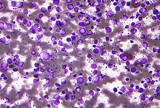
Primary effusion lymphoma (PEL) is classified as a diffuse large B cell lymphoma. It is a rare malignancy of plasmablastic cells that occurs in individuals that are infected with the Kaposi's sarcoma-associated herpesvirus. Plasmablasts are immature plasma cells, i.e. lymphocytes of the B-cell type that have differentiated into plasmablasts but because of their malignant nature do not differentiate into mature plasma cells but rather proliferate excessively and thereby cause life-threatening disease. In PEL, the proliferating plasmablastoid cells commonly accumulate within body cavities to produce effusions, primarily in the pleural, pericardial, or peritoneal cavities, without forming a contiguous tumor mass. In rare cases of these cavitary forms of PEL, the effusions develop in joints, the epidural space surrounding the brain and spinal cord, and underneath the capsule which forms around breast implants. Less frequently, individuals present with extracavitary primary effusion lymphomas, i.e., solid tumor masses not accompanied by effusions. The extracavitary tumors may develop in lymph nodes, bone, bone marrow, the gastrointestinal tract, skin, spleen, liver, lungs, central nervous system, testes, paranasal sinuses, muscle, and, rarely, inside the vasculature and sinuses of lymph nodes. As their disease progresses, however, individuals with the classical effusion-form of PEL may develop extracavitary tumors and individuals with extracavitary PEL may develop cavitary effusions.

<span class="mw-page-title-main">Robert Yarchoan</span>

Robert Yarchoan is a medical researcher who played an important role in the development of the first effective drugs for AIDS. He is the Chief of the HIV and AIDS Malignancy Branch in the NCI and also coordinates HIV/AIDS malignancy research throughout the NCI as director of the Office of HIV and AIDS Malignancy (OHAM).

<span class="mw-page-title-main">Patrick S. Moore</span>

Patrick S. Moore is an Irish and American virologist and epidemiologist who co-discovered together with his wife, Yuan Chang, two different human viruses causing the AIDS-related cancer Kaposi's sarcoma and the skin cancer Merkel cell carcinoma. The couple met while in medical school together and were married in 1989 while they pursued fellowships at different universities.

<span class="mw-page-title-main">Yuan Chang</span> American virologist and pathologist

Yuan Chang is a Taiwanese-American virologist and pathologist who co-discovered together with her husband, Patrick S. Moore, the Kaposi's sarcoma-associated herpesvirus (KSHV) and Merkel cell polyomavirus, two of the seven known human oncoviruses.

<span class="mw-page-title-main">Human herpesvirus 6</span> Informal grouping of viruses which caused human herpesvirus 6 Infection

Human herpesvirus 6 (HHV-6) is the common collective name for human betaherpesvirus 6A (HHV-6A) and human betaherpesvirus 6B (HHV-6B). These closely related viruses are two of the nine known herpesviruses that have humans as their primary host.

<span class="mw-page-title-main">Diffuse large B-cell lymphoma</span> Type of blood cancer

Diffuse large B-cell lymphoma (DLBCL) is a cancer of B cells, a type of lymphocyte that is responsible for producing antibodies. It is the most common form of non-Hodgkin lymphoma among adults, with an annual incidence of 7–8 cases per 100,000 people per year in the US and UK. This cancer occurs primarily in older individuals, with a median age of diagnosis at ~70 years, although it can occur in young adults and, in rare cases, children. DLBCL can arise in virtually any part of the body and, depending on various factors, is often a very aggressive malignancy. The first sign of this illness is typically the observation of a rapidly growing mass or tissue infiltration that is sometimes associated with systemic B symptoms, e.g. fever, weight loss, and night sweats.

The latency-associated nuclear antigen (LANA-1) or latent nuclear antigen is a Kaposi's sarcoma-associated herpesvirus (KSHV) latent protein initially found by Moore and colleagues as a speckled nuclear antigen present in primary effusion lymphoma cells that reacts with antibodies from patients with KS. It is the most immunodominant KSHV protein identified by Western-blotting as 222–234 kDa double bands migrate slower than the predicted molecular weight. LANA has been suspected of playing a crucial role in modulating viral and cellular gene expression. It is commonly used as an antigen in blood tests to detect antibodies in persons that have been exposed to KSHV.

<span class="mw-page-title-main">Kaposi's sarcoma</span> Cancer of the skin, integumentary lymph nodes, or other organs

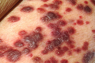
Kaposi's sarcoma (KS) is a type of cancer that can form masses in the skin, in lymph nodes, in the mouth, or in other organs. The skin lesions are usually painless, purple and may be flat or raised. Lesions can occur singly, multiply in a limited area, or may be widespread. Depending on the sub-type of disease and level of immune suppression, KS may worsen either gradually or quickly. Except for Classical KS where there is generally no immune suppression, KS is caused by a combination of immune suppression and infection by Human herpesvirus 8.

Siltuximab is a chimeric monoclonal antibody. It binds to interleukin-6. Siltuximab has been investigated for the treatment of neoplastic diseases: metastatic renal cell cancer, prostate cancer, other types of cancer, and for Castleman's disease.

Human betaherpesvirus 7 (HHV-7) is one of nine known members of the Herpesviridae family that infects humans. HHV-7 is a member of Betaherpesvirinae, a subfamily of the Herpesviridae that also includes HHV-6 and Cytomegalovirus. HHV-7 often acts together with HHV-6, and the viruses together are sometimes referred to by their genus, Roseolovirus. HHV-7 was first isolated in 1990 from CD4+ T cells taken from peripheral blood lymphocytes.

<span class="mw-page-title-main">Signs and symptoms of HIV/AIDS</span>

The stages of HIV infection are acute infection, latency, and AIDS. Acute infection lasts for several weeks and may include symptoms such as fever, swollen lymph nodes, inflammation of the throat, rash, muscle pain, malaise, and mouth and esophageal sores. The latency stage involves few or no symptoms and can last anywhere from two weeks to twenty years or more, depending on the individual. AIDS, the final stage of HIV infection, is defined by low CD4+ T cell counts, various opportunistic infections, cancers, and other conditions.

Large B-cell lymphoma arising in HHV8-associated multicentric Castleman's disease is a type of large B-cell lymphoma, recognized in the WHO 2008 classification. It is sometimes called the plasmablastic form of multicentric Castleman disease. It has sometimes been confused with plasmablastic lymphoma in the literature, although that is a dissimilar specific entity. It has variable CD20 expression and unmutated immunoglobulin variable region genes.

<span class="mw-page-title-main">Unicentric Castleman disease</span> Medical condition

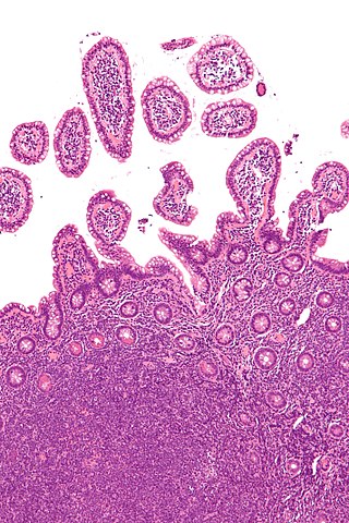
Unicentric Castleman disease is a subtype of Castleman disease, a group of lymphoproliferative disorders characterized by lymph node enlargement, characteristic features on microscopic analysis of enlarged lymph node tissue, and a range of symptoms and clinical findings.

<span class="mw-page-title-main">Idiopathic multicentric Castleman disease</span> Medical condition

Idiopathic multicentric Castleman disease (iMCD) is a subtype of Castleman disease (also known as giant lymph node hyperplasia, lymphoid hamartoma, or angiofollicular lymph node hyperplasia), a group of lymphoproliferative disorders characterized by lymph node enlargement, characteristic features on microscopic analysis of enlarged lymph node tissue, and a range of symptoms and clinical findings.

Epstein–Barr virus–associated lymphoproliferative diseases are a group of disorders in which one or more types of lymphoid cells, i.e. B cells, T cells, NK cells, and histiocytic-dendritic cells, are infected with the Epstein–Barr virus (EBV). This causes the infected cells to divide excessively, and is associated with the development of various non-cancerous, pre-cancerous, and cancerous lymphoproliferative disorders (LPDs). These LPDs include the well-known disorder occurring during the initial infection with the EBV, infectious mononucleosis, and the large number of subsequent disorders that may occur thereafter. The virus is usually involved in the development and/or progression of these LPDs although in some cases it may be an "innocent" bystander, i.e. present in, but not contributing to, the disease.

References

  1. 1 2 3 4 5 Bower M, Newsom-Davis T, Naresh K, Merchant S, Lee B, Gazzard B, Stebbing J, Nelson M (June 2011). "Clinical Features and Outcome in HIV-Associated Multicentric Castleman's Disease". Journal of Clinical Oncology. 29 (18): 2481–6. CiteSeerX   10.1.1.1019.8222 . doi:10.1200/JCO.2010.34.1909. PMID   21555697.
  2. 1 2 3 Dossier A, Meignin V, Fieschi C, Boutboul D, Oksenhendler E, Galicier L (March 2013). "Human herpesvirus 8-related Castleman disease in the absence of HIV infection". Clinical Infectious Diseases. 56 (6): 833–42. doi: 10.1093/cid/cis1009 . PMID   23223599.
  3. Oksenhendler E, Boulanger E, Galicier L, Du MQ, Dupin N, Diss TC, Hamoudi R, Daniel MT, Agbalika F, Boshoff C, Clauvel JP, Isaacson PG, Meignin V (April 2002). "High incidence of Kaposi sarcoma-associated herpesvirus-related non-Hodgkin lymphoma in patients with HIV infection and multicentric Castleman disease". Blood. 99 (7): 2331–6. doi: 10.1182/blood.V99.7.2331 . PMID   11895764.
  4. 1 2 Fajgenbaum DC, Shilling D (February 2018). "Castleman Disease Pathogenesis". Hematology/Oncology Clinics of North America. 32 (1): 11–21. doi:10.1016/j.hoc.2017.09.002. PMID   29157613.
  5. Rabkin CS, Schulz TF, Whitby D, Lennette ET, Magpantay LI, Chatlynne L, Biggar RJ (August 1998). "Interassay correlation of human herpesvirus 8 serologic tests. HHV-8 Interlaboratory Collaborative Group". The Journal of Infectious Diseases. 178 (2): 304–9. doi:10.1086/515649. PMID   9697708.
  6. 1 2 Szalat R, Munshi NC (February 2018). "Diagnosis of Castleman Disease". Hematology/Oncology Clinics of North America. 32 (1): 53–64. doi:10.1016/j.hoc.2017.09.005. PMID   29157619.
  7. Fajgenbaum DC, Uldrick TS, Bagg A, Frank D, Wu D, Srkalovic G, et al. (March 2017). "International, evidence-based consensus diagnostic criteria for HHV-8-negative/idiopathic multicentric Castleman disease". Blood. 129 (12): 1646–1657. doi:10.1182/blood-2016-10-746933. PMC   5364342 . PMID   28087540.
  8. Oksenhendler E, Carcelain G, Aoki Y, Boulanger E, Maillard A, Clauvel JP, Agbalika F (September 2000). "High levels of human herpesvirus 8 viral load, human interleukin-6, interleukin-10, and C reactive protein correlate with exacerbation of multicentric castleman disease in HIV-infected patients". Blood. 96 (6): 2069–73. doi:10.1182/blood.V96.6.2069. PMID   10979949.
  9. Barker R, Kazmi F, Stebbing J, Ngan S, Chinn R, Nelson M, O'Doherty M, Bower M (April 2009). "FDG-PET/CT imaging in the management of HIV-associated multicentric Castleman's disease". European Journal of Nuclear Medicine and Molecular Imaging. 36 (4): 648–52. doi:10.1007/s00259-008-0998-4. PMID   19050873. S2CID   21596522.
  10. Dupin, N.; Diss, T. L.; Kellam, P.; Tulliez, M.; Du, M. Q.; Sicard, D.; Weiss, R. A.; Isaacson, P. G.; Boshoff, C. (2000-02-15). "HHV-8 is associated with a plasmablastic variant of Castleman disease that is linked to HHV-8-positive plasmablastic lymphoma". Blood. 95 (4): 1406–1412. doi:10.1182/blood.V95.4.1406.004k26_1406_1412. ISSN   0006-4971. PMID   10666218.
  11. Dupin N, Diss TL, Kellam P, Tulliez M, Du MQ, Sicard D, Weiss RA, Isaacson PG, Boshoff C (February 2000). "HHV-8 is associated with a plasmablastic variant of Castleman disease that is linked to HHV-8-positive plasmablastic lymphoma". Blood. 95 (4): 1406–12. doi:10.1182/blood.V95.4.1406.004k26_1406_1412. PMID   10666218.
  12. 1 2 3 4 Bower M (November 2010). "How I treat HIV-associated multicentric Castleman disease". Blood. 116 (22): 4415–21. doi:10.1182/blood-2010-07-290213. PMID   20688959.
  13. Powles T, Stebbing J, Bazeos A, Hatzimichael E, Mandalia S, Nelson M, Gazzard B, Bower M (April 2009). "The role of immune suppression and HHV-8 in the increasing incidence of HIV-associated multicentric Castleman's disease". Annals of Oncology. 20 (4): 775–9. doi: 10.1093/annonc/mdn697 . PMID   19179554.
  14. Castleman, B.; Iverson, L.; Menendez, V. P. (July 1956). "Localized mediastinal lymphnode hyperplasia resembling thymoma". Cancer. 9 (4): 822–830. doi: 10.1002/1097-0142(195607/08)9:4<822::AID-CNCR2820090430>3.0.CO;2-4 . ISSN   0008-543X. PMID   13356266.
  15. Soulier J, Grollet L, Oksenhendler E, Cacoub P, Cazals-Hatem D, Babinet P, d'Agay MF, Clauvel JP, Raphael M, Degos L (August 1995). "Kaposi's sarcoma-associated herpesvirus-like DNA sequences in multicentric Castleman's disease". Blood. 86 (4): 1276–80. doi: 10.1182/blood.V86.4.1276.bloodjournal8641276 . PMID   7632932.
  16. Fajgenbaum, David C.; Ruth, Jason R.; Kelleher, Dermot; Rubenstein, Arthur H. (April 2016). "The collaborative network approach: a new framework to accelerate Castleman's disease and other rare disease research". The Lancet. Haematology. 3 (4): e150–152. doi:10.1016/S2352-3026(16)00007-7. ISSN   2352-3026. PMID   27063967.