Unicentric Castleman disease

Last updated
Unicentric Castleman disease
Other namesGiant lymph node hyperplasia, lymphoid hamartoma, angiofollicular lymph node hyperplasia
Castleman disease - high mag.jpg
Micrograph of lymph node biopsy demonstrating hyaline vascular features consistent with Castleman disease
Specialty Hematology, immunology, rheumatology, pathology
Diagnostic method Based on patient history, physical exam, laboratory testing, medical imaging, histopathology
FrequencyApproximately 5000-6000 new cases per year in the United States

Unicentric Castleman disease is a subtype of Castleman disease (also known as giant lymph node hyperplasia, lymphoid hamartoma, or angiofollicular lymph node hyperplasia), a group of lymphoproliferative disorders characterized by lymph node enlargement, characteristic features on microscopic analysis of enlarged lymph node tissue, and a range of symptoms and clinical findings.

Contents

People with unicentric Castleman disease (UCD) have an enlarged lymph node or multiple enlarged lymph nodes in a single lymph node region. It is the most common subtype of Castleman disease, symptoms are typically mild, abnormalities on blood tests are uncommon, organ dysfunction is uncommon, and surgical treatment is curative in the majority of patients. The cause of UCD is not known.

Castleman disease is named after Benjamin Castleman, who first described the disease in 1956. The Castleman Disease Collaborative Network is the largest organization focused on the disease and is involved in research, awareness, and patient support.

Signs and symptoms

In approximately half of patients, UCD presents without symptoms; however, people with the disease may experience enlarged lymph nodes in a single lymph node region or report symptoms related to compression of neighboring structures by enlarged lymph nodes, such as difficulty breathing and pain or pressure in the abdomen or chest.[ citation needed ] [1]

Systemic symptoms (fever, night sweats, weight loss, fatigue) are seen in up to half of UCD patients. [2]

Some symptoms that are commonly seen in HHV-8-associated MCD and iMCD like extravascular fluid accumulation (peripheral edema, ascites, pleural effusions), and enlargement of the liver and/or spleen are uncommon in UCD. [3]

Associated diseases

UCD is associated with increased risk of paraneoplastic pemphigus [3] as well as bronchiolitis obliterans (BO), AA amyloidosis, vascular neoplasms (e.g., FDC sarcoma), and possibly lymphomas. [4]

Causes

UCD has no known causes or risk factors. Cases of Castleman disease running in families have been reported; however, no causative genetic variants have been identified [5]

Mechanism

The mechanism of UCD is poorly understood. Most published research supports a growth of abnormal immune system cells (neoplasm) as the most likely cause of UCD, but this has not been conclusively demonstrated or fully characterized. Other proposed mechanisms include viral infections and autoimmune processes. Because surgical removal of affected lymph nodes in UCD is typically curative and recurrence is rare, it is believed that the pathologic process is limited to affected lymph nodes. Unlike HHV-8-associated MCD, which is caused by the HHV-8 virus, UCD has not been associated with HHV-8 infection1

When findings typically seen in MCD, such as systemic symptoms and laboratory abnormalities, they are likely related to increased levels of molecules that stimulate the immune system (cytokines), such as interleukin 6 (IL-6). [5] Systemic symptoms and laboratory abnormalities may be associated with the presence of plasmacytic features on microscopic analysis of affected lymph node tissue. [6]

There have been no reported cases of UCD transforming into iMCD.[ citation needed ]

Diagnosis

UCD is diagnosed based on patient history, physical exam, laboratory testing, radiologic imaging, and microscopic analysis (histology) of biopsied tissue from an enlarged lymph node.[ citation needed ]

There are no widely accepted diagnostic criteria for UCD; however, diagnosis generally requires enlargement of lymph nodes limited to a single region of lymph nodes (typically confirmed with radiologic imaging), biopsy of an enlarged lymph node demonstrating characteristic features of Castleman disease, and exclusion of other diseases that can mimic UCD. [3]

Classification

Castleman disease describes a group of at least 3 distinct disorders—unicentric Castleman disease (UCD), human herpesvirus 8 associated multicentric Castleman disease (HHV-8-associated MCD), and idiopathic multicentric Castleman disease (iMCD). Identifying the correct subtype of the disease is important, as the three disorders vary significantly in symptoms, clinical findings, disease mechanism, treatment approach, and prognosis. [7]

Medical imaging

Radiologic imaging shows an enlarged lymph node or multiple enlarged lymph nodes in a single region, which are typically 18F-fluorodoxyglucose (FDG) avid on positron-emission tomography (PET). [3]

Laboratory testing

Laboratory testing is typically normal, including blood counts, metabolic tests, and inflammatory markers; however, in some people with UCD, laboratory testing may show abnormalities more commonly seen in HHV-8-associated MCD or iMCD. These abnormal tests include elevated C-Reactive Protein, decreased hemoglobin levels (anemia), low albumin levels, elevated creatinine (kidney dysfunction), increased immunoglobulin levels, abnormal platelet counts, and elevations of molecules involved in inflammation (cytokines), such as interleukin 6 (IL-6). [3]

Pathology

The microscopic appearance (histology) of biopsied tissue from an enlarged lymph node must demonstrate a constellation of features consistent with Castleman disease. There are three patterns of characteristic histologic features associated with UCD: [7]

UCD most commonly demonstrates hyaline vascular features; however, plasmacytic features or a mixture of both hyaline vascular and plasmacytic features may also be seen in UCD lymph nodes. [6] The clinical utility of subtyping Castleman disease by histologic features is uncertain, as histologic subtypes do not consistently predict disease severity or treatment response. [7]

Staining with latency-associated nuclear antigen (LANA-1), a marker for HHV-8 infection, must be negative to diagnose UCD. [3]

Diseases to be excluded

Diagnosis of UCD requires ruling out other diseases that can present with enlarged lymph nodes limited to a single region of lymph nodes and histologic findings similar to UCD on microscopic analysis of biopsied lymph nodes. This include infectious causes, such as toxoplasma lymphadenitis, and cancers, including Hodgkin lymphoma, follicular dendritic cell sarcoma, and plasmacytoma. [3]

Treatment

Due to the rarity of the disease, data regarding treatment is limited to observational case series and case reports. No randomized trials have been conducted comparing treatment options for UCD.[ citation needed ]

Complete surgical removal of the enlarged lymph node or region of lymph nodes is considered the gold standard treatment for UCD and is typically curative, with resolution of symptoms and lab abnormalities attributable to the disease. [8]

Occasionally, surgical removal of an enlarged lymph node may be prohibitively high risk at the time of diagnosis due to large size or proximity to critical structures. This is more common with lymph nodes located deep in the chest, which may be close to major airways and blood vessels. In these cases, chemotherapy, immunosuppressive medications, catheter embolization of blood vessels supplying the lymph node, and/or radiation therapy may be used to shrink the involved lymph nodes, potentially reducing the risk of surgery to acceptable levels. If surgical risk remains too high after treatment to shrink the enlarged lymph node, treatments used to reduce the size of the lymph node may be continued to control symptoms related to the disease. [9]

Follow-up

After initiation of treatment, patients are regularly evaluated for recurrence of disease and response to treatment with laboratory testing and radiologic imaging. [10]

Prognosis

Most people with UCD who undergo complete surgical resection of enlarged lymph nodes achieve long-term disease-free survival, with an observed ten-year mortality of 4% in the largest case series to date. [8]

Epidemiology

There are approximately 5000-6000 new cases of UCD diagnosed per year in the United States, making it the most common form of Castleman disease. [11] UCD can occur at any age, but the median age at presentation is approximately 35 years old. There is a slightly increased incidence of UCD in women. [8]

There have been no published epidemiologic studies of Castleman disease outside of the United States; however, there is no evidence of increased or decreased incidence of Castleman disease in specific regions or ethnicities.[ citation needed ]

History

Castleman disease was first described by Dr. Benjamin Castleman in 1956. [12] World Castleman Disease Day was established in 2018 and is held every year on July 23.[ citation needed ]

Organizations

The Castleman Disease Collaborative Network was founded in 2012 and is the largest organization focused on Castleman disease. It is a global collaborative network involved in research, awareness, and patient support. [13]

Related Research Articles

<span class="mw-page-title-main">Mycobacterial cervical lymphadenitis</span> Human medical condition

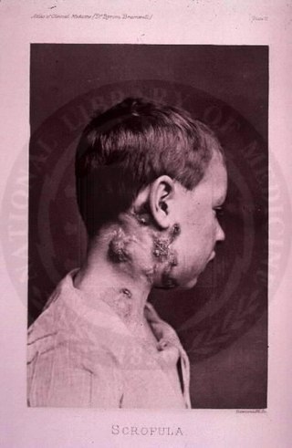
The disease mycobacterial cervical lymphadenitis, also known as scrofula and historically as king's evil, involves a lymphadenitis of the cervical lymph nodes associated with tuberculosis as well as nontuberculous (atypical) mycobacteria.

<span class="mw-page-title-main">Castleman disease</span> Group of lymphoproliferative disorders

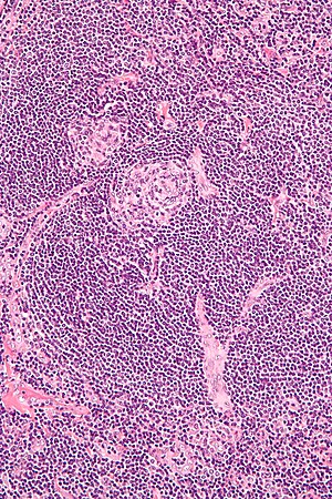
Castlemandisease (CD) describes a group of rare lymphoproliferative disorders that involve enlarged lymph nodes, and a broad range of inflammatory symptoms and laboratory abnormalities. Whether Castleman disease should be considered an autoimmune disease, cancer, or infectious disease is currently unknown.

<span class="mw-page-title-main">Tumors of the hematopoietic and lymphoid tissues</span> Tumors that affect the blood, bone marrow, lymph, and lymphatic system

Tumors of the hematopoietic and lymphoid tissues or tumours of the haematopoietic and lymphoid tissues are tumors that affect the blood, bone marrow, lymph, and lymphatic system. Because these tissues are all intimately connected through both the circulatory system and the immune system, a disease affecting one will often affect the others as well, making aplasia, myeloproliferation and lymphoproliferation closely related and often overlapping problems. While uncommon in solid tumors, chromosomal translocations are a common cause of these diseases. This commonly leads to a different approach in diagnosis and treatment of hematological malignancies. Hematological malignancies are malignant neoplasms ("cancer"), and they are generally treated by specialists in hematology and/or oncology. In some centers "hematology/oncology" is a single subspecialty of internal medicine while in others they are considered separate divisions. Not all hematological disorders are malignant ("cancerous"); these other blood conditions may also be managed by a hematologist.

<span class="mw-page-title-main">Lymphadenopathy</span> Disease of lymph nodes

Lymphadenopathy or adenopathy is a disease of the lymph nodes, in which they are abnormal in size or consistency. Lymphadenopathy of an inflammatory type is lymphadenitis, producing swollen or enlarged lymph nodes. In clinical practice, the distinction between lymphadenopathy and lymphadenitis is rarely made and the words are usually treated as synonymous. Inflammation of the lymphatic vessels is known as lymphangitis. Infectious lymphadenitis affecting lymph nodes in the neck is often called scrofula.

<span class="mw-page-title-main">POEMS syndrome</span> Paraneoplastic syndrome

POEMS syndrome is a rare paraneoplastic syndrome caused by a clone of aberrant plasma cells. The name POEMS is an acronym for some of the disease's major signs and symptoms, as is PEP.

<span class="mw-page-title-main">Invasive carcinoma of no special type</span> Medical condition

Invasive carcinoma of no special type (NST) is also referred to as invasive ductal carcinoma or infiltrating ductal carcinoma(IDC) and invasive ductal carcinoma, not otherwise specified (NOS). Each of these terms represents to the same disease entity, but for international audiences this article will use invasive carcinoma NST because it is the preferred term of the World Health Organization (WHO).

<span class="mw-page-title-main">Lymphangioma</span> Malformations of the lymphatic system characterized by lesions that are thin-walled cysts

Lymphangiomas are malformations of the lymphatic system characterized by lesions that are thin-walled cysts; these cysts can be macroscopic, as in a cystic hygroma, or microscopic. The lymphatic system is the network of vessels responsible for returning to the venous system excess fluid from tissues as well as the lymph nodes that filter this fluid for signs of pathogens. These malformations can occur at any age and may involve any part of the body, but 90% occur in children less than 2 years of age and involve the head and neck. These malformations are either congenital or acquired. Congenital lymphangiomas are often associated with chromosomal abnormalities such as Turner syndrome, although they can also exist in isolation. Lymphangiomas are commonly diagnosed before birth using fetal ultrasonography. Acquired lymphangiomas may result from trauma, inflammation, or lymphatic obstruction.

<span class="mw-page-title-main">Anaplastic thyroid cancer</span> Medical condition

Anaplastic thyroid cancer (ATC), also known as anaplastic thyroid carcinoma, is an aggressive form of thyroid cancer characterized by uncontrolled growth of cells in the thyroid gland. This form of cancer generally carries a very poor prognosis due to its aggressive behavior and resistance to cancer treatments. The cells of anaplastic thyroid cancer are highly abnormal and usually no longer resemble the original thyroid cells and have poor differentiation.

Lymphoid hyperplasia is the rapid proliferation of normal lymphocytic cells that resemble lymph tissue which may occur with bacterial or viral infections. The growth is termed hyperplasia which may result in enlargement of various tissue including an organ, or cause a cutaneous lesion.

<span class="mw-page-title-main">Kimura's disease</span> Medical condition

Kimura's disease is a benign rare chronic inflammatory disorder. Its primary symptoms are subdermal lesions in the head or neck or painless unilateral inflammation of cervical lymph nodes.

Siltuximab is a chimeric monoclonal antibody. It binds to interleukin-6. Siltuximab has been investigated for the treatment of neoplastic diseases: metastatic renal cell cancer, prostate cancer, other types of cancer, and for Castleman's disease.

<span class="mw-page-title-main">Marginal zone B-cell lymphoma</span> Group of lymphomas

Marginal zone B-cell lymphomas, also known as marginal zone lymphomas (MZLs), are a heterogeneous group of lymphomas that derive from the malignant transformation of marginal zone B-cells. Marginal zone B cells are innate lymphoid cells that normally function by rapidly mounting IgM antibody immune responses to antigens such as those presented by infectious agents and damaged tissues. They are lymphocytes of the B-cell line that originate and mature in secondary lymphoid follicles and then move to the marginal zones of mucosa-associated lymphoid tissue, the spleen, or lymph nodes. Mucosa-associated lymphoid tissue is a diffuse system of small concentrations of lymphoid tissue found in various submucosal membrane sites of the body such as the gastrointestinal tract, mouth, nasal cavity, pharynx, thyroid gland, breast, lung, salivary glands, eye, skin and the human spleen.

<span class="mw-page-title-main">Cervical lymphadenopathy</span> Disease of the lymph nodes

Cervical lymphadenopathy refers to lymphadenopathy of the cervical lymph nodes. The term lymphadenopathy strictly speaking refers to disease of the lymph nodes, though it is often used to describe the enlargement of the lymph nodes. Similarly, the term lymphadenitis refers to inflammation of a lymph node, but often it is used as a synonym of lymphadenopathy.

<span class="mw-page-title-main">Follicular hyperplasia</span> Medical condition of lymphatic cells

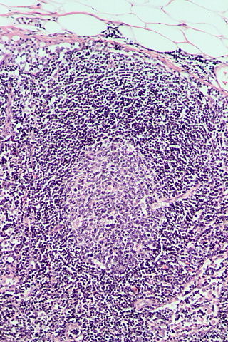
Follicular hyperplasia (FH) is a type of lymphoid hyperplasia and is classified as a lymphadenopathy, which means a disease of the lymph nodes. It is caused by a stimulation of the B cell compartment and by abnormal cell growth of secondary follicles. This typically occurs in the cortex without disrupting the lymph node capsule. The follicles are pathologically polymorphous, are often contrasting and varying in size and shape. Follicular hyperplasia is distinguished from follicular lymphoma in its polyclonality and lack of bcl-2 protein expression, whereas follicular lymphoma is monoclonal, and expresses bcl-2.

Large B-cell lymphoma arising in HHV8-associated multicentric Castleman's disease is a type of large B-cell lymphoma, recognized in the WHO 2008 classification. It is sometimes called the plasmablastic form of multicentric Castleman disease. It has sometimes been confused with plasmablastic lymphoma in the literature, although that is a dissimilar specific entity. It has variable CD20 expression and unmutated immunoglobulin variable region genes.

Diffuse idiopathic pulmonary neuroendocrine cell hyperplasia (DIPNECH) is a diffuse parenchymal lung disease which often presents with symptoms of cough and shortness of breath. The pathological definition published by the World Health Organization is “a generalized proliferation of scattered single cells, small nodules, or linear proliferations of pulmonary neuroendocrine (PNE) cells that may be confined to the bronchial and bronchiolar epithelium.” The true prevalence of this disease is not known. To date, just under 200 cases have been reported in the literature. However, with an increase in recognition of this disease by radiologists and pulmonologists, the number of cases has been increasing. DIPNECH predominantly affects middle-aged women with slowly progressive lung obstruction. DIPNECH is usually discovered in one of two ways: 1) as an unexpected finding following a lung surgery; or 2) by evaluation of a patient in a pulmonary clinic with longstanding, unexplained symptoms.

<span class="mw-page-title-main">HHV-8-associated MCD</span> Disease subtype

Human herpesvirus 8 associated multicentric Castleman disease is a subtype of Castleman disease, a group of rare lymphoproliferative disorders characterized by lymph node enlargement, characteristic features on microscopic analysis of enlarged lymph node tissue, and a range of symptoms and clinical findings.

<span class="mw-page-title-main">Idiopathic multicentric Castleman disease</span> Medical condition

Idiopathic multicentric Castleman disease (iMCD) is a subtype of Castleman disease (also known as giant lymph node hyperplasia, lymphoid hamartoma, or angiofollicular lymph node hyperplasia), a group of lymphoproliferative disorders characterized by lymph node enlargement, characteristic features on microscopic analysis of enlarged lymph node tissue, and a range of symptoms and clinical findings.

The Castleman Disease Collaborative Network (CDCN) is an organization focused on research and awareness of Castleman disease. It was founded in 2012 and has used a collaborative network approach to advance several research studies on Castleman disease.

Epstein–Barr virus–associated lymphoproliferative diseases are a group of disorders in which one or more types of lymphoid cells, i.e. B cells, T cells, NK cells, and histiocytic-dendritic cells, are infected with the Epstein–Barr virus (EBV). This causes the infected cells to divide excessively, and is associated with the development of various non-cancerous, pre-cancerous, and cancerous lymphoproliferative disorders (LPDs). These LPDs include the well-known disorder occurring during the initial infection with the EBV, infectious mononucleosis, and the large number of subsequent disorders that may occur thereafter. The virus is usually involved in the development and/or progression of these LPDs although in some cases it may be an "innocent" bystander, i.e. present in, but not contributing to, the disease.

References

  1. Khanna, Sudhansoo; Singh, Rana Sandip; Goyal, Priyanka; Mohan, Harsh; Bhaker, Poonam; Rana, Shaleen; Sanga, Sunil (2022-02-15). "Incidentally detected Castleman disease of the thorax and its surgical management- A Case Report". www.researchsquare.com. doi:10.21203/rs.3.rs-1318275/v1 . Retrieved 2022-05-16.
  2. Sarmiento Bustamante, Mateo; Shyamsundar, Saishravan; Coren, Freda R.; Bagg, Adam; Srkalovic, Gordan; Alapat, Daisy; van Rhee, Frits; Lim, Megan S.; Lechowicz, Mary Jo; Brandstadter, Joshua D.; Pierson, Sheila K.; Fajgenbaum, David C. (November 2023). "Ongoing symptoms following complete surgical excision in unicentric Castleman disease". American Journal of Hematology. 98 (11): E334–E337. doi: 10.1002/ajh.27065 . ISSN   1096-8652. PMID   37635628. S2CID   261232121.
  3. 1 2 3 4 5 6 7 Szalat R, Munshi NC (February 2018). "Diagnosis of Castleman Disease". Hematology/Oncology Clinics of North America. 32 (1): 53–64. doi:10.1016/j.hoc.2017.09.005. PMID   29157619.
  4. van Rhee, Frits; Oksenhendler, Eric; Srkalovic, Gordan; Voorhees, Peter; Lim, Megan; Dispenzieri, Angela; Ide, Makoto; Parente, Sophia; Schey, Stephen; Streetly, Matthew; Wong, Raymond; Wu, David; Maillard, Ivan; Brandstadter, Joshua; Munshi, Nikhil (2020-12-07). "International evidence-based consensus diagnostic and treatment guidelines for unicentric Castleman disease". Blood Advances. 4 (23): 6039–6050. doi:10.1182/bloodadvances.2020003334. ISSN   2473-9529. PMC   7724917 . PMID   33284946.
  5. 1 2 Fajgenbaum DC, Shilling D (February 2018). "Castleman Disease Pathogenesis". Hematology/Oncology Clinics of North America. 32 (1): 11–21. doi:10.1016/j.hoc.2017.09.002. PMID   29157613.
  6. 1 2 Keller AR, Hochholzer L, Castleman B (March 1972). "Hyaline-vascular and plasma-cell types of giant lymph node hyperplasia of the mediastinum and other locations". Cancer. 29 (3): 670–83. doi:10.1002/1097-0142(197203)29:3<670::aid-cncr2820290321>3.0.co;2-#. PMID   4551306. S2CID   196375825.
  7. 1 2 3 Fajgenbaum DC, Uldrick TS, Bagg A, Frank D, Wu D, Srkalovic G, et al. (March 2017). "International, evidence-based consensus diagnostic criteria for HHV-8-negative/idiopathic multicentric Castleman disease". Blood. 129 (12): 1646–1657. doi:10.1182/blood-2016-10-746933. PMC   5364342 . PMID   28087540.
  8. 1 2 3 Talat N, Belgaumkar AP, Schulte KM (April 2012). "Surgery in Castleman's disease: a systematic review of 404 published cases". Annals of Surgery. 255 (4): 677–84. doi:10.1097/SLA.0b013e318249dcdc. PMID   22367441. S2CID   7553851.
  9. Soumerai JD, Sohani AR, Abramson JS (October 2014). "Diagnosis and management of Castleman disease". Cancer Control. 21 (4): 266–78. doi: 10.1177/107327481402100403 . PMID   25310208.
  10. Casper C (April 2005). "The aetiology and management of Castleman disease at 50 years: translating pathophysiology to patient care". British Journal of Haematology. 129 (1): 3–17. doi: 10.1111/j.1365-2141.2004.05311.x . PMID   15801951.
  11. Munshi N, Mehra M, van de Velde H, Desai A, Potluri R, Vermeulen J (May 2015). "Use of a claims database to characterize and estimate the incidence rate for Castleman disease". Leukemia & Lymphoma. 56 (5): 1252–60. doi: 10.3109/10428194.2014.953145 . PMID   25120049.
  12. Castleman, B.; Iverson, L.; Menendez, V. P. (July 1956). "Localized mediastinal lymphnode hyperplasia resembling thymoma". Cancer. 9 (4): 822–830. doi: 10.1002/1097-0142(195607/08)9:4<822::aid-cncr2820090430>3.0.co;2-4 . ISSN   0008-543X. PMID   13356266.
  13. Fajgenbaum, David C.; Ruth, Jason R.; Kelleher, Dermot; Rubenstein, Arthur H. (April 2016). "The collaborative network approach: a new framework to accelerate Castleman's disease and other rare disease research". The Lancet. Haematology. 3 (4): e150–152. doi:10.1016/S2352-3026(16)00007-7. ISSN   2352-3026. PMID   27063967.