High-grade prostatic intraepithelial neoplasia

Last updated
High-grade prostatic intraepithelial neoplasia
Other namesProstatic intraepithelial neoplasia
High-grade prostatic intraepithelial neoplasia high mag.jpg
Micrograph showing high-grade prostatic intraepithelial neoplasia. H&E stain.
Specialty Urology

High-grade prostatic intraepithelial neoplasia (HGPIN) is an abnormality of prostatic glands and believed to precede the development of prostate adenocarcinoma (the most common form of prostate cancer). [1] [2]

Contents

It may be referred to simply as prostatic intraepithelial neoplasia (PIN). It is considered to be a pre-malignancy, or carcinoma in situ, of the prostatic glands.

Signs and symptoms

HGPIN in isolation is asymptomatic. It is typically discovered in prostate biopsies taken to rule-out prostate cancer and very frequently seen in prostates removed for prostate cancer.[ citation needed ]

Relation to prostate cancer

There are several reasons why PIN is the most likely prostate cancer precursor. [3] PIN is more common in men with prostate cancer. High grade PIN can be found in 85 to 100% of radical prostatectomy specimens, [4] nearby or even in connection with prostate cancer. It tends to occur in the peripheral zone of the prostate. With age, it becomes increasingly multifocal, like prostate cancer. Molecular analysis has shown that high grade PIN and prostate cancer share many genetic abnormalities. [5]

The risk for men with high grade PIN of being diagnosed with prostate cancer after repeat biopsy has decreased since the introduction of biopsies at more than six locations (traditional sextant biopsies). [3]

Histology

Histopathology of high-grade prostatic intraepithelial neoplasia (HGPIN) with typical features, H&E stain. Histopathology of high-grade prostatic intraepithelial neoplasia (HGPIN), annotated.jpg
Histopathology of high-grade prostatic intraepithelial neoplasia (HGPIN) with typical features, H&E stain.
High-grade prostatic intraepithelial neoplasia. High-grade prostatic intraepithelial neoplasia low mag.jpg
High-grade prostatic intraepithelial neoplasia.

HGPIN typically has one of four different histologic patterns: [2]

Its cytologic features are that of prostatic adenocarcinoma:

Microscopically, PIN is a collection of irregular, atypical epithelial cells. The architecture of the glands and ducts remains normal. The epithelial cells proliferate and crowding results in a pseudo-multilayer appearance. They remain fully contained within a prostate acinus (the berry-shaped termination of a gland, where the secretion is produced) or duct. The latter can be demonstrated with special staining techniques (immunohistochemistry for cytokeratins) to identify the basal cells forming the supporting layer of the acinus. In prostate cancer, the abnormal cells spread beyond the boundaries of the acinus and form clusters without basal cells. In HGPIN, the basal cell layer is disrupted but present. PIN is primarily found in the peripheral zone of the prostate (75-80%), rarely in the transition zone (10-15%) and very rarely in the central zone (5%), a distribution that parallels the zonal distribution for prostate carcinoma. [7]

Because it is thought to be a premalignant state, PIN is often considered the prostate equivalent of what is called carcinoma in situ (localized cancer) in other organs. However, PIN differs from carcinoma in situ in that it may remain unchanged or even spontaneously regress.[ citation needed ]

Several architectural variants of PIN have been described, and many cases have multiple patterns. The main ones are tufting, micropapillary, cribriform, and flat. Although these different appearances may cause confusion with other conditions, they have not been found to be of clinical importance. Rarer types are signet-ring-cell, small-cell-neuroendocrine, mucinous, foamy, inverted, and with squamous differentiation. [3]

Diagnosis

HGPIN is diagnosed from tissue by a pathologist, which may come from:[ citation needed ]

Blood tests for prostate specific antigen (PSA), digital rectal examination, ultrasound scanning of the prostate via the rectum, fine needle aspiration or medical imaging studies (such as magnetic resonance imaging) are not useful for diagnosing HGPIN.[ citation needed ]

Treatment

HGPIN in isolation does not require treatment. In prostate biopsies it is not predictive of prostate cancer in one year if the prostate was well-sampled, i.e. if there were 8 or more cores. [8]

The exact timing of repeat biopsies remains an area of controversy, as the time required for, and probability of HGPIN transformations to prostate cancer are not well understood.[ citation needed ]

Prognosis

On a subsequent biopsy, given a history of a HGPIN diagnosis, the chance of finding prostatic adenocarcinoma is approximately 30%. [9]

History

PIN was historically subdivided into different stages, based on the level of cell atypia. PIN was formerly classified as PIN 1, 2 or 3, in order of increasing cell irregularities. Nowadays, PIN 1 is referred to as low grade PIN, and PIN 2 and PIN 3 are grouped together as high grade PIN. [10] Only high grade PIN has been shown to be a risk factor for prostate cancer. Because low grade PIN has no significance and does not require repeat biopsies or treatment, it is not mentioned in pathology reports. As such, PIN has become synonymous with high grade PIN.[ citation needed ]

See also

Related Research Articles

<span class="mw-page-title-main">Adenocarcinoma</span> Medical condition

Adenocarcinoma is a type of cancerous tumor that can occur in several parts of the body. It is defined as neoplasia of epithelial tissue that has glandular origin, glandular characteristics, or both. Adenocarcinomas are part of the larger grouping of carcinomas, but are also sometimes called by more precise terms omitting the word, where these exist. Thus invasive ductal carcinoma, the most common form of breast cancer, is adenocarcinoma but does not use the term in its name—however, esophageal adenocarcinoma does to distinguish it from the other common type of esophageal cancer, esophageal squamous cell carcinoma. Several of the most common forms of cancer are adenocarcinomas, and the various sorts of adenocarcinoma vary greatly in all their aspects, so that few useful generalizations can be made about them.

<span class="mw-page-title-main">Cervical intraepithelial neoplasia</span> Medical condition

Cervical intraepithelial neoplasia (CIN), also known as cervical dysplasia, is the abnormal growth of cells on the surface of the cervix that could potentially lead to cervical cancer. More specifically, CIN refers to the potentially precancerous transformation of cells of the cervix.

<span class="mw-page-title-main">Prostatic acid phosphatase</span> Human protein

Prostatic acid phosphatase (PAP), also prostatic specific acid phosphatase (PSAP), is an enzyme produced by the prostate. It may be found in increased amounts in men who have prostate cancer or other diseases.

<span class="mw-page-title-main">Grading (tumors)</span> Measure of the cell appearance in tumors and other neoplasms

In pathology, grading is a measure of the cell appearance in tumors and other neoplasms. Some pathology grading systems apply only to malignant neoplasms (cancer); others apply also to benign neoplasms. The neoplastic grading is a measure of cell anaplasia in the sampled tumor and is based on the resemblance of the tumor to the tissue of origin. Grading in cancer is distinguished from staging, which is a measure of the extent to which the cancer has spread.

<span class="mw-page-title-main">Gleason grading system</span> Score given to a prostate cancer based on its microscopic appearance

The Gleason grading system is used to help evaluate the prognosis of men with prostate cancer using samples from a prostate biopsy. Together with other parameters, it is incorporated into a strategy of prostate cancer staging which predicts prognosis and helps guide therapy. A Gleason score is given to prostate cancer based upon its microscopic appearance. Cancers with a higher Gleason score are more aggressive and have a worse prognosis. Pathological scores range from 2 to 10, with higher numbers indicating greater risks and higher mortality. The system is widely accepted and used for clinical decision making even as it is recognised that certain biomarkers, like ACP1 expression, might yield higher predictive value for future disease course.

<span class="mw-page-title-main">Endometrial intraepithelial neoplasia</span>

Endometrial intraepithelial neoplasia (EIN) is a premalignant lesion of the uterine lining that predisposes to endometrioid endometrial adenocarcinoma. It is composed of a collection of abnormal endometrial cells, arising from the glands that line the uterus, which have a tendency over time to progress to the most common form of uterine cancer—endometrial adenocarcinoma, endometrioid type.

<span class="mw-page-title-main">Vulvar cancer</span> Cancer involving the vulva

Vulvar cancer is a cancer of the vulva, the outer portion of the female genitals. It most commonly affects the labia majora. Less often, the labia minora, clitoris, or vaginal glands are affected. Symptoms include a lump, itchiness, changes in the skin, or bleeding from the vulva.

<span class="mw-page-title-main">Endometrial hyperplasia</span> Medical condition

Endometrial hyperplasia is a condition of excessive proliferation of the cells of the endometrium, or inner lining of the uterus.

<span class="mw-page-title-main">NKX3-1</span> Protein-coding gene in the species Homo sapiens

Homeobox protein Nkx-3.1, also known as NKX3-1, NKX3, BAPX2, NKX3A and NKX3.1 is a protein that in humans is encoded by the NKX3-1 gene located on chromosome 8p. NKX3-1 is a prostatic tumor suppressor gene.

<span class="mw-page-title-main">Salivary gland tumour</span> Medical condition

Salivary gland tumours, also known as mucous gland adenomas or neoplasms, are tumours that form in the tissues of salivary glands. The salivary glands are classified as major or minor. The major salivary glands consist of the parotid, submandibular, and sublingual glands. The minor salivary glands consist of 800 to 1000 small mucus-secreting glands located throughout the lining of the oral cavity. Patients with these types of tumours may be asymptomatic.

<span class="mw-page-title-main">Lobular carcinoma in situ</span> Medical condition

Lobular carcinoma in situ (LCIS) is an incidental microscopic finding with characteristic cellular morphology and multifocal tissue patterns. The condition is a laboratory diagnosis and refers to unusual cells in the lobules of the breast. The lobules and acini of the terminal duct-lobular unit (TDLU), the basic functional unit of the breast, may become distorted and undergo expansion due to the abnormal proliferation of cells comprising the structure. These changes represent a spectrum of atypical epithelial lesions that are broadly referred to as lobular neoplasia (LN).

In urologic pathology, atypical small acinar proliferation, is a collection of small prostatic glands, on prostate biopsy, whose significance is uncertain and cannot be determined to be benign or malignant.

The Bethesda system (TBS), officially called The Bethesda System for Reporting Cervical Cytology, is a system for reporting cervical or vaginal cytologic diagnoses, used for reporting Pap smear results. It was introduced in 1988 and revised in 1991, 2001, and 2014. The name comes from the location of the conference, sponsored by the National Institutes of Health, that established the system.

<span class="mw-page-title-main">Perineural invasion</span> Biological process

In pathology, perineural invasion, abbreviated PNI, refers to the invasion of cancer to the space surrounding a nerve. It is common in head and neck cancer, prostate cancer and colorectal cancer.

<span class="mw-page-title-main">Fetal adenocarcinoma</span> Rare subtype of pulmonary adenocarcinoma

Fetal adenocarcinoma (FA) of the lung is a rare subtype of pulmonary adenocarcinoma that exhibits tissue architecture and cell characteristics that resemble fetal lung tissue upon microscopic examination. It is currently considered a variant of solid adenocarcinoma with mucin production.

Microglandular hyperplasia (MGH) of the cervix is an epithelial benign abnormality (lesion) associated with gland proliferation. It can terminate in mature squamous metaplasia, and it is suspected reserve cells are involved in this process, perhaps in the form of reserve cell hyperplasia with glandular differentiation.

<span class="mw-page-title-main">Squamous-cell carcinoma</span> Carcinoma that derives from squamous epithelial cells

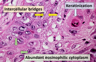
Squamous-cell carcinoma (SCC), also known as epidermoid carcinoma, comprises a number of different types of cancer that begin in squamous cells. These cells form on the surface of the skin, on the lining of hollow organs in the body, and on the lining of the respiratory and digestive tracts.

A histopathologic diagnosis of prostate cancer is the discernment of whether there is a cancer in the prostate, as well as specifying any subdiagnosis of prostate cancer if possible. The histopathologic subdiagnosis of prostate cancer has implications for the possibility and methodology of any subsequent Gleason scoring. The most common histopathological subdiagnosis of prostate cancer is acinar adenocarcinoma, constituting 93% of prostate cancers. The most common form of acinar adenocarcinoma, in turn, is "adenocarcinoma, not otherwise specified", also termed conventional, or usual acinar adenocarcinoma.

<span class="mw-page-title-main">Histopathology of colorectal adenocarcinoma</span> Analysis of tissue from a biopsy or surgery to identify colorectal cancer characteristics

The histopathology of colorectal cancer of the adenocarcinoma type involves analysis of tissue taken from a biopsy or surgery. A pathology report contains a description of the microscopical characteristics of the tumor tissue, including both tumor cells and how the tumor invades into healthy tissues and finally if the tumor appears to be completely removed. The most common form of colon cancer is adenocarcinoma, constituting between 95% and 98% of all cases of colorectal cancer. Other, rarer types include lymphoma, adenosquamous and squamous cell carcinoma. Some subtypes have been found to be more aggressive.

<span class="mw-page-title-main">Invasive cribriform carcinoma of the breast</span> Medical condition

Invasive cribriform carcinoma of the breast (ICCB), also termed invasive cribriform carcinoma, is a rare type of breast cancer that accounts for 0.3% to 0.6% of all carcinomas in the breast. It originates in a lactiferous duct as opposed to the lobules that form the alveoli in the breasts' mammary glands. ICCB was first described by Dixon and colleagues in 1983 as a tumor that on microscopic histopathological inspection had a cribriform pattern, i.e. a tissue pattern consisting of numerous "Swiss cheese"-like open spaces and/or sieve-like small holes. The latest edition (2019) of the World Health Organization (2019) termed these lesions invasive cribriform carcinomas indicating that by definition they must have a component that invades out of their ducts of origin into adjacent tissues. In situ ductal cancers that have a cribriform histopathology are regarded as belonging to the group of ductal carcinoma in situ tumors.

References

  1. Montironi R, Mazzucchelli R, Lopez-Beltran A, Cheng L, Scarpelli M (June 2007). "Mechanisms of disease: high-grade prostatic intraepithelial neoplasia and other proposed preneoplastic lesions in the prostate". Nat Clin Pract Urol. 4 (6): 321–32. doi:10.1038/ncpuro0815. PMID   17551536. S2CID   31478798.
  2. 1 2 Bostwick DG, Qian J (March 2004). "High-grade prostatic intraepithelial neoplasia". Mod. Pathol. 17 (3): 360–79. doi: 10.1038/modpathol.3800053 . PMID   14739906.
  3. 1 2 3 Montironi R, Mazzucchelli R, Lopez-Beltran A, Cheng L, Scarpelli M (June 2007). "Mechanisms of disease: high-grade prostatic intraepithelial neoplasia and other proposed preneoplastic lesions in the prostate". Nat Clin Pract Urol. 4 (6): 321–32. doi:10.1038/ncpuro0815. PMID   17551536. S2CID   31478798.
  4. Godoy G, Taneja SS (2008). "Contemporary clinical management of isolated high-grade prostatic intraepithelial neoplasia". Prostate Cancer Prostatic Dis. 11 (1): 20–31. doi: 10.1038/sj.pcan.4501014 . PMID   17909565.
  5. Hughes C, Murphy A, Martin C, Sheils O, O'Leary J (July 2005). "Molecular pathology of prostate cancer". J. Clin. Pathol. 58 (7): 673–84. doi:10.1136/jcp.2002.003954. PMC   1770715 . PMID   15976331.
  6. Image by Mikael Häggström, MD. Reference for features:
    - Margaret Sanders, M.B.B.Ch., Murali Varma, M.B.B.S. "High grade prostatic intraepithelial neoplasia (HGPIN)". Pathology Outlines.{{cite web}}: CS1 maint: multiple names: authors list (link) Last author update: 23 February 2021
  7. Ayala, AG; Ro, JY (August 2007). "Prostatic intraepithelial neoplasia: recent advances". Archives of Pathology & Laboratory Medicine. 131 (8): 1257–66. doi:10.5858/2007-131-1257-PINRA. PMID   17683188.
  8. Herawi, M.; Kahane, H.; Cavallo, C.; Epstein, JI. (Jan 2006). "Risk of prostate cancer on first re-biopsy within 1 year following a diagnosis of high grade prostatic intraepithelial neoplasia is related to the number of cores sampled". J Urol. 175 (1): 121–4. doi:10.1016/S0022-5347(05)00064-9. PMID   16406886.
  9. Leite KR, Camara-Lopes LH, Cury J, Dall'oglio MF, Sañudo A, Srougi M (June 2008). "Prostate cancer detection at rebiopsy after an initial benign diagnosis: results using sextant extended prostate biopsy". Clinics. 63 (3): 339–42. doi:10.1590/S1807-59322008000300009. PMC   2664245 . PMID   18568243.
  10. Montironi R, Mazzucchelli R, Algaba F, Lopez-Beltran A (September 2000). "Morphological identification of the patterns of prostatic intraepithelial neoplasia and their importance". J. Clin. Pathol. 53 (9): 655–65. doi:10.1136/jcp.53.9.655. PMC   1731241 . PMID   11041054.