Imaging particle analysis

Last updated

Imaging particle analysis is a technique for making particle measurements using digital imaging, one of the techniques defined by the broader term particle size analysis. The measurements that can be made include particle size, particle shape (morphology or shape analysis and grayscale or color, as well as distributions (graphs) of statistical population measurements.

Contents

Description and history

Imaging particle analysis uses the techniques common to image analysis or image processing for the analysis of particles. Particles are defined here per particle size analysis as particulate solids, and thereby not including atomic or sub-atomic particles. Furthermore, this article is limited to real images (optically formed), as opposed to "synthetic" (computed) images (computed tomography, confocal microscopy, SIM and other super resolution microscopy techniques, etc.).

Given the above, the primary method for imaging particle analysis is using optical microscopy. While optical microscopes have been around and used for particle analysis since the 1600s, [1] the "analysis" in the past has been accomplished by humans using the human visual system. As such, much of this analysis is subjective, or qualitative in nature. Even when some sort of qualitative tools are available, such as a measuring reticle in the microscope, it has still required a human to determine and record those measurements.

Beginning in the late 1800s [2] with the availability of photographic plates, it became possible to capture microscope images permanently on film or paper, making measurements easier to acquire by simply using a scaled ruler on the hard copy image. While this significantly speeded up the acquisition of particle measurements, it was still a tedious, labor-intensive process, which not only made it difficult to measure statistically significant particle populations, but also still introduced some degree of human error to the process.

Finally, beginning roughly in the late 1970s, CCD digital sensors for capturing images and computers which could process those images, began to revolutionize the process by using digital imaging. Although the actual algorithms for performing digital image processing had been around for some time, it was not until the significant computing power needed to perform these analyses became available at reasonable prices that digital imaging techniques could be brought to bear in the mainstream. The first dynamic imaging particle analysis system was patented in 1982. [3] As faster computing resources became available at lowered costs, the task of making measurements from microscope images of particles could now be performed automatically by machine without human intervention, making it possible to measure significantly larger numbers of particles in much less time.

Image acquisition methods

The basic process by which imaging particle analysis is carried out is as follows:

  1. A digital camera captures an image of the field of view in the optical system.
  2. A gray scale thresholding process is used to perform image segmentation, segregating out the particles from the background, creating a binary image of each particle. [4] [5] [6]
  3. Digital image processing techniques are used to perform image analysis operations, resulting in morphological and grey-scale measurements to be stored for each particle. [7]
  4. The measurements saved for each particle are then used to generate image population statistics, [8] or as inputs to algorithms for filtering and sorting the particles into groups of similar types. In some systems, sophisticated pattern recognition techniques [9] [10] may also be employed in order to separate different particle types contained in a heterogeneous sample.

Imaging particle analyzers can be subdivided into two distinct types, static and dynamic, based upon the image acquisition methods. While the basic principles are the same, the methods of image acquisition are different in nature, and each has advantages and disadvantages.

Static imaging particle analysis

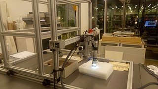
Static image acquisition is the most common form. Almost all microscopes can be easily adapted to accept a digital camera via a C mount adaptor. This type of set-up is often referred to as a digital microscope, although many systems using that name are used only for displaying an image on a monitor.

The sample is prepared on a microscope slide which is placed on the microscope stage. Once the sample has been focused on, then an image can be acquired and displayed on the monitor. If it is a digital camera or a frame grabber is present, the image can now be saved in digital format, and image processing algorithms can be used to isolate particles in the field of view and measure them. [11] [12]

In static image acquisition only one field of view image is captured at a time. If the user wishes to image other portions of the same sample on the slide, they can use the X-Y positioning hardware (typically composed of two linear stages on the microscope to move to a different area of the slide. Care must be taken to insure that two images do not overlap so as not to count and measure the same particles more than once.

The major drawback to static image acquisition is that it is time consuming, both in sample preparation (getting the sample onto the slide with proper dilution if necessary), and in multiple movements of the stage in order to be able to acquire a statistically significant number of particles to count/measure. Computer-controlled X-Y positioning stages are sometimes used in these systems to speed the process up and to reduce the amount of operator intervention, but it is still a time consuming process, and the motorized stages can be expensive due to the level of precision required when working at high magnification. [13]

The major advantages to static particle imaging systems are the use of standard microscope systems and simplicity of depth of field considerations. Since these systems can be made from any standard optical microscope, they may be a lower cost approach for people who already have microscopes. More important, though, is that microscope-based systems have less depth of field issues generally versus dynamic imaging systems. This is because the sample is placed on a microscope slide, and then usually covered with a cover slip, thus limiting the plane containing the particles relative to the optical axis. This means that more particles will be in acceptable focus at high magnifications. [13]

Dynamic imaging particle analysis

Diagram showing flow-through architecture for dynamic imaging particle analysis. Basic flow through diag on white.png
Diagram showing flow-through architecture for dynamic imaging particle analysis.

In Dynamic image acquisition, large amounts of sample are imaged by moving the sample past the microscope optics and using high speed flash illumination to effectively "freeze" the motion of the sample. The flash is synchronized with a high shutter speed in the camera to further prevent motion blur. In a dry particle system, the particles are dispensed from a shaker table and fall by gravity past the optical system. In fluid imaging particle analysis systems, the liquid is passed across the optical axis by use of a narrow flow cell as shown at right.

Diagram showing the flow cell cross-section perpendicular to the optical axis in a dynamic imaging particle analyzer. Credit: Fluid Imaging Technologies, Inc. Flow cell Cross Section.png
Diagram showing the flow cell cross-section perpendicular to the optical axis in a dynamic imaging particle analyzer. Credit: Fluid Imaging Technologies, Inc.

The flow cell is characterized by its depth perpendicular to the optical axis, as shown in the second diagram on right. In order to keep the particles in focus, the flow depth is restricted so that the particles remain in a plane of best focus perpendicular to the optical axis. This is similar in concept to the effect of the microscope slide plus cover slip in a static imaging system. Since depth of field decreases exponentially with increasing magnification, the depth of the flow cell must be narrowed significantly with higher magnifications.

The major drawback to dynamic image acquisition is that the flow cell depth must be limited as described above. This means that, in general, particles larger in size than the flow cell depth can not be allowed in the sample being processed, because they will probably clog the system. So the sample will typically have to be filtered to remove particles larger than the flow cell depth prior to being evaluated. If it is desired to look at a very wide range of particle size, this may mean that the sample would have to be fractionated into smaller size range components, and run with different magnification/flow cell combinations. [13]

The major advantage to dynamic image acquisition is that it enables acquiring and measuring particles at significantly higher speed, typically on the order of 10,000 particles/minute or greater. This means that statistically significant populations can be analyzed in far shorter time periods than previously possible with manual microscopy or even static imaging particle analysis. In this sense, dynamic imaging particle analysis systems combine the speed typical of particle counters with the discriminatory capabilities of microscopy. [13]

Dynamic imaging particle analysis is used in aquatic microorganism research to analyze phytoplankton, zooplankton, and other aquatic microorganisms ranging from 2 um to 5 mm in size. Dynamic imaging particle analysis is also biopharmaceutical research to characterize and analyze particles ranging from 300 nm to 5mm in size.

Micro-flow imaging

Micro-flow imaging (MFI) is a particle analysis technique that uses flow microscopy to quantify particles contained in a solution based on size. This technique is used in the biopharmaceutical industry to characterize subvisible particles from approximately 1 μm to >50 μm. [14]


Related Research Articles

Microscopy Viewing of objects which are too small to be seen with the naked eye

Microscopy is the technical field of using microscopes to view objects and areas of objects that cannot be seen with the naked eye. There are three well-known branches of microscopy: optical, electron, and scanning probe microscopy, along with the emerging field of X-ray microscopy.

Microscope Scientific instrument

A microscope is a laboratory instrument used to examine objects that are too small to be seen by the naked eye. Microscopy is the science of investigating small objects and structures using a microscope. Microscopic means being invisible to the eye unless aided by a microscope.

Scanning electron microscope Type of electron microscope

A scanning electron microscope (SEM) is a type of electron microscope that produces images of a sample by scanning the surface with a focused beam of electrons. The electrons interact with atoms in the sample, producing various signals that contain information about the surface topography and composition of the sample. The electron beam is scanned in a raster scan pattern, and the position of the beam is combined with the intensity of the detected signal to produce an image. In the most common SEM mode, secondary electrons emitted by atoms excited by the electron beam are detected using a secondary electron detector. The number of secondary electrons that can be detected, and thus the signal intensity, depends, among other things, on specimen topography. Some SEMs can achieve resolutions better than 1 nanometer.

Optical microscope Microscope that uses visible light

The optical microscope, also referred to as a light microscope, is a type of microscope that commonly uses visible light and a system of lenses to generate magnified images of small objects. Optical microscopes are the oldest design of microscope and were possibly invented in their present compound form in the 17th century. Basic optical microscopes can be very simple, although many complex designs aim to improve resolution and sample contrast.

Atomic force microscopy Type of microscopy

Atomic force microscopy (AFM) or scanning force microscopy (SFM) is a very-high-resolution type of scanning probe microscopy (SPM), with demonstrated resolution on the order of fractions of a nanometer, more than 1000 times better than the optical diffraction limit.

Microscope image processing is a broad term that covers the use of digital image processing techniques to process, analyze and present images obtained from a microscope. Such processing is now commonplace in a number of diverse fields such as medicine, biological research, cancer research, drug testing, metallurgy, etc. A number of manufacturers of microscopes now specifically design in features that allow the microscopes to interface to an image processing system.

Total internal reflection fluorescence microscope

A total internal reflection fluorescence microscope (TIRFM) is a type of microscope with which a thin region of a specimen, usually less than 200 nanometers can be observed.

Confocal microscopy

Confocal microscopy, most frequently confocal laser scanning microscopy (CLSM) or laser confocal scanning microscopy (LCSM), is an optical imaging technique for increasing optical resolution and contrast of a micrograph by means of using a spatial pinhole to block out-of-focus light in image formation. Capturing multiple two-dimensional images at different depths in a sample enables the reconstruction of three-dimensional structures within an object. This technique is used extensively in the scientific and industrial communities and typical applications are in life sciences, semiconductor inspection and materials science.

Micrograph Process for producing pictures with a microscope

A micrograph or photomicrograph is a photograph or digital image taken through a microscope or similar device to show a magnified image of an object. This is opposed to a macrograph or photomacrograph, an image which is also taken on a microscope but is only slightly magnified, usually less than 10 times. Micrography is the practice or art of using microscopes to make photographs.

Metallography

Metallography is the study of the physical structure and components of metals, by using microscopy.

Profilometer

A profilometer is a measuring instrument used to measure a surface's profile, in order to quantify its roughness. Critical dimensions as step, curvature, flatness are computed from the surface topography.

Digital holography refers to the acquisition and processing of holograms with a digital sensor array , typically a CCD camera or a similar device. Image rendering, or reconstruction of object data is performed numerically from digitized interferograms. Digital holography offers a means of measuring optical phase data and typically delivers three-dimensional surface or optical thickness images. Several recording and processing schemes have been developed to assess optical wave characteristics such as amplitude, phase, and polarization state, which make digital holography a very powerful method for metrology applications .

Chemical imaging is the analytical capability to create a visual image of components distribution from simultaneous measurement of spectra and spatial, time information. Hyperspectral imaging measures contiguous spectral bands, as opposed to multispectral imaging which measures spaced spectral bands.

Particle size analysis

Particle size analysis, particle size measurement, or simply particle sizing, is the collective name of the technical procedures, or laboratory techniques which determines the size range, and/or the average, or mean size of the particles in a powder or liquid sample.

Digital microscope

A digital microscope is a variation of a traditional optical microscope that uses optics and a digital camera to output an image to a monitor, sometimes by means of software running on a computer. A digital microscope often has its own in-built LED light source, and differs from an optical microscope in that there is no provision to observe the sample directly through an eyepiece. Since the image is focused on the digital circuit, the entire system is designed for the monitor image. The optics for the human eye are omitted.

Hirox

Hirox (ハイロックス) is a lens company in Tokyo, Japan that created the first digital microscope in 1985. This company is now known as Hirox Co Ltd. Hirox's main industry is digital microscopes, but still makes the lenses for a variety of items including rangefinders.

Cytometry

Cytometry is the measurement of the characteristics of cells. Variables that can be measured by cytometric methods include cell size, cell count, cell morphology, cell cycle phase, DNA content, and the existence or absence of specific proteins on the cell surface or in the cytoplasm. Cytometry is used to characterize and count blood cells in common blood tests such as the complete blood count. In a similar fashion, cytometry is also used in cell biology research and in medical diagnostics to characterize cells in a wide range of applications associated with diseases such as cancer and AIDS.

Digital holographic microscopy

Digital holographic microscopy (DHM) is digital holography applied to microscopy. Digital holographic microscopy distinguishes itself from other microscopy methods by not recording the projected image of the object. Instead, the light wave front information originating from the object is digitally recorded as a hologram, from which a computer calculates the object image by using a numerical reconstruction algorithm. The image forming lens in traditional microscopy is thus replaced by a computer algorithm. Other closely related microscopy methods to digital holographic microscopy are interferometric microscopy, optical coherence tomography and diffraction phase microscopy. Common to all methods is the use of a reference wave front to obtain amplitude (intensity) and phase information. The information is recorded on a digital image sensor or by a photodetector from which an image of the object is created (reconstructed) by a computer. In traditional microscopy, which do not use a reference wave front, only intensity information is recorded and essential information about the object is lost.

Live-cell imaging

Live-cell imaging is the study of living cells using time-lapse microscopy. It is used by scientists to obtain a better understanding of biological function through the study of cellular dynamics. Live-cell imaging was pioneered in first decade of the 21st century. One of the first time-lapse microcinematographic films of cells ever made was made by Julius Ries, showing the fertilization and development of the sea urchin egg. Since then, several microscopy methods have been developed to study living cells in greater detail with less effort. A newer type of imaging using quantum dots have been used, as they are shown to be more stable. The development of holotomographic microscopy has disregarded phototoxicity and other staining-derived disadvantages by implementing digital staining based on cells’ refractive index.

Characterization of nanoparticles Measurement of physical and chemical properties of nanoparticles

The characterization of nanoparticles is a branch of nanometrology that deals with the characterization, or measurement, of the physical and chemical properties of nanoparticles. Nanoparticles measure less than 100 nanometers in at least one of their external dimensions, and are often engineered for their unique properties. Nanoparticles are unlike conventional chemicals in that their chemical composition and concentration are not sufficient metrics for a complete description, because they vary in other physical properties such as size, shape, surface properties, crystallinity, and dispersion state.

References

  1. Jabez Hogg (1887). The Microscope: Its History, Construction, and Application: Being a Familiar Introduction to the Use of the Instrument, and the Study of Microscopical Science (12th ed.). G. Routledge and Sons. p. 8.
  2. Gaston Tissandier (1877). A History and Handbook of Photography. Sampson, Low, Marston, Low, & Searle. p. 1.
  3. U.S. Patent 4,338,024
  4. Gonzalez, Rafael C.; Woods, Richard E. (2002). Digital Image Processing. Pearson Education. pp. 595–611. ISBN   978-8178086293.
  5. Sankur, Bulent (2004). "Survey over image thresholding techniques and quantitative performance evaluation". Journal of Electronic Imaging. 13 (1): 146. Bibcode:2004JEI....13..146S. doi:10.1117/1.1631315. ISSN   1017-9909.
  6. Otsu, Nobuyuki (1979). "A Threshold Selection Method from Gray-Level Histograms". IEEE Transactions on Systems, Man, and Cybernetics. 9 (1): 62–66. doi:10.1109/TSMC.1979.4310076. ISSN   0018-9472.
  7. Carter, R M; Yan, Y (2005). "Measurement of particle shape using digital imaging techniques". Journal of Physics: Conference Series. 15 (1): 177–182. Bibcode:2005JPhCS..15..177C. doi: 10.1088/1742-6596/15/1/030 . ISSN   1742-6588.
  8. Pouli, T.; Cunningham, D; Reinhard, E. "Image Statistics and their Applications in Computer Graphics (2010)" (PDF). Eurographics, State of the Art. Archived from the original (PDF) on 1 April 2011. Retrieved 2 January 2014.
  9. Rosenfeld, A. (1981). "Image pattern recognition". Proceedings of the IEEE. 69 (5): 596–605. doi:10.1109/PROC.1981.12027. ISSN   0018-9219. S2CID   13410801.
  10. Young, T. Y. (1986). Handbook of Pattern Recognition and Image Processing . Academic Press. ISBN   978-0127745602.
  11. Russ, J.C. (1990). Computer-Assisted Microscopy: The Measurement and Analysis of Images. Springer US. ISBN   978-1-4612-7868-9.
  12. Hazelwood, Kristin L.; Olenych, Scott G.; Griffin, John D.; Cathcart, Judith A.; Davidson, Michael W. (2007). "Entering the Portal: Understanding the Digital Image Recorded Through a Microscope". In Shorte, Spencer L.; Frischknecht, Friedrich (eds.). Imaging Cellular and Molecular Biological Functions . Springer. pp.  3–43. ISBN   978-3-540-71330-2.
  13. 1 2 3 4 Brown, L. "Dynamic Versus Static Image Acquisition in Particle Imaging". www.particleimaging.com. Archived from the original on 3 January 2014. Retrieved 2 January 2014.
  14. Sharma, DK; King, D; Oma, P; Merchant, C (2010). "Micro-flow imaging: flow microscopy applied to sub-visible particulate analysis in protein formulations". AAPS J. 12 (3): 455–64. doi:10.1208/s12248-010-9205-1. PMC   2895433 . PMID   20517661.