Juvenile hormones (JHs) are a group of acyclic sesquiterpenoids that regulate many aspects of insect physiology. The first discovery of a JH was by Vincent Wigglesworth. JHs regulate development, reproduction, diapause, and polyphenisms.

Epoxide hydrolases (EH's), also known as epoxide hydratases, are enzymes that metabolize compounds that contain an epoxide residue; they convert this residue to two hydroxyl residues through a dihydroxylation reaction to form diol products. Several enzymes possess EH activity. Microsomal epoxide hydrolase, soluble epoxide hydrolase, and the more recently discovered but not as yet well defined functionally, epoxide hydrolase 3 (EH3) and epoxide hydrolase 4 (EH4) are structurally closely related isozymes. Other enzymes with epoxide hydrolase activity include leukotriene A4 hydrolase, Cholesterol-5,6-oxide hydrolase, MEST (gene) (Peg1/MEST), and Hepoxilin-epoxide hydrolase. The hydrolases are distinguished from each other by their substrate preferences and, directly related to this, their functions.
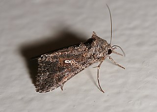
The cabbage looper is a medium-sized moth in the family Noctuidae, a family commonly referred to as owlet moths. Its common name comes from its preferred host plants and distinctive crawling behavior. Cruciferous vegetables, such as cabbage, bok choy, and broccoli, are its main host plant; hence, the reference to cabbage in its common name. The larva is called a looper because it arches its back into a loop when it crawls.

Monoacylglycerol lipase, also known as MAG lipase, acylglycerol lipase, MAGL, MGL or MGLL is an enzyme that, in humans, is encoded by the MGLL gene. MAGL is a 33-kDa, membrane-associated member of the serine hydrolase superfamily and contains the classical GXSXG consensus sequence common to most serine hydrolases. The catalytic triad has been identified as Ser122, His269, and Asp239.

Myrosinase is a family of enzymes involved in plant defense against herbivores. The three-dimensional structure has been elucidated and is available in the PDB.
In enzymology, a hepoxilin-epoxide hydrolase is an enzyme that catalyzes the conversion of the epoxyalcohol metabolites arachidonic acid, hepoxilin A3 and hepoxilin B3 to their tri-hydroxyl products, trioxolin A3 and trioxilin B3, respectively. These reactions in general inactivate the two biologically active hepoxilins.

Leukotriene A4 hydrolase, also known as LTA4H is a human gene. The protein encoded by this gene is a bifunctional enzyme which converts leukotriene A4 to leukotriene B4 and acts as an aminopeptidase.
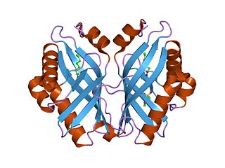
In enzymology, a limonene-1,2-epoxide hydrolase (EC 3.3.2.8) is an enzyme that catalyzes the chemical reaction

In enzymology, a microsomal epoxide hydrolase (mEH) is an enzyme that catalyzes the hydrolysis reaction between an epoxide and water to form a diol.

In enzymology, juvenile hormone esterase (JH esterase) is an enzyme that catalyzes the hydrolysis of juvenile hormone. For example, the juvenile hormone II (found in Lepidoptera):

The alpha/beta hydrolase superfamily is superfamily of hydrolytic enzymes of widely differing phylogenetic origin and catalytic function that share a common fold. The core of each enzyme is an alpha/beta-sheet, containing 8 beta strands connected by 6 alpha helices. The enzymes are believed to have diverged from a common ancestor, retaining little obvious sequence similarity, but preserving the arrangement of the catalytic residues. All have a catalytic triad, the elements of which are borne on loops, which are the best-conserved structural features of the fold.
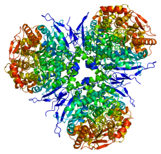
Liver carboxylesterase 1 also known as carboxylesterase 1 is an enzyme that in humans is encoded by the CES1 gene. The protein is also historically known as serine esterase 1 (SES1), monocyte esterase and cholesterol ester hydrolase (CEH). Three transcript variants encoding three different isoforms have been found for this gene. The various protein products from isoform a, b and c range in size from 568, 567 and 566 amino acids long, respectively.
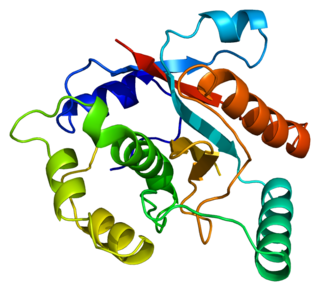
Ubiquitin carboxyl-terminal hydrolase isozyme L3 is an enzyme that in humans is encoded by the UCHL3 gene.
The pheromone biosynthesis activation neuropeptide (PBAN) is a neurohormone that activates the biosynthesis of pheromones in moths. Female moths release PBAN into their hemolymph during the scotophase to stimulate the biosynthesis of the unique pheromone that will attract the conspecific males. PBAN release is drastically reduced after mating, contributing to the loss in female receptivity. In Agrotis ipsilon, it has been shown that the Juvenile Hormone helps induce release of PBAN which goes on to influence pheromone production and responsiveness in females and males, respectively. In the Helicoverpa assulta, the circadian rhythm of pheromone production is closely associated with PBAN release.
In molecular biology, the haemolymph juvenile hormone-binding protein (JHPB) family of proteins consists of several insect specific haemolymph juvenile hormone binding proteins. Juvenile hormone (JH) has a profound effect on insects. It regulates embryogenesis, maintains the status quo of larva development and stimulates reproductive maturation in the adult forms. JH is transported from the sites of its synthesis to target tissues by a haemolymph carrier called juvenile hormone-binding protein (JHBP). JHBP protects the JH molecules from hydrolysis by non-specific esterases present in the insect haemolymph. The crystal structure of the JHBP from Galleria mellonella shows an unusual fold consisting of a long alpha-helix wrapped in a much curved antiparallel beta-sheet. The folding pattern for this structure closely resembles that found in some tandem-repeat mammalian lipid-binding and bactericidal permeability-increasing proteins, with a similar organisation of the major cavity and a disulfide bond linking the long helix and the beta-sheet. It would appear that JHBP forms two cavities, only one of which, the one near the N- and C-termini, binds the hormone; binding induces a conformational change, of unknown significance.

Soluble epoxide hydrolase (sEH) is a bifunctional enzyme that in humans is encoded by the EPHX2 gene. sEH is a member of the epoxide hydrolase family. This enzyme, found in both the cytosol and peroxisomes, binds to specific epoxides and converts them to the corresponding diols. A different region of this protein also has lipid-phosphate phosphatase activity. Mutations in the EPHX2 gene have been associated with familial hypercholesterolemia.
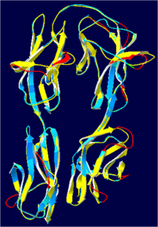
Hemolin is an immunoglobulin-like protein exclusively found in Lepidoptera. It was first discovered in immune-challenged pupae of Hyalophora cecropia and Manduca sexta.

Epoxide hydrolase 3, encoded by the EPHX3 gene, is the third defined isozyme in a set of epoxide hydrolase isozymes, i.e. the epoxide hydrolases. This set includes the Microsomal epoxide hydrolase ; the epoxide hydrolase 2 ; and the far less well defined enzymatically, epoxide hydrolase 4. All four enzyme contain an Alpha/beta hydrolase fold suggesting that they have Hydrolysis activity. EH1, EH2, and EH3 have been shown to have such activity in that they add water to epoxides of unsaturated fatty acids to form vicinal cis products; the activity of EH4 has not been reported. The former three EH's differ in subcellular location, tissue expression patterns, substrate preferences, and thereby functions. These functions include limiting the biologically actions of certain fatty acid epoxides, increasing the toxicity of other fatty acid epoxides, and contributing to the metabolism of drugs and other xenobiotics.
The conjugate (10S,11S) JH diol phosphate is the product of a two-step enzymatic process: conversion of JH to JH diol and then addition of a phosphate group to C10. The enzyme responsible for the phosphorylation of JH diol is JH diol kinase (JHDK), which was first characterized from the Malpighian tubules of early fifth instars of M. sexta. JHDK was discovered when an analysis of JH I metabolites in vivo yielded, in addition to the expected metabolites, a very polar JH I conjugate that was subsequently identified as JH I diol phosphate. Maxwell et al. showed JHDK to contain 3 potential calcium binding sites, and a single ATP-Mg2+ binding site (p-loop). The modeled structure contains nine helices, one beta sheet, and 10 loops. JHDK is also present in the silkworm, where it also functions as homodimer. It lacks a JH response element; Li et al. (2005). It has a high degree of identity to M. sexta JHDK. Later Uno et al. (2007) characterized the A. mellifera enzyme in a proteomic study. It has 183 amino acid residues. More recently, Zeng et al. (2015) have characterized JHDK from Spodoptera litura. It also has 183 amino acid residues, just as does the B. mori enzyme. These enzymes all have high sequence similarity. The M. sexta enzyme contains 184 residues.
Michael R. Kanost is a University Distinguished Professor in the Department of Biochemistry and Molecular Biophysics at Kansas State University.


