Lipid bilayer phase behavior

Last updated

One property of a lipid bilayer is the relative mobility (fluidity) of the individual lipid molecules and how this mobility changes with temperature. This response is known as the phase behavior of the bilayer. Broadly, at a given temperature a lipid bilayer can exist in either a liquid or a solid phase. The solid phase is commonly referred to as a “gel” phase. All lipids have a characteristic temperature at which they undergo a transition (melt) from the gel to liquid phase. In both phases the lipid molecules are constrained to the two dimensional plane of the membrane, but in liquid phase bilayers the molecules diffuse freely within this plane. Thus, in a liquid bilayer a given lipid will rapidly exchange locations with its neighbor millions of times a second and will, through the process of a random walk, migrate over long distances. [1]

Contents

Motion constraints

In contrast to this large in-plane mobility, it is very difficult for lipid molecules to flip-flop from one side of the lipid bilayer to the other. In a phosphatidylcholine-based bilayer this process typically occurs over a timescale of weeks. [2] This discrepancy can be understood in terms of the basic structure of the bilayer. For a lipid to flip from one leaflet to the other, its hydrated headgroup must cross the hydrophobic core of the bilayer, an energetically unfavorable process. Unlike liquid phase bilayers, the lipids in a gel phase bilayer are locked in place and exhibit neither flip-flop nor lateral mobility. Due to this limited mobility, gel bilayers lack an important property of liquid bilayers: the ability to reseal small holes. Liquid phase bilayers can spontaneously heal small voids, much the same way a film of oil on water could flow in to fill a gap. This functionality is one of the reasons that cell membranes are usually composed of fluid phase bilayers. Motion constraints on lipids in lipid bilayers are also imposed by presence of proteins in biological membranes, especially so in the annular lipid shell 'attached' to surface of integral membrane proteins.

Physical origins

Diagram showing the effect of unsaturated lipids on a bilayer. The lipids with an unsaturated tail (blue) disrupt the packing of those with only saturated tails (black). The resulting bilayer has more free space and is consequently more permeable to water and other small molecules. Lipid unsaturation effect.svg
Diagram showing the effect of unsaturated lipids on a bilayer. The lipids with an unsaturated tail (blue) disrupt the packing of those with only saturated tails (black). The resulting bilayer has more free space and is consequently more permeable to water and other small molecules.

The phase behavior of lipid bilayers is largely determined by the strength of the attractive Van der Waals interactions between adjacent lipid molecules. The extent of this interaction is in turn governed by how long the lipid tails are and how well they can pack together. Longer tailed lipids have more area over which to interact, increasing the strength of this interaction and consequently decreasing the lipid mobility. Thus, at a given temperature, a short-tailed lipid will be more fluid than an otherwise identical long-tailed lipid. [3] Another way of expressing this would be to say that the gel to liquid phase transition temperature increases with increasing number of carbons in the lipid alkane chains. Saturated phosphatidylcholine lipids with tails longer than 14 carbons are solid at room temperature, while those with fewer than 14 are liquid. This phenomenon is analogous to the fact that paraffin wax, which is composed of long alkanes, is solid at room temperature, while octane (gasoline), a short alkane, is liquid.

Aside from chain length, transition temperature can also be affected by the degree of unsaturation of the lipid tails. An unsaturated double bond can produce a kink in the alkane chain, disrupting the regular periodic structure. This disruption creates extra free space within the bilayer which allows additional flexibility in the adjacent chains. It is this disruption of packing that leads to lower transition temperatures with increasing double bonds. [3] This is a particularly powerful effect; decreasing the overall chain length by one carbon usually alters the transition temperature of a lipid by ten degrees Celsius or less, but adding a single double bond can decrease the transition temperature by fifty degrees or more (see table). An example of this effect can be noted in everyday life as butter, which has a large percentage saturated fats, is solid at room temperature while vegetable oil, which is mostly unsaturated, is liquid.

Transition temperature (in °C) as a function of tail length and saturation. All data are for lipids with PC headgroups and two identical tails. [4]
Tail LengthDouble BondsTransition Temperature
120-1
14023
16041
18055
20066
22075
24080
1811
182-53
183-60

Mixed systems

Bilayers need not be composed of a single type of lipid and, in fact, most natural membranes are a complex mixture of different lipid molecules. Such mixtures often exhibit properties intermediate to their components, but are also capable of a phenomenon not seen in single component systems: phase separation. If some of the components are liquid at a given temperature while others are in the gel phase, the two phases can coexist in spatially separated populations. This phase separation plays a critical role in biochemical phenomena because membrane components such as proteins can partition into one or the other phase [5] and thus be locally concentrated or activated.

Cholesterol

The chemical structure of cholesterol, which differs greatly from a standard phospholipid. Cholesterol.svg
The chemical structure of cholesterol, which differs greatly from a standard phospholipid.

The presence of cholesterol exerts a profound but complicated influence on lipid bilayer properties because of its unique physical characteristics. Although it is a lipid, cholesterol bears little resemblance to a phospholipid. The hydrophilic domain of cholesterol is quite small, consisting of a single hydroxyl group. Adjacent to this hydroxyl group is a rigid planar structure composed of several fused rings. At the opposite end of the ring structure is a short single chain tail. It has been known for decades that the addition of cholesterol to a fluid phase bilayer decreases its permeability to water. [6] [7] The mode of this interaction has more recently been shown to be due to cholesterol intercalating between lipid molecules, filling in free space and decreasing the flexibility of surrounding lipid chains. [8] This interaction also increases the mechanical rigidity of fluid membrane lipid bilayers [9] and decreases their lateral diffusion coefficient. [10] In contrast, the addition of cholesterol to gel phase bilayers disrupts local packing order, increasing the diffusion coefficient [10] and decreasing the elastic modulus. Interactions of cholesterol with multi-component systems are even more complicated, as these can result in intricate phase diagrams. [11] One lipid-cholesterol system that has recently been studied intently is the lipid raft. Lipid rafts are cholesterol-enriched gel domains that have been potentially implicated in certain cell signaling processes, [12] but the subject remains controversial, with some researchers doubting even their existence in vivo. [13]

Lipid polymorphism

Example of lipid polymorphism as bilayer (le), reverse spherical micelles (M) and reverse hexagonal cylinders H-II phase (H) in negatively stained transmission electron micrograph of spinach thylakoid lipid-water dispersions. HexLamMic phases.jpg
Example of lipid polymorphism as bilayer (le), reverse spherical micelles (M) and reverse hexagonal cylinders H-II phase (H) in negatively stained transmission electron micrograph of spinach thylakoid lipid-water dispersions.

Mixed lipid liposomes can undergo changes into different phase dispersion structures, called lipid polymorphisms, for example, spherical micelles, lipid bilayer lamellae and hexagonal phase cylinders, depending on physical and chemical changes in their microenvironment. [14] Phase transition temperature of liposomes and biological membranes can be measured using calorimetry, magnetic resonance spectroscopy and other techniques. [15]

See also

Related Research Articles

<span class="mw-page-title-main">Biological membrane</span> Enclosing or separating membrane in organisms acting as selective semi-permeable barrier

A biological membrane, biomembrane or cell membrane is a selectively permeable membrane that separates the interior of a cell from the external environment or creates intracellular compartments by serving as a boundary between one part of the cell and another. Biological membranes, in the form of eukaryotic cell membranes, consist of a phospholipid bilayer with embedded, integral and peripheral proteins used in communication and transportation of chemicals and ions. The bulk of lipids in a cell membrane provides a fluid matrix for proteins to rotate and laterally diffuse for physiological functioning. Proteins are adapted to high membrane fluidity environment of the lipid bilayer with the presence of an annular lipid shell, consisting of lipid molecules bound tightly to the surface of integral membrane proteins. The cell membranes are different from the isolating tissues formed by layers of cells, such as mucous membranes, basement membranes, and serous membranes.

<span class="mw-page-title-main">Phospholipid</span> Class of lipids

Phospholipids are a class of lipids whose molecule has a hydrophilic "head" containing a phosphate group and two hydrophobic "tails" derived from fatty acids, joined by an alcohol residue. Marine phospholipids typically have omega-3 fatty acids EPA and DHA integrated as part of the phospholipid molecule. The phosphate group can be modified with simple organic molecules such as choline, ethanolamine or serine.

<span class="mw-page-title-main">Lipid bilayer</span> Membrane of two layers of lipid molecules

The lipid bilayer is a thin polar membrane made of two layers of lipid molecules. These membranes are flat sheets that form a continuous barrier around all cells. The cell membranes of almost all organisms and many viruses are made of a lipid bilayer, as are the nuclear membrane surrounding the cell nucleus, and membranes of the membrane-bound organelles in the cell. The lipid bilayer is the barrier that keeps ions, proteins and other molecules where they are needed and prevents them from diffusing into areas where they should not be. Lipid bilayers are ideally suited to this role, even though they are only a few nanometers in width, because they are impermeable to most water-soluble (hydrophilic) molecules. Bilayers are particularly impermeable to ions, which allows cells to regulate salt concentrations and pH by transporting ions across their membranes using proteins called ion pumps.

<span class="mw-page-title-main">Peripheral membrane protein</span> Membrane proteins that adhere temporarily to membranes with which they are associated

Peripheral membrane proteins, or extrinsic membrane proteins, are membrane proteins that adhere only temporarily to the biological membrane with which they are associated. These proteins attach to integral membrane proteins, or penetrate the peripheral regions of the lipid bilayer. The regulatory protein subunits of many ion channels and transmembrane receptors, for example, may be defined as peripheral membrane proteins. In contrast to integral membrane proteins, peripheral membrane proteins tend to collect in the water-soluble component, or fraction, of all the proteins extracted during a protein purification procedure. Proteins with GPI anchors are an exception to this rule and can have purification properties similar to those of integral membrane proteins.

<span class="mw-page-title-main">Fluid mosaic model</span> Describe the fluid mosaic model of plasma membrane

The fluid mosaic model explains various characteristics regarding the structure of functional cell membranes. According to this biological model, there is a lipid bilayer in which protein molecules are embedded. The phospholipid bilayer gives fluidity and elasticity to the membrane. Small amounts of carbohydrates are also found in the cell membrane. The biological model, which was devised by Seymour Jonathan Singer and Garth L. Nicolson in 1972, describes the cell membrane as a two-dimensional liquid that restricts the lateral diffusion of membrane components. Such domains are defined by the existence of regions within the membrane with special lipid and protein cocoon that promote the formation of lipid rafts or protein and glycoprotein complexes. Another way to define membrane domains is the association of the lipid membrane with the cytoskeleton filaments and the extracellular matrix through membrane proteins. The current model describes important features relevant to many cellular processes, including: cell-cell signaling, apoptosis, cell division, membrane budding, and cell fusion. The fluid mosaic model is the most acceptable model of the plasma membrane. In this definition of the cell membrane, its main function is to act as a barrier between the contents inside the cell and the extracellular environment.

<span class="mw-page-title-main">Lipid raft</span>

The plasma membranes of cells contain combinations of glycosphingolipids, cholesterol and protein receptors organised in glycolipoprotein lipid microdomains termed lipid rafts. Their existence in cellular membranes remains somewhat controversial. It has been proposed that they are specialized membrane microdomains which compartmentalize cellular processes by serving as organising centers for the assembly of signaling molecules, allowing a closer interaction of protein receptors and their effectors to promote kinetically favorable interactions necessary for the signal transduction. Lipid rafts influence membrane fluidity and membrane protein trafficking, thereby regulating neurotransmission and receptor trafficking. Lipid rafts are more ordered and tightly packed than the surrounding bilayer, but float freely within the membrane bilayer. Although more common in the cell membrane, lipid rafts have also been reported in other parts of the cell, such as the Golgi apparatus and lysosomes.

<span class="mw-page-title-main">Sphingomyelin</span> Class of chemical compounds

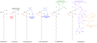
Sphingomyelin is a type of sphingolipid found in animal cell membranes, especially in the membranous myelin sheath that surrounds some nerve cell axons. It usually consists of phosphocholine and ceramide, or a phosphoethanolamine head group; therefore, sphingomyelins can also be classified as sphingophospholipids. In humans, SPH represents ~85% of all sphingolipids, and typically make up 10–20 mol % of plasma membrane lipids.

<span class="mw-page-title-main">Pulmonary surfactant</span> Complex of phospholipids and proteins

Pulmonary surfactant is a surface-active complex of phospholipids and proteins formed by type II alveolar cells. The proteins and lipids that make up the surfactant have both hydrophilic and hydrophobic regions. By adsorbing to the air-water interface of alveoli, with hydrophilic head groups in the water and the hydrophobic tails facing towards the air, the main lipid component of surfactant, dipalmitoylphosphatidylcholine (DPPC), reduces surface tension.

Regarding biological membranes, the liquid ordered phase is a liquid crystalline phase of a lipid bilayer, and is of significant biological importance. It occurs in many lipid mixtures combining cholesterol with a phospholipid and/or sphingolipids e.g. sphingomyelin. This phase has been related to lipid rafts that may exist in plasma membranes.

<span class="mw-page-title-main">Dipalmitoylphosphatidylcholine</span> Chemical compound

Dipalmitoylphosphatidylcholine (DPPC) is a phospholipid (and a lecithin) consisting of two C16 palmitic acid groups attached to a phosphatidylcholine head-group.

<span class="mw-page-title-main">Surfactin</span> Chemical compound

Surfactin is a cyclic lipopeptide, commonly used as an antibiotic for its capacity as a surfactant. It is an amphiphile capable of withstanding hydrophilic and hydrophobic environments. The Gram-positive bacterial species Bacillus subtilis produces surfactin for its antibiotic effects against competitors. Surfactin showcases antibacterial, antiviral, antifungal, and hemolytic effects.

In biology, membrane fluidity refers to the viscosity of the lipid bilayer of a cell membrane or a synthetic lipid membrane. Lipid packing can influence the fluidity of the membrane. Viscosity of the membrane can affect the rotation and diffusion of proteins and other bio-molecules within the membrane, there-by affecting the functions of these things.

<span class="mw-page-title-main">Membrane lipid</span> Lipid molecules on cell membrane

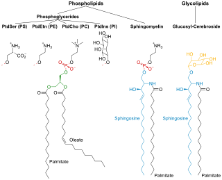
Membrane lipids are a group of compounds which form the lipid bilayer of the cell membrane. The three major classes of membrane lipids are phospholipids, glycolipids, and cholesterol. Lipids are amphiphilic: they have one end that is soluble in water ('polar') and an ending that is soluble in fat ('nonpolar'). By forming a double layer with the polar ends pointing outwards and the nonpolar ends pointing inwards membrane lipids can form a 'lipid bilayer' which keeps the watery interior of the cell separate from the watery exterior. The arrangements of lipids and various proteins, acting as receptors and channel pores in the membrane, control the entry and exit of other molecules and ions as part of the cell's metabolism. In order to perform physiological functions, membrane proteins are facilitated to rotate and diffuse laterally in two dimensional expanse of lipid bilayer by the presence of a shell of lipids closely attached to protein surface, called annular lipid shell.

<span class="mw-page-title-main">Lipid polymorphism</span>

Polymorphism in biophysics is the ability of lipids to aggregate in a variety of ways, giving rise to structures of different shapes, known as "phases". This can be in the form of spheres of lipid molecules (micelles), pairs of layers that face one another, a tubular arrangement (hexagonal), or various cubic phases. More complicated aggregations have also been observed, such as rhombohedral, tetragonal and orthorhombic phases.

Protein–lipid interaction is the influence of membrane proteins on the lipid physical state or vice versa.

<span class="mw-page-title-main">Lipid bilayer fusion</span>

In membrane biology, fusion is the process by which two initially distinct lipid bilayers merge their hydrophobic cores, resulting in one interconnected structure. If this fusion proceeds completely through both leaflets of both bilayers, an aqueous bridge is formed and the internal contents of the two structures can mix. Alternatively, if only one leaflet from each bilayer is involved in the fusion process, the bilayers are said to be hemifused. In hemifusion, the lipid constituents of the outer leaflet of the two bilayers can mix, but the inner leaflets remain distinct. The aqueous contents enclosed by each bilayer also remain separated.

A model lipid bilayer is any bilayer assembled in vitro, as opposed to the bilayer of natural cell membranes or covering various sub-cellular structures like the nucleus. They are used to study the fundamental properties of biological membranes in a simplified and well-controlled environment, and increasingly in bottom-up synthetic biology for the construction of artificial cells. A model bilayer can be made with either synthetic or natural lipids. The simplest model systems contain only a single pure synthetic lipid. More physiologically relevant model bilayers can be made with mixtures of several synthetic or natural lipids.

<span class="mw-page-title-main">Ethanol-induced non-lamellar phases in phospholipids</span>

The presence of ethanol can lead to the formations of non-lamellar phases also known as non-bilayer phases. Ethanol has been recognized as being an excellent solvent in an aqueous solution for inducing non-lamellar phases in phospholipids. The formation of non-lamellar phases in phospholipids is not completely understood, but it is significant that this amphiphilic molecule is capable of doing so. The formation of non-lamellar phases is significant in biomedical studies which include drug delivery, the transport of polar and non-polar ions using solvents capable of penetrating the biomembrane, increasing the elasticity of the biomembrane when it is being disrupted by unwanted substances and functioning as a channel or transporter of biomaterial.

<span class="mw-page-title-main">Cell membrane</span> Biological membrane that separates the interior of a cell from its outside environment

The cell membrane is a biological membrane that separates and protects the interior of a cell from the outside environment. The cell membrane consists of a lipid bilayer, made up of two layers of phospholipids with cholesterols interspersed between them, maintaining appropriate membrane fluidity at various temperatures. The membrane also contains membrane proteins, including integral proteins that span the membrane and serve as membrane transporters, and peripheral proteins that loosely attach to the outer (peripheral) side of the cell membrane, acting as enzymes to facilitate interaction with the cell's environment. Glycolipids embedded in the outer lipid layer serve a similar purpose. The cell membrane controls the movement of substances in and out of a cell, being selectively permeable to ions and organic molecules. In addition, cell membranes are involved in a variety of cellular processes such as cell adhesion, ion conductivity, and cell signalling and serve as the attachment surface for several extracellular structures, including the cell wall and the carbohydrate layer called the glycocalyx, as well as the intracellular network of protein fibers called the cytoskeleton. In the field of synthetic biology, cell membranes can be artificially reassembled.

<span class="mw-page-title-main">Laurdan</span> Chemical compound

Laurdan is an organic compound which is used as a fluorescent dye when applied to fluorescence microscopy. It is used to investigate membrane qualities of the phospholipid bilayers of cell membranes. One of its most important characteristics is its sensitivity to membrane phase transitions as well as other alterations to membrane fluidity such as the penetration of water.

References

  1. H. C. Berg, "Random Walks in Biology". Extended Paperback Ed. ed. 1993, Princeton, NJ: Princeton University Press.
  2. R. Homan and H. J. Pownall."Transbilayer diffusion of phospholipids: dependence on headgroup structure and acyl chain length." Biochimica et Biophysica Acta 938. (1988) 155 -166.
  3. 1 2 W. Rawicz, K. C. Olbrich, T. McIntosh, D. Needham and E. Evans."Effect of chain length and unsaturation on elasticity of lipid bilayers." Biophysical Journal. 79. (2000) 328-39.
  4. J. R. Silvius. Thermotropic Phase Transitions of Pure Lipids in Model Membranes and Their Modifications by Membrane Proteins. John Wiley & Sons, Inc., New York. (1982)
  5. Dietrich, C.; Volovyk, Z. N.; Levi, M.; Thompson, N. L.; Jacobson, K. (2001). "Partitioning of Thy-1, GM1, and cross-linked phospholipid analogs into lipid rafts reconstituted in supported model membrane monolayers". Proceedings of the National Academy of Sciences. 98 (19): 10642–10647. doi: 10.1073/pnas.191168698 . ISSN   0027-8424. PMC   58519 . PMID   11535814.
  6. Corvera, E.; Mouritsen, O. G.; Singer, M. A.; Zuckermann, M. J. (1992). "The permeability and the effect of acyl chain length for phospholipid bilayers containing cholesterol". Biochimica et Biophysica Acta. 1107 (2): 261–270. doi:10.1016/0005-2736(92)90413-g. PMID   1504071.
  7. Needham, D.; Nunn, R. S. (1990). "Elastic deformation and failure of lipid bilayer membranes containing cholesterol". Biophysical Journal. 58 (4): 997–1009. Bibcode:1990BpJ....58..997N. doi:10.1016/s0006-3495(90)82444-9. PMC   1281045 . PMID   2249000.
  8. Bhattacharya, S.; Haldar, S. (2000). "Interactions between cholesterol and lipids in bilayer membranes: Role of lipid headgroup and hydrocarbon chain-backbone linkage". Biochimica et Biophysica Acta (BBA) - Biomembranes. 1467 (1): 39–53. doi: 10.1016/s0005-2736(00)00196-6 . PMID   10930507.
  9. D. Boal, "Mechanics of the Cell". 2002, Cambridge, UK: Cambridge University Press
  10. 1 2 Rubenstein, J. L.; Smith, B. A.; McConnell, H. M. (1979). "Lateral diffusion in binary mixtures of cholesterol and phosphatidylcholines". Proceedings of the National Academy of Sciences of the United States of America. 76 (1): 15–18. Bibcode:1979PNAS...76...15R. doi: 10.1073/pnas.76.1.15 . PMC   382866 . PMID   284326.
  11. Konyakhina, TM; Wu, J; Mastroianni, JD; Heberle, FA; Feigenson, GW (September 2013). "Phase diagram of a 4-component lipid mixture: DSPC/DOPC/POPC/chol". Biochimica et Biophysica Acta (BBA) - Biomembranes. 1828 (9): 2204–14. doi:10.1016/j.bbamem.2013.05.020. PMC   3738200 . PMID   23747294.
  12. Dietrich, C.; Bagatolli, L. A.; Volovyk, Z. N.; Thompson, N. L.; Levi, M.; Jacobson, K.; Gratton, E. (2001). "Lipid rafts reconstituted in model membranes". Biophysical Journal. 80 (3): 1417–1428. Bibcode:2001BpJ....80.1417D. doi:10.1016/s0006-3495(01)76114-0. PMC   1301333 . PMID   11222302.
  13. Munro, S. (2003). "Lipid rafts: elusive or illusive?". Cell. 115 (4): 377–388. doi: 10.1016/s0092-8674(03)00882-1 . PMID   14622593. S2CID   14947495.
  14. YashRoy, R.C. (1994). "Destabilisation of lamellar dispersions of thylakoid membrane lipids by sucrose". Biochimica et Biophysica Acta (BBA) - Lipids and Lipid Metabolism. 1212 (1): 129–133. doi:10.1016/0005-2760(94)90198-8. PMID   8155722.
  15. YashRoy, R.C. (1990). "Determination of membrane lipid phase transition temperature from 13C NMR intensities". Journal of Biochemical and Biophysical Methods. 20 (4): 353–356. doi:10.1016/0165-022x(90)90097-v. PMID   2365951.