Parieto-frontal integration theory

Last updated

The parieto-frontal integration theory (P-FIT) considers intelligence to relate to how well different brain regions integrate to form intelligent behaviors. The theory proposes that large scale brain networks connect brain regions, including regions within frontal, parietal, temporal, and cingulate cortices, underlie the biological basis of human intelligence. These regions, which overlap significantly with the task-positive network, allow the brain to communicate and exchange information efficiently with one another. Support for this theory is primarily based on neuroimaging evidence, with support from lesion studies. The P-FIT is influential in that it explains the majority of current neuroimaging findings, as well as increasing empirical support for cognition being the result of large-scale brain networks, rather than numerous domain-specific processes or modules. [1] A 2010 review of the neuroscience of intelligence described P-FIT as "the best available answer to the question of where in the brain intelligence resides". [2]

Brain organ that serves as the center of the nervous system in all vertebrate and most invertebrate animals

The brain is an organ that serves as the center of the nervous system in all vertebrate and most invertebrate animals. The brain is located in the head, usually close to the sensory organs for senses such as vision. The brain is the most complex organ in a vertebrate's body. In a human, the cerebral cortex contains approximately 14–16 billion neurons, and the estimated number of neurons in the cerebellum is 55–70 billion. Each neuron is connected by synapses to several thousand other neurons. These neurons communicate with one another by means of long protoplasmic fibers called axons, which carry trains of signal pulses called action potentials to distant parts of the brain or body targeting specific recipient cells.

A scientific theory is an explanation of an aspect of the natural world that can be repeatedly tested and verified in accordance with the scientific method, using accepted protocols of observation, measurement, and evaluation of results. Where possible, theories are tested under controlled conditions in an experiment. In circumstances not amenable to experimental testing, theories are evaluated through principles of abductive reasoning. Established scientific theories have withstood rigorous scrutiny and embody scientific knowledge.

Large scale brain networks are collections of widespread brain regions showing functional connectivity by statistical analysis of the fMRI BOLD signal or other signal fluctuations. An emerging paradigm in neuroscience is that cognitive tasks are performed not by individual brain regions working in isolation, but by networks consisting of several discrete brain regions that are said to be "functionally connected" due to tightly coupled activity. Functional connectivity may be measured as long-range synchronization of the EEG, MEG, or other dynamic brain signals. Synchronized brain regions may also be identified using spatial independent component analysis. The set of identified brain areas that are linked together in a large-scale network varies with cognitive function. When the cognitive state is not explicit, the large scale brain network is a resting state network (RSN). As a physical system with graph-like properties, a large scale brain network has both nodes and edges, and cannot be identified simply by the co-activation of brain areas. In recent decades, the analysis of brain networks was made feasible by advances in imaging techniques as well as new tools from graph theory and dynamical systems. Large scale brain networks are identified by their function, and provide a coherent framework for understanding cognition by offering a neural model of how different cognitive functions emerge when different sets of brain regions join together as self-organized coalitions. Disruptions in activity in various networks have been implicated in neuropsychiatric disorders such as depression, Alzheimer's, autism spectrum disorder, schizophrenia and bipolar disorder.

Contents

The theory

General intelligence requires specific brain regions and incorporates:

Temporal lobe part of the brain

The temporal lobe is involved in processing sensory input into derived meanings for the appropriate retention of visual memory, language comprehension, and emotion association.

Parietal lobe part of the brain

The parietal lobe is one of the four major lobes of the cerebral cortex in the brain of mammals. The parietal lobe is positioned above the temporal lobe and behind the frontal lobe and central sulcus.

Cerebral cortex Part of a mammals brain

The cerebral cortex, also known as the cerebral mantle, is the outer layer of neural tissue of the cerebrum of the brain, in humans and other mammals. It is separated into two cortices, by the longitudinal fissure that divides the cerebrum into the left and right cerebral hemispheres. The two hemispheres are joined beneath the cortex by the corpus callosum. The cerebral cortex is the largest site of neural integration in the central nervous system. It plays a key role in memory, attention, perception, awareness, thought, language, and consciousness.

This theory proposes that greater general intelligence in individuals results from the greater communication efficiency between the dorsolateral prefrontal cortex, parietal lobe, anterior cingulate cortex, and specific temporal and parietal cortex regions.

Prefrontal cortex part of brain

In mammalian brain anatomy, the prefrontal cortex (PFC) is the cerebral cortex which covers the front part of the frontal lobe. The PFC contains the Brodmann areas BA8, BA9, BA10, BA11, BA12, BA13, BA14, BA24, BA25, BA32, BA44, BA45, BA46, and BA47.

Anterior cingulate cortex brain region

The anterior cingulate cortex (ACC) is the frontal part of the cingulate cortex that resembles a "collar" surrounding the frontal part of the corpus callosum. It consists of Brodmann areas 24, 32, and 33.

Neuroimaging evidence

Jung and Haier (2007)

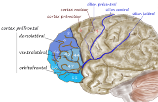
Jung and Haier (2007) proposed the P-FIT in a review of 37 neuroimaging studies with a total of 1,557 participants. The review included only neuroimaging techniques with high spatial resolution to examine the structural and functional correlates of intelligence. Across the structural neuroimaging studies (using voxel-based morphometry, magnetic resonance spectroscopy, and diffusion tensor imaging), Jung and Haier found that the full scale IQ scores from the Wechsler Intelligence scales correlated with frontal and parietal regions in more than 40% of 11 studies. [3] More than 30% of studies using full-scale IQ as their intelligence measure correlated with left cingulate as well as both left and right frontal regions. However, there were no observed structural correlations between regions in the temporal or occipital lobes with any of the intelligence scales. The authors attribute this contradictory finding to the task-dependency of relationships between intellectual performance and these brain regions.

Rex Eugene Jung is an American psychologist who has researched on the neural basis of human intelligence and creativity. He is an assistant professor at the University of New Mexico, where he is the director of neuropsychological services. Jung is also a practicing psychologist at his private clinic.

Richard J. Haier is an American psychologist best known for his work on the neural basis of human intelligence psychometrics, general intelligence, and sex and intelligence.

The Wechsler Adult Intelligence Scale (WAIS) is an IQ test designed to measure intelligence and cognitive ability in adults and older adolescents. The original WAIS was published in February 1955 by David Wechsler, as a revision of the Wechsler–Bellevue Intelligence Scale, released in 1939. It is currently in its fourth edition (WAIS-IV) released in 2008 by Pearson, and is the most widely used IQ test, for both adults and older adolescents, in the world. Data collection for the next version began in 2016 and the test is projected to publish in 2021.

Across functional studies, the authors found that more than 40% of the studies, included in the review, found correlations between bilateral activations in the frontal and occipital cortices and intelligence. In these studies, activation in the left hemisphere was usually significantly higher than that of the right hemisphere. Similarly, bilateral cortical areas in the occipital lobe, such as BA (Brodmann area) 19 were activated during reasoning tasks in more than 40% of studies. Here left activation tended also to be greater than activation in the right hemisphere. [3]

Brodmann area

A Brodmann area is a region of the cerebral cortex, in the human or other primate brain, defined by its cytoarchitecture, or histological structure and organization of cells.

Across the functional imaging studies reviewed, the parietal lobe was consistently involved in reasoning tasks, with BA 7 activated in more than 70% of studies and BA 40 activation was observed in more than 60% of studies. [3]

In recognition of the correlational nature of neuroimaging, the authors complement their neuroimaging review with a shorter review of evidence from lesion studies and imaging genomics regarding the biological basis of intelligence. The authors conclude that the lesion evidence supports a P-FIT theory of intelligence. Further, based on the imaging genomic studies reviewed, the authors suggest a mediating role of ASPM and microcephalin genes in the relationship between volumes of gray and white matter of the areas implicated in the P-FIT theory.

Further structural imaging evidence

Haier et al. (2009) provided further neuroimaging evidence for the P-FIT by investigating the correlation between g and gray matter volume. This was in order to see whether psychometric g is consistently related to a certain neural substrate, or a neuro-g. The authors argue that previous studies examining the neural correlates of g have mostly used indirect measures of g, render the findings of these studies as inconclusive. [4] The scores of 6292 participants on eight cognitive tests were used to derive g, and a small subset of 40 participants were also scanned using voxel-based morphometry. The evidence indicates that the neural correlates of g depend on part on the type of test used to derive g, despite evidence indicating that g derived from different tests tap onto the same underlying psychometric construct. [5] The authors suggest that this may, in part, explain some of the variance in the neuroimaging findings reviewed by Jung and Haier (2007).

In the same year, a study by Colom and colleagues also measured the gray matter correlates of g in a sample of 100 healthy Spanish adults. Similar to Haier et al. (2009), a direct measure of g was derived from a battery measuring fluid, crystallized, and spatial aspects of intelligence. [6] Although finding some differences between the P-FIT theory and their results, the authors conclude that their findings support the P-FIT theory. The identified inconsistencies include voxel clusters in the frontal eye fields, the inferior and middle temporal gyrus, areas which are involved in planning complex movements, high-level visual processing, respectively. [6]

Functional imaging evidence

Vakhtin et al. (2014) determined to identify functional networks relating to fluid intelligence, as measured by both the standard and advanced versions of Raven’s Progressive Matrices test. Using fMRI, Vakhtin et al. found a discrete set of networks associated with fluid reasoning, including the dorsolateral cortex, inferior and parietal lobule, anterior cingulate, as well as temporal and occipital regions. [7] The authors argue that this is “broadly consistent” [7] with the P-FIT theory. The authors scanned 79 American university students three times each, wherein one session was at ‘resting state’, and in the other two the participants were asked to complete problems taken from Raven’s Standard and Advanced Progressive Matrices. Attentional, cognitive, sensorimotor, visual, and default-mode networks were activated during the reasoning task.

Evidence from lesion studies

The majority of studies providing lesion evidence to the P-FIT theory use voxel-based lesion symptom mapping, a method in which scores on an intelligence test battery are compared between participants with and without a lesion at every voxel. This allows for the identification of regions with a causal role in performance on test measures, as it maps where brain damage can impact performance. [8]

Gläscher et al. (2010) explored whether g has distinct neural substrates, or whether it is related to global neural properties such as total brain volume. Using voxel-based lesion symptom mapping, Gläscher et al. (2010) found significant relationships between g scores and regions in primarily the left hemisphere, and major white matter tract regions in temporal, parietal, and inferior frontal areas. [9] Only one brain area was unique to g, which was Brodmann Area 10 in the left frontal pole. The remaining areas activated by g were shared with subtests of the Wechsler Adult Intelligence Scale (WAIS), the test battery used to calculate g.

A study of 182 male veterans from the Phase 3 Vietnam Head Injury Study registry causally identifies several areas implicated in the P-FIT theory. [10] Barbey, Colom, Solomon, Krueger, and Forbes (2012) use voxel-based lesion symptom mapping to identify regions that interfere with performance on the WAIS and the Delis-Kaplan executive function system. The authors only include the five measures from the Delis-Kaplan system that are known to be especially sensitive to frontal lobe damage. The findings indicate that g, calculated from the WAIS test battery, shared neural substrates with several WAIS subtests, such as Verbal Comprehension, Working Memory, Perceptual Organization, and Processing Speed. The areas implicated are known to be involved in language processing, working memory, spatial processing, and motor processing, as well as major white matter tracts, including the arcuate fasciculus which connects temporal, parietal, and inferior frontal regions. The frontal and parietal lobes were found to be critical for executive control processes, which was demonstrated by significantly worse performance on specific executive functioning subtests in participants with damage to frontal and parietal regions, as well as the white matter tracts connecting these regions, such as the superior fronto-occipital fasciculus.

Issues with the theory

There is little published criticism of the P-FIT, and it stands as the best current model for the biological basis of human intelligence. [2] Nevertheless, questions remain regarding the biological functioning of intelligence. A review of the methods used to identify large-scale networks involved in cognition highlights the importance of multi-dimensional context in understanding the neural bases of cognitive processes. [1] Although this review does not directly criticize the P-FIT, the authors caution that structural imaging and lesion studies, although helpful in implicating specific regions in processes, do little to elucidate the dynamical nature of cognitive processes. Furthermore, a review of the neuroscience of intelligence emphasizes the need of studies to consider the different cognitive and neural strategies individuals may use in completing cognitive tasks. [2]

Compatibility with other biological correlates of intelligence

The P-FIT is highly compatible with the neural efficiency hypothesis, and is supported by evidence of the relationship between white matter integrity and intelligence. For example, a study indicates that white matter integrity provides the neural basis for the rapid processing of information, which is considered central to general intelligence. [11]

Related Research Articles

Bilateral cingulotomy is a form of psychosurgery, introduced in 1948 as an alternative to lobotomy. Today it is mainly used in the treatment of depression and obsessive-compulsive disorder. In the early years of the twenty-first century it was used in Russia to treat addiction. It is also used in the treatment of chronic pain. The objective of this procedure is the severing of the supracallosal fibres of the cingulum bundle, which pass through the anterior cingulate gyrus.

Frontal lobe part of the brain

The frontal lobe is the largest of the four major lobes of the brain in mammals, and is located at the front of each hemisphere. It is separated from the parietal lobe by a groove between tissues called the central sulcus, and from the temporal lobe by a deeper groove called the lateral sulcus. The most anterior rounded part of the frontal lobe is known as the frontal pole, one of the three poles of the cerebrum.

A mental image or mental picture is an experience that, on most occasions, significantly resembles the experience of perceiving some object, event, or scene, but occurs when the relevant object, event, or scene is not actually present to the senses. There are sometimes episodes, particularly on falling asleep and waking up (hypnopompic), when the mental imagery, being of a rapid, phantasmagoric and involuntary character, defies perception, presenting a kaleidoscopic field, in which no distinct object can be discerned. Mental imagery can sometimes produce the same effects as would be produced by the behavior or experience imagined.

A mirror neuron is a neuron that fires both when an animal acts and when the animal observes the same action performed by another. Thus, the neuron "mirrors" the behavior of the other, as though the observer were itself acting. Such neurons have been directly observed in primate species. Birds have been shown to have imitative resonance behaviors and neurological evidence suggests the presence of some form of mirroring system. In humans, brain activity consistent with that of mirror neurons has been found in the premotor cortex, the supplementary motor area, the primary somatosensory cortex and the inferior parietal cortex.

Visual memory

Visual memory describes the relationship between perceptual processing and the encoding, storage and retrieval of the resulting neural representations. Visual memory occurs over a broad time range spanning from eye movements to years in order to visually navigate to a previously visited location. Visual memory is a form of memory which preserves some characteristics of our senses pertaining to visual experience. We are able to place in memory visual information which resembles objects, places, animals or people in a mental image. The experience of visual memory is also referred to as the mind's eye through which we can retrieve from our memory a mental image of original objects, places, animals or people. Visual memory is one of several cognitive systems, which are all interconnected parts that combine to form the human memory. Types of palinopsia, the persistence or recurrence of a visual image after the stimulus has been removed, is a dysfunction of visual memory.

Neuroscience and intelligence refers to the various neurological factors that are partly responsible for the variation of intelligence within a species or between different species. A large amount of research in this area has been focused on the neural basis of human intelligence. Historic approaches to study the neuroscience of intelligence consisted of correlating external head parameters, for example head circumference, to intelligence. Post-mortem measures of brain weight and brain volume have also been used. More recent methodologies focus on examining correlates of intelligence within the living brain using techniques such as magnetic resonance imaging (MRI), functional MRI (fMRI), electroencephalography (EEG), positron emission tomography and other non-invasive measures of brain structure and activity.

Lobes of the brain part of the cerebral cortex

The lobes of the brain were originally a purely anatomical classification, but have been shown also to be related to different brain functions. The cerebrum, the largest portion of the human brain, is divided into lobes, but so is the cerebellum. If not specified, the expression "lobes of the brain" refers to the cerebrum.

Posterior cingulate cortex

The posterior cingulate cortex (PCC) is the caudal part of the cingulate cortex, located posterior to the anterior cingulate cortex. This is the upper part of the "limbic lobe". The cingulate cortex is made up of an area around the midline of the brain. Surrounding areas include the retrosplenial cortex and the precuneus.

Stanislas Dehaene French neuroscientist

Stanislas Dehaene is a French author and cognitive neuroscientist whose research centers on a number of topics, including numerical cognition, the neural basis of reading and the neural correlates of consciousness. As of 2017, he is a professor at the Collège de France and, since 1989, the director of INSERM Unit 562, "Cognitive Neuroimaging".

Neural correlates of consciousness Bodily components, such as electrical signals, correlating to consciousness and thinking

The neural correlates of consciousness (NCC) constitute the minimal set of neuronal events and mechanisms sufficient for a specific conscious percept. Neuroscientists use empirical approaches to discover neural correlates of subjective phenomena; that is, neural changes which necessarily and regularly correlate with a specific experience. The set should be minimal because, under the assumption that the brain is sufficient to give rise to any given conscious experience, the question is which of its components is necessary to produce it.

Recognition memory, a subcategory of declarative memory, is the ability to recognize previously encountered events, objects, or people. When the previously experienced event is reexperienced, this environmental content is matched to stored memory representations, eliciting matching signals.

The biology of obsessive-compulsive disorder(OCD) refers biologically based theories about the mechanism of OCD. Cognitive models generally fall into the category of executive dysfunction or modulatory control. Neuroanatomically, functional and structural neuroimaging studies implicate the prefrontal cortex (PFC), basal ganglia (BG), insula, and posterior cingulate cortex (PCC). Genetic and neurochemical studies implicate glutamate and monoamine neurotransmitters.

The neuroscience of music is the scientific study of brain-based mechanisms involved in the cognitive processes underlying music. These behaviours include music listening, performing, composing, reading, writing, and ancillary activities. It also is increasingly concerned with the brain basis for musical aesthetics and musical emotion. Scientists working in this field may have training in cognitive neuroscience, neurology, neuroanatomy, psychology, music theory, computer science, and other relevant fields.

Functional specialization (brain)

Functional specialization suggests that different areas in the brain are specialized for different functions.

Emotional lateralization is the asymmetrical representation of emotional control and processing in the brain. There is evidence for the lateralization of other brain functions as well.

Brain activity and meditation

Meditation and its effect on brain activity and the central nervous system became a focus of collaborative research in neuroscience, psychology and neurobiology during the latter half of the 20th century. Research on meditation sought to define and characterize various practices. Meditation’s effect on the brain can be broken up into two categories: state changes and trait changes, respectively alterations in brain activities during the act of meditating and changes that are the outcome of long-term practice.

Kathleen McDermott is Professor of Psychological and Brain Sciences at Washington University in St. Louis. She is known for her research on how human memory is encoded and retrieved, with a specific interest in how false memories develop. In collaboration with Henry L. (Roddy) Roediger III, she developed the Deese-Roediger-McDermott paradigm used to study the phenomenon of memory illusions. McDermott received the 2004-2005 F.J. McGuigan Young Investigator Prize for research on memory from the American Psychological Foundation and the American Psychological Association's Science Directorate. She was recognized by the Association for Psychological Science as a Rising Star in 2007.

Meditation and Pain is the study of the physiological mechanisms underlying meditation-specifically its neural components- that implicate it in the reduction of pain perception.

Social cognitive neuroscience is the scientific study of the biological processes underpinning social cognition. Specifically, it uses the tools of neuroscience to study "the mental mechanisms that create, frame, regulate, and respond to our experience of the social world". Social cognitive neuroscience uses the epistemological foundations of cognitive neuroscience, and is closely related to social neuroscience. Social cognitive neuroscience employs human neuroimaging, typically using functional magnetic resonance imaging (fMRI). Human brain stimulation techniques such as transcranial magnetic stimulation and transcranial direct-current stimulation are also used. In nonhuman animals, direct electrophysiological recordings and electrical stimulation of single cells and neuronal populations are utilized for investigating lower-level social cognitive processes.

References

  1. 1 2 Bressler, S. L., & Menon, V. (2010). Large-scale brain networks in cognition: emerging methods and principles. Trends in Cognitive Sciences, 14(6), 277–290. doi:10.1016/j.tics.2010.04.004
  2. 1 2 3 Deary, I. J., Penke, L., & Johnson, W. (2010). The neuroscience of human intelligence differences. Nature Reviews Neuroscience, 11(3), 201-211. [doi:10.1038/nrn2793]
  3. 1 2 3 Jung, R. E., & Haier, R. J. (2007). The parieto-frontal integration theory (P-FIT) of intelligence: converging neuroimaging evidence. Behavioral and Brain Sciences, 30, 135–187.
  4. Haier, R. J., Colom, R., Schroeder, D. H., Condon, C. A., Tang, C., Eaves, E., & Head, K. (2009). Gray matter and intelligence factors: is there a neuro-g? Intelligence, 37(2), 136-144. doi:10.1016/j.intell.2008.10.011
  5. Johnson, W., te Nijenhuis, J., & Bouchard, T. J. (2008). Still just 1 g: Consistent results from five test batteries. Intelligence, 36, 81−95
  6. 1 2 Colom, R., Haier, R. J., Head, K., Alvarez-Linera, J., Ouiroga, M. A., Shih, P. C., & Jung, R. E. (2009). Gray matter correlates of fluid, crystallized, and spatial intelligence: testing the P-FIT model. Intelligence, 37, 124–135. [doi:10.1016/j.intell.2008.07.007]
  7. 1 2 Vakhtin, A. A., Ryman, S. G., Flores, R. A., & Jung, R. E. (2014). Functional brain networks contributing to the parieto-frontal integration theory of intelligence. NeuroImage, 103, 349–354. doi:10.1016/j.neuroimage.2014.09.055
  8. Deary, I. J. (2012). Intelligence. Annual Review of Psychology, 63(1), 453-482. doi:10.1146/annurev-psych-120710-100353
  9. Gläscher, J., Rudrauf, D., Colom, R., Paul, L. K., Tranel, D., Damasio, H., & Adolphs, R. (2010). Distributed neural system for general intelligence revealed by lesion mapping. Proceedings of the National Academy of Sciences of the United States of America, 107(10), 4705-4709. doi:10.1093/scan/nss124
  10. Barbey, A. K., Colom, R., Solomon, J., Krueger, F., & Forbes, C. (2012). An integrative architecture for general intelligence and executive function revealed by lesion mapping. Brain, 135, 1154-1164. doi:10.1093/brain/aws021
  11. Penke, L., Muñoz Maniega, S., Bastin, M. E., Valdés Hernández, M. C., Murray, C., Royle, N. A., … Deary, I. J. (2012). Brain white matter tract integrity as a neural foundation for general intelligence. Molecular Psychiatry, 17, 1026–1030. doi:10.1038/mp.2012.66