
The tongue is a muscular organ in the mouth of a typical tetrapod. It manipulates food for chewing and swallowing as part of the digestive process, and is the primary organ of taste. The tongue's upper surface (dorsum) is covered by taste buds housed in numerous lingual papillae. It is sensitive and kept moist by saliva and is richly supplied with nerves and blood vessels. The tongue also serves as a natural means of cleaning the teeth. A major function of the tongue is the enabling of speech in humans and vocalization in other animals.

The uvula, also known as the palatine uvula, is a conic projection from the back edge of the middle of the soft palate, composed of connective tissue containing a number of racemose glands, and some muscular fibers. It also contains many serous glands, which produce thin saliva. It is only found in humans.

The soft palate is, in mammals, the soft tissue constituting the back of the roof of the mouth. The soft palate is part of the palate of the mouth; the other part is the hard palate. The soft palate is distinguished from the hard palate at the front of the mouth in that it does not contain bone.

The voiced pharyngeal approximant or fricative is a type of consonantal sound, used in some spoken languages. The symbol in the International Phonetic Alphabet that represents this sound is ⟨ʕ⟩, and the equivalent X-SAMPA symbol is ?\. Epiglottals and epiglotto-pharyngeals are often mistakenly taken to be pharyngeal.

Pharyngeal slits are filter-feeding organs found among deuterostomes. Pharyngeal slits are repeated openings that appear along the pharynx caudal to the mouth. With this position, they allow for the movement of water in the mouth and out the pharyngeal slits. It is postulated that this is how pharyngeal slits first assisted in filter-feeding, and later, with the addition of gills along their walls, aided in respiration of aquatic chordates. These repeated segments are controlled by similar developmental mechanisms. Some hemichordate species can have as many as 200 gill slits. Pharyngeal clefts resembling gill slits are transiently present during the embryonic stages of tetrapod development. The presence of pharyngeal arches and clefts in the neck of the developing human embryo famously led Ernst Haeckel to postulate that "ontogeny recapitulates phylogeny"; this hypothesis, while false, contains elements of truth, as explored by Stephen Jay Gould in Ontogeny and Phylogeny. However, it is now accepted that it is the vertebrate pharyngeal pouches and not the neck slits that are homologous to the pharyngeal slits of invertebrate chordates. Pharyngeal arches, pouches, and clefts are, at some stage of life, found in all chordates. One theory of their origin is the fusion of nephridia which opened both on the outside and the gut, creating openings between the gut and the environment.

The lips are a horizontal pair of soft appendages attached to the jaws and are the most visible part of the mouth of many animals, including humans. Vertebrate lips are soft, movable and serve to facilitate the ingestion of food and the articulation of sound and speech. Human lips are also a somatosensory organ, and can be an erogenous zone when used in kissing and other acts of intimacy.

The facial artery is a branch of the external carotid artery that supplies structures of the superficial face.
A neurogenic placode is an area of thickening of the epithelium in the embryonic head ectoderm layer that gives rise to neurons and other structures of the sensory nervous system.

The pharyngeal arches, also known as visceral arches, are structures seen in the embryonic development of vertebrates that are recognisable precursors for many structures. In fish, the arches are known as the branchial arches, or gill arches.
In the embryonic development of vertebrates, pharyngeal pouches form on the endodermal side between the pharyngeal arches. The pharyngeal grooves form the lateral ectodermal surface of the neck region to separate the arches.

The pterygoid processes of the sphenoid, one on either side, descend perpendicularly from the regions where the body and the greater wings of the sphenoid bone unite.

Augmentation pharyngoplasty is a kind of plastic surgery for the pharynx when the tissue at the back of the mouth is not able to close properly. It is typically used to correct speech problems in children with cleft palate. It may also be used to correct problems from a tonsillectomy or because of degenerative diseases. After the surgery, patients have an easier time pronouncing certain sounds, such as 'p' and 't', and the voice may have a less nasal sound.
The cervical sinus is a structure formed during embryonic development. It is a deep depression found on each side of the neck. It is formed as the second pharyngeal arch grows faster than the other pharyngeal arches, so they become covered. The first pharyngeal arch also grows slightly faster. It may fail to obliterate, forming a branchial cleft cyst or fistula, which is prone to infection.
The lesser palatine arteries are arteries of the head. It is a branch of the descending palatine artery. They supply the palatine tonsils and the soft palate.

A branchial cleft cyst or simply branchial cyst is a cyst as a swelling in the upper part of neck anterior to sternocleidomastoid. It can, but does not necessarily, have an opening to the skin surface, called a fistula. The cause is usually a developmental abnormality arising in the early prenatal period, typically failure of obliteration of the second, third, and fourth branchial cleft, i.e. failure of fusion of the second branchial arches and epicardial ridge in lower part of the neck. Branchial cleft cysts account for almost 20% of neck masses in children. Less commonly, the cysts can develop from the first, third, or fourth clefts, and their location and the location of associated fistulas differs accordingly.
The pharyngeal apparatus is an embryological structure.

Hypernasal speech is a disorder that causes abnormal resonance in a human's voice due to increased airflow through the nose during speech. It is caused by an open nasal cavity resulting from an incomplete closure of the soft palate and/or velopharyngeal sphincter. In normal speech, nasality is referred to as nasalization and is a linguistic category that can apply to vowels or consonants in a specific language. The primary underlying physical variable determining the degree of nasality in normal speech is the opening and closing of a velopharyngeal passage way between the oral vocal tract and the nasal vocal tract. In the normal vocal tract anatomy, this opening is controlled by lowering and raising the velum or soft palate, to open or close, respectively, the velopharyngeal passageway.

Branchiosauridae is an extinct family of small amphibamiform temnospondyls with external gills and an overall juvenile appearance. The family has been characterized by hundreds of well-preserved specimens from the Permo-Carboniferous of Middle Europe. Specimens represent well defined ontogenetic stages and thus the taxon has been described to display paedomorphy (perennibranchiate). However, more recent work has revealed branchiosaurid taxa that display metamorphosing trajectories. The name Branchiosauridae refers to the retention of gills.
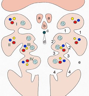
The face and neck development of the human embryo refers to the development of the structures from the third to eighth week that give rise to the future head and neck. They consist of three layers, the ectoderm, mesoderm and endoderm, which form the mesenchyme, neural crest and neural placodes. The paraxial mesoderm forms structures named somites and somitomeres that contribute to the development of the floor of the brain and voluntary muscles of the craniofacial region. The lateral plate mesoderm consists of the laryngeal cartilages. The three tissue layers give rise to the pharyngeal apparatus, formed by six pairs of pharyngeal arches, a set of pharyngeal pouches and pharyngeal grooves, which are the most typical feature in development of the head and neck. The formation of each region of the face and neck is due to the migration of the neural crest cells which come from the ectoderm. These cells determine the future structure to develop in each pharyngeal arch. Eventually, they also form the neurectoderm, which forms the forebrain, midbrain and hindbrain, cartilage, bone, dentin, tendon, dermis, pia mater and arachnoid mater, sensory neurons, and glandular stroma.
