Pyruvate dehydrogenase (lipoamide) alpha 1

Last updated
PDHA1
Protein PDHA1 PDB 1ni4.png
Available structures
PDB Ortholog search: PDBe RCSB
Identifiers
Aliases PDHA1 , PDHA, PDHCE1A, PHE1A, PDHAD, Pyruvate dehydrogenase (lipoamide) alpha 1, pyruvate dehydrogenase alpha 1, pyruvate dehydrogenase E1 alpha 1 subunit, pyruvate dehydrogenase E1 subunit alpha 1
External IDs OMIM: 300502 MGI: 97532 HomoloGene: 121491 GeneCards: PDHA1
Orthologs
SpeciesHumanMouse
Entrez
Ensembl
UniProt
RefSeq (mRNA)

NM_000284
NM_001173454
NM_001173455
NM_001173456

NM_008810

RefSeq (protein)

NP_000275
NP_001166925
NP_001166926
NP_001166927

NP_032836

Location (UCSC) Chr X: 19.34 – 19.36 Mb Chr X: 158.91 – 158.92 Mb
PubMed search [3] [4]
Wikidata
View/Edit Human View/Edit Mouse

Pyruvate dehydrogenase E1 component subunit alpha, somatic form, mitochondrial is an enzyme that in humans is encoded by the PDHA1 gene.The pyruvate dehydrogenase complex is a nuclear-encoded mitochondrial matrix multienzyme complex that provides the primary link between glycolysis and the tricarboxylic acid (TCA) cycle by catalyzing the irreversible conversion of pyruvate into acetyl-CoA. The PDH complex is composed of multiple copies of 3 enzymes: E1 (PDHA1); dihydrolipoyl transacetylase (DLAT) (E2; EC 2.3.1.12); and dihydrolipoyl dehydrogenase (DLD) (E3; EC 1.8.1.4). The E1 enzyme is a heterotetramer of 2 alpha and 2 beta subunits. The E1-alpha subunit contains the E1 active site and plays a key role in the function of the PDH complex. [5]

Contents

Structure

The PDHA1 gene has about 17 kilobase pairs; it contains 11 exons, which range from 61 to 174 base pairs, and introns, whose sizes range from 600 base pairs to 5.7 kilobase pairs. The splice donor and acceptor sites present within the gene all conform to GT/AC rule of splicing. The DNA sequence in the transcription initiation site is very GC-rich. There is a "TATA box"-like sequence and a "CAAT present upstream from the cap site. There are also several sets of repeats, sequences resembling the Sp1 transcription factor binding site, and two cAMP receptor binding sites upstream of the cap. [6]

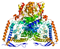
The preliminary peptide encoded by this gene was 29 amino acids at the very start of the sequence that correspond to a typical mitochondrial targeting leader sequence. The remaining 361 amino acids, starting at the N terminus with phenylalanine, represent the mature mitochondrial E1 alpha peptide. [7] Two of these mature PDHA proteins come together with two PDHB proteins to form a heterotetrameric E1 subunit. Crystal Structures allowed for a model in which the enzyme undergoes a 2-A shuttle-like motion of its heterodimers to perform the catalysis. [8] The protein encoded by the human PDHA1 gene is part of the pyruvate dehydrogenase multienzyme complex. The entire human complex is 9.5 MDa in size, and has been described as 60-meric, meaning there are over 60 components that are assembled to make the entire complex. These subunits are conserved across many species, as the function of this complex is essential for the generation of ATP for all eukaryotes. [9] Each component is responsible for the catalysis of one step in this pathway; this complex exists for the purpose of channeling the intermediates of each reaction to the next enzyme, thus greatly increasing the rate of reaction. [10]

Function

The pyruvate dehydrogenase complex is responsible for the oxidative decarboxylation of pyruvate, with the final product being Acetyl CoA. Overall the complex catalyzes five reactions, with the overall reaction being:

Pyruvate + CoA + NAD+ → acetyl-CoA + CO2

There are three different coenzymes required throughout the 5 steps that this complex carries out: thiamine pyrophosphate (TPP), lipoamide, and coenzyme A. This step is only one of the central metabolic pathway carried out by eukaryotes, in which glucose is oxidized to form carbon dioxide, water, and ATP. The E1 complex specifically uses the TPP cofactor to cleave the Calpha-C(=O) bond of pyruvate, and then transfer the acetyl group to the TPP coenzyme, thus resulting in an intermediate, hydroxylethyl-Tpp*E1, and producing CO2. The thiazolium ring on the TPP is ideal for adding to carbonyl groups and acting as an electron sink, or a group that can pull electrons from a reaction and stabilize an electron-deficient intermediate. [10]

Regulation

The activity of the PDH complex in mammalian tissues is largely determined by the phosphorylation of certain subunits within the complex. As such, the absolute amounts of site-specific kinases and phosphates expressed in the mitochondria directly affect PDH activity. [11] Specifically, a tyrosine reside, Tyr-301, can be phosphorylated on PDHA1 and therefore inhibit its activity; this is done by blocking the substrate binding of pyruvate. This phosphorylation is in tandem with phosphorylation of the Ser-293 residue. [12] Phosphorylation of another site, Ser-264, can regulate function. A phosphorylated serine residue at this position prevents TPP from ordering two loops within E1 that contain three phosphorylation sites. The disorder is the direct result of steric clash between the new phosphate group at Ser-264 and another serine, Ser-266, which destabilizes the hydrogen-bonding network that keeps the loops in place. Overall, this disorder results in the inactivation of the catalytic machinery. [13]

The PDH complex can be regulated in a variety of conditions. The PDHA1 subunit has been shown to be regulated by free fatty acids during bouts of exercise. The presence of free fatty acids increases the level of phosphorylation, thereby decreasing PDH activity. During exercise, however, these effects are overruled, and there is a much higher level of dephosphorylated PDHA1 in the cells. [14] In certain muscles, such as the triceps, the metabolic enzyme profile seems to directly affect the level of PDH activity, which can result in higher levels of lactate in muscles with these characteristics. [15]

Clinical significance

At least 71 disease-causing mutations in this gene have been discovered. [16] Mutations in the PDHA1 gene have been known to cause one form of pyruvate dehydrogenase deficiency. Pyruvate dehydrogenase deficiency is characterized by the buildup of a chemical called lactic acid in the body and a variety of neurological problems. Signs and symptoms of this condition usually first appear shortly after birth, and they can vary widely among affected individuals. The most common feature is a potentially life-threatening buildup of lactic acid (lactic acidosis), which can cause nausea, vomiting, severe breathing problems, and an abnormal heartbeat. People with pyruvate dehydrogenase deficiency usually have neurological problems as well. Most have delayed development of mental abilities and motor skills such as sitting and walking. Other neurological problems can include intellectual disability, seizures, weak muscle tone (hypotonia), poor coordination, and difficulty walking. Some affected individuals have abnormal brain structures, such as underdevelopment of the tissue connecting the left and right halves of the brain (corpus callosum), wasting away (atrophy) of the exterior part of the brain known as the cerebral cortex, or patches of damaged tissue (lesions) on some parts of the brain. Because of the severe health effects, many individuals with pyruvate dehydrogenase deficiency do not survive past childhood, although some may live into adolescence or adulthood. [5] Most cases result from mutations in this gene, and countless types of mutations have been identified. The mutations in this gene range from missense mutations, in which one nucleotide is substituted, to much larger deletions of many nucleotide base pairs. [17] [18] [19]

While most of the clinical presentation is homogenous with the general disease, some cases have been identified in which there was focal dilatation of the right lateral ventricle with unilateral periventricular leukomalacia (PVL) with subependymal cyst. [20]

In women, this deficiency can be much harder to detect. This is because of the chance that there will be a skewed X inactivation pattern enzyme measurement in fibroblasts, meaning that the enzyme activity measurement may not be entirely accurate. Because the clinical presentation of this disorder overlaps heavily with deficiencies in oxidative phosphorylation, it is recommended to perform a detailed biochemical analysis on a muscle biopsy in females with a suspected pyruvate dehydrogenase deficiency, followed by molecular genetic analysis of the PDHA1 gene. [21]

Interactive pathway map

Click on genes, proteins and metabolites below to link to respective articles. [§ 1]

[[File:
WP534.png go to articlego to articlego to articlego to articlego to articlego to articlego to articlego to articlego to articlego to articlego to articlego to articlego to articlego to articlego to articlego to articlego to articlego to articlego to articlego to articlego to articlego to articlego to articlego to articlego to articlego to articlego to articlego to articlego to articlego to articlego to articlego to articlego to articlego to articlego to articlego to articlego to articlego to articlego to articlego to articlego to articlego to articlego to articlego to articlego to articlego to articlego to articlego to articlego to articlego to articlego to articlego to articlego to articlego to articlego to articlego to articlego to articlego to articlego to articlego to articlego to articlego to articlego to articlego to articlego to articlego to articlego to articlego to articlego to articlego to articlego to articlego to article
[[
]]
[[
]]
[[
]]
[[
]]
[[
]]
[[
]]
[[
]]
[[
]]
[[
]]
[[
]]
[[
]]
[[
]]
[[
]]
[[
]]
[[
]]
[[
]]
[[
]]
[[
]]
[[
]]
[[
]]
[[
]]
[[
]]
[[
]]
[[
]]
[[
]]
[[
]]
[[
]]
[[
]]
[[
]]
[[
]]
[[
]]
[[
]]
[[
]]
[[
]]
[[
]]
[[
]]
[[
]]
[[
]]
[[
]]
[[
]]
[[
]]
[[
]]
[[
]]
[[
]]
[[
]]
[[
]]
[[
]]
[[
]]
[[
]]
[[
]]
[[
]]
[[
]]
[[
]]
[[
]]
[[
]]
[[
]]
[[
]]
[[
]]
[[
]]
[[
]]
[[
]]
[[
]]
[[
]]
[[
]]
[[
]]
[[
]]
[[
]]
[[
]]
[[
]]
[[
]]
[[
]]
[[
]]
[[
]]
[[
]]
[[
]]
WP534.png go to articlego to articlego to articlego to articlego to articlego to articlego to articlego to articlego to articlego to articlego to articlego to articlego to articlego to articlego to articlego to articlego to articlego to articlego to articlego to articlego to articlego to articlego to articlego to articlego to articlego to articlego to articlego to articlego to articlego to articlego to articlego to articlego to articlego to articlego to articlego to articlego to articlego to articlego to articlego to articlego to articlego to articlego to articlego to articlego to articlego to articlego to articlego to articlego to articlego to articlego to articlego to articlego to articlego to articlego to articlego to articlego to articlego to articlego to articlego to articlego to articlego to articlego to articlego to articlego to articlego to articlego to articlego to articlego to articlego to articlego to articlego to article
|alt=Glycolysis and Gluconeogenesis edit]]
Glycolysis and Gluconeogenesis edit
  1. The interactive pathway map can be edited at WikiPathways: "GlycolysisGluconeogenesis_WP534".

Related Research Articles

<span class="mw-page-title-main">Pyruvate dehydrogenase complex</span> Three-enzyme complex responsible for pyruvate decarboxylation

Pyruvate dehydrogenase complex (PDC) is a complex of three enzymes that converts pyruvate into acetyl-CoA by a process called pyruvate decarboxylation. Acetyl-CoA may then be used in the citric acid cycle to carry out cellular respiration, and this complex links the glycolysis metabolic pathway to the citric acid cycle. Pyruvate decarboxylation is also known as the "pyruvate dehydrogenase reaction" because it also involves the oxidation of pyruvate.

<span class="mw-page-title-main">Pyruvate dehydrogenase lipoamide kinase isozyme 1</span> Protein-coding gene in the species Homo sapiens

Pyruvate dehydrogenase lipoamide kinase isozyme 1, mitochondrial is an enzyme that in humans is encoded by the PDK1 gene. It codes for an isozyme of pyruvate dehydrogenase kinase (PDK).

The branched-chain α-ketoacid dehydrogenase complex is a multi-subunit complex of enzymes that is found on the mitochondrial inner membrane. This enzyme complex catalyzes the oxidative decarboxylation of branched, short-chain alpha-ketoacids. BCKDC is a member of the mitochondrial α-ketoacid dehydrogenase complex family comprising pyruvate dehydrogenase and alpha-ketoglutarate dehydrogenase, key enzymes that function in the Krebs cycle.

Pyruvate dehydrogenase deficiency is a rare neurodegenerative disorder associated with abnormal mitochondrial metabolism. PDCD is a genetic disease resulting from mutations in one of the components of the pyruvate dehydrogenase complex (PDC). The PDC is a multi-enzyme complex that plays a vital role as a key regulatory step in the central pathways of energy metabolism in the mitochondria. The disorder shows heterogeneous characteristics in both clinical presentation and biochemical abnormality.

<span class="mw-page-title-main">Dihydrolipoyl transacetylase</span>

Dihydrolipoyl transacetylase is an enzyme component of the multienzyme pyruvate dehydrogenase complex. The pyruvate dehydrogenase complex is responsible for the pyruvate decarboxylation step that links glycolysis to the citric acid cycle. This involves the transformation of pyruvate from glycolysis into acetyl-CoA which is then used in the citric acid cycle to carry out cellular respiration.

<span class="mw-page-title-main">Pyruvate dehydrogenase</span> Class of enzymes

Pyruvate dehydrogenase is an enzyme that catalyzes the reaction of pyruvate and a lipoamide to give the acetylated dihydrolipoamide and carbon dioxide. The conversion requires the coenzyme thiamine pyrophosphate.

<span class="mw-page-title-main">Pyruvate dehydrogenase kinase</span> Class of enzymes

Pyruvate dehydrogenase kinase is a kinase enzyme which acts to inactivate the enzyme pyruvate dehydrogenase by phosphorylating it using ATP.

<span class="mw-page-title-main">Dihydrolipoamide dehydrogenase</span> Protein-coding gene in the species Homo sapiens

Dihydrolipoamide dehydrogenase (DLD), also known as dihydrolipoyl dehydrogenase, mitochondrial, is an enzyme that in humans is encoded by the DLD gene. DLD is a flavoprotein enzyme that oxidizes dihydrolipoamide to lipoamide.

<span class="mw-page-title-main">E3 binding protein</span> Protein-coding gene in the species Homo sapiens

E3 binding protein also known as pyruvate dehydrogenase protein X component, mitochondrial is a protein that in humans is encoded by the PDHX gene. The E3 binding protein is a component of the pyruvate dehydrogenase complex found only in eukaryotes. Defects in this gene are a cause of pyruvate dehydrogenase deficiency which results in neurological dysfunction and lactic acidosis in infancy and early childhood. This protein is also a minor antigen for antimitochondrial antibodies. These autoantibodies are present in nearly 95% of patients with primary biliary cholangitis, an autoimmune disease of the liver. In primary biliary cholangitis, activated T lymphocytes attack and destroy epithelial cells in the bile duct where this protein is abnormally distributed and overexpressed. Primary biliary cholangitis eventually leads to liver failure.

<span class="mw-page-title-main">Pyruvate dehydrogenase phosphatase</span> Protein-coding gene in the species Homo sapiens

Pyruvate dehydrogenase phosphatase catalytic subunit 1, also known as protein phosphatase 2C, is an enzyme that in humans is encoded by the PDP1 gene. PDPC 1 is an enzyme which serves to reverse the effects of pyruvate dehydrogenase kinase upon pyruvate dehydrogenase, activating pyruvate dehydrogenase.

<span class="mw-page-title-main">Lactate dehydrogenase</span> Class of enzymes

Lactate dehydrogenase (LDH or LD) is an enzyme found in nearly all living cells. LDH catalyzes the conversion of pyruvate to lactate and back, as it converts NAD+ to NADH and back. A dehydrogenase is an enzyme that transfers a hydride from one molecule to another.

<span class="mw-page-title-main">PDK4</span> Protein-coding gene in the species Homo sapiens

Pyruvate dehydrogenase lipoamide kinase isozyme 4, mitochondrial (PDK4) is an enzyme that in humans is encoded by the PDK4 gene. It codes for an isozyme of pyruvate dehydrogenase kinase.

<span class="mw-page-title-main">BCKDHA</span> Protein-coding gene in the species Homo sapiens

A 2-oxoisovalerate dehydrogenase subunit alpha, mitochondrial is an enzyme that in humans is encoded by the BCKDHA gene.

<span class="mw-page-title-main">PDK2</span> Protein-coding gene in the species Homo sapiens

Pyruvate dehydrogenase kinase isoform 2 (PDK2) also known as pyruvate dehydrogenase lipoamide kinase isozyme 2, mitochondrial is an enzyme that in humans is encoded by the PDK2 gene. PDK2 is an isozyme of pyruvate dehydrogenase kinase.

<span class="mw-page-title-main">BCKDHB</span> Protein-coding gene in the species Homo sapiens

2-Oxoisovalerate dehydrogenase subunit beta, mitochondrial is an enzyme that in humans is encoded by the BCKDHB gene.

<span class="mw-page-title-main">ETFB</span> Protein-coding gene in humans

The human ETFB gene encodes the Electron-transfer-flavoprotein, beta subunit, also known as ETF-β. Together with Electron-transfer-flavoprotein, alpha subunit, encoded by the 'ETFA' gene, it forms the heterodimeric Electron transfer flavoprotein (ETF). The native ETF protein contains one molecule of FAD and one molecule of AMP, respectively.

<span class="mw-page-title-main">PDK3</span> Protein-coding gene in the species Homo sapiens

Pyruvate dehydrogenase lipoamide kinase isozyme 3, mitochondrial is an enzyme that in humans is encoded by the PDK3 gene. It codes for an isozyme of pyruvate dehydrogenase kinase.The pyruvate dehydrogenase (PDH) complex is a nuclear-encoded mitochondrial multienzyme complex that catalyzes the overall conversion of pyruvate to acetyl-CoA and CO2. It provides the primary link between glycolysis and the tricarboxylic acid (TCA) cycle, and thus is one of the major enzymes responsible for the regulation of glucose metabolism. The enzymatic activity of PDH is regulated by a phosphorylation/dephosphorylation cycle, and phosphorylation results in inactivation of PDH. The protein encoded by this gene is one of the four pyruvate dehydrogenase kinases that inhibits the PDH complex by phosphorylation of the E1 alpha subunit. This gene is predominantly expressed in the heart and skeletal muscles. Alternatively spliced transcript variants encoding different isoforms have been found for this gene.

<span class="mw-page-title-main">NDUFS1</span> Protein-coding gene in the species Homo sapiens

NADH-ubiquinone oxidoreductase 75 kDa subunit, mitochondrial (NDUFS1) is an enzyme that in humans is encoded by the NDUFS1 gene. The encoded protein, NDUFS1, is the largest subunit of complex I, located on the inner mitochondrial membrane, and is important for mitochondrial oxidative phosphorylation. Mutations in this gene are associated with complex I deficiency.

<span class="mw-page-title-main">Pyruvate dehydrogenase (lipoamide) alpha 2</span> Protein-coding gene in the species Homo sapiens

Pyruvate dehydrogenase (lipoamide) alpha 2, also known as pyruvate dehydrogenase E1 component subunit alpha, testis-specific form, mitochondrial or PDHE1-A type II, is an enzyme that in humans is encoded by the PDHA2 gene.

<span class="mw-page-title-main">Pyruvate dehydrogenase (lipoamide) beta</span> Protein-coding gene in the species Homo sapiens

Pyruvate dehydrogenase (lipoamide) beta, also known as pyruvate dehydrogenase E1 component subunit beta, mitochondrial or PDHE1-B is an enzyme that in humans is encoded by the PDHB gene. The pyruvate dehydrogenase (PDH) complex is a nuclear-encoded mitochondrial multienzyme complex that catalyzes the overall conversion of pyruvate to acetyl-CoA and CO2, and provides the primary link between glycolysis and the tricarboxylic acid (TCA) cycle. The PDH complex is composed of multiple copies of three enzymatic components: pyruvate dehydrogenase (E1), dihydrolipoamide acetyltransferase (E2) and lipoamide dehydrogenase (E3). The E1 enzyme is a heterotetramer of two alpha and two beta subunits. This gene encodes the E1 beta subunit. Mutations in this gene are associated with pyruvate dehydrogenase E1-beta deficiency.

References

  1. 1 2 3 GRCh38: Ensembl release 89: ENSG00000131828 - Ensembl, May 2017
  2. 1 2 3 GRCm38: Ensembl release 89: ENSMUSG00000031299 - Ensembl, May 2017
  3. "Human PubMed Reference:". National Center for Biotechnology Information, U.S. National Library of Medicine.
  4. "Mouse PubMed Reference:". National Center for Biotechnology Information, U.S. National Library of Medicine.
  5. 1 2 "Entrez Gene: PDHA1 pyruvate dehydrogenase (lipoamide) alpha 1".
  6. Maragos C, Hutchison WM, Hayasaka K, Brown GK, Dahl HH (Jul 1989). "Structural organization of the gene for the E1 alpha subunit of the human pyruvate dehydrogenase complex". The Journal of Biological Chemistry. 264 (21): 12294–8. doi: 10.1016/S0021-9258(18)63857-8 . PMID   2745444.
  7. Ho L, Wexler ID, Liu TC, Thekkumkara TJ, Patel MS (Jul 1989). "Characterization of cDNAs encoding human pyruvate dehydrogenase alpha subunit". Proceedings of the National Academy of Sciences of the United States of America. 86 (14): 5330–4. Bibcode:1989PNAS...86.5330H. doi: 10.1073/pnas.86.14.5330 . PMC   297615 . PMID   2748588.
  8. Ciszak EM, Korotchkina LG, Dominiak PM, Sidhu S, Patel MS (Jun 2003). "Structural basis for flip-flop action of thiamin pyrophosphate-dependent enzymes revealed by human pyruvate dehydrogenase". The Journal of Biological Chemistry. 278 (23): 21240–6. doi: 10.1074/jbc.M300339200 . hdl: 2060/20030106063 . PMID   12651851.
  9. Hiromasa Y, Fujisawa T, Aso Y, Roche TE (Feb 2004). "Organization of the cores of the mammalian pyruvate dehydrogenase complex formed by E2 and E2 plus the E3-binding protein and their capacities to bind the E1 and E3 components". The Journal of Biological Chemistry. 279 (8): 6921–33. doi: 10.1074/jbc.M308172200 . PMID   14638692.
  10. 1 2 Voet DJ, Voet JG, Pratt CW (2010). "Chapter 17, Citric Acid Cycle". Principles of Biochemistry (4th ed.). Wiley. p. 550. ISBN   978-0-470-23396-2.
  11. Kolobova E, Tuganova A, Boulatnikov I, Popov KM (Aug 2001). "Regulation of pyruvate dehydrogenase activity through phosphorylation at multiple sites". The Biochemical Journal. 358 (Pt 1): 69–77. doi:10.1042/0264-6021:3580069. PMC   1222033 . PMID   11485553.
  12. Fan J, Kang HB, Shan C, Elf S, Lin R, Xie J, Gu TL, Aguiar M, Lonning S, Chung TW, Arellano M, Khoury HJ, Shin DM, Khuri FR, Boggon TJ, Kang S, Chen J (Sep 2014). "Tyr-301 phosphorylation inhibits pyruvate dehydrogenase by blocking substrate binding and promotes the Warburg effect". The Journal of Biological Chemistry. 289 (38): 26533–41. doi: 10.1074/jbc.M114.593970 . PMC   4176253 . PMID   25104357.
  13. Kato M, Wynn RM, Chuang JL, Tso SC, Machius M, Li J, Chuang DT (Dec 2008). "Structural basis for inactivation of the human pyruvate dehydrogenase complex by phosphorylation: role of disordered phosphorylation loops". Structure. 16 (12): 1849–59. doi:10.1016/j.str.2008.10.010. PMC   2849990 . PMID   19081061.
  14. Pilegaard H, Birk JB, Sacchetti M, Mourtzakis M, Hardie DG, Stewart G, Neufer PD, Saltin B, van Hall G, Wojtaszewski JF (Nov 2006). "PDH-E1alpha dephosphorylation and activation in human skeletal muscle during exercise: effect of intralipid infusion". Diabetes. 55 (11): 3020–7. doi: 10.2337/db06-0152 . PMID   17065338.
  15. Kiilerich K, Birk JB, Damsgaard R, Wojtaszewski JF, Pilegaard H (Jan 2008). "Regulation of PDH in human arm and leg muscles at rest and during intense exercise". American Journal of Physiology. Endocrinology and Metabolism. 294 (1): E36–42. doi:10.1152/ajpendo.00352.2007. PMID   17957032.
  16. Šimčíková D, Heneberg P (December 2019). "Refinement of evolutionary medicine predictions based on clinical evidence for the manifestations of Mendelian diseases". Scientific Reports. 9 (1): 18577. Bibcode:2019NatSR...918577S. doi:10.1038/s41598-019-54976-4. PMC   6901466 . PMID   31819097.
  17. Giribaldi G, Doria-Lamba L, Biancheri R, Severino M, Rossi A, Santorelli FM, Schiaffino C, Caruso U, Piemonte F, Bruno C (May 2012). "Intermittent-relapsing pyruvate dehydrogenase complex deficiency: a case with clinical, biochemical, and neuroradiological reversibility". Developmental Medicine and Child Neurology. 54 (5): 472–6. doi: 10.1111/j.1469-8749.2011.04151.x . PMID   22142326.
  18. Magner M, Vinšová K, Tesařová M, Hájková Z, Hansíková H, Wenchich L, Ješina P, Smolka V, Adam T, Vaněčková M, Zeman J, Honzík T (2011). "Two patients with clinically distinct manifestation of pyruvate dehydrogenase deficiency due to mutations in PDHA1 gene". Prague Medical Report. 112 (1): 18–28. PMID   21470495.
  19. Imbard A, Boutron A, Vequaud C, Zater M, de Lonlay P, de Baulny HO, Barnerias C, Miné M, Marsac C, Saudubray JM, Brivet M (Dec 2011). "Molecular characterization of 82 patients with pyruvate dehydrogenase complex deficiency. Structural implications of novel amino acid substitutions in E1 protein". Molecular Genetics and Metabolism. 104 (4): 507–16. doi:10.1016/j.ymgme.2011.08.008. PMID   21914562.
  20. Sharma R, Sharrard MJ, Connolly DJ, Mordekar SR (May 2012). "Unilateral periventricular leukomalacia in association with pyruvate dehydrogenase deficiency". Developmental Medicine and Child Neurology. 54 (5): 469–71. doi:10.1111/j.1469-8749.2011.04108.x. PMID   21895644. S2CID   10729481.
  21. Willemsen M, Rodenburg RJ, Teszas A, van den Heuvel L, Kosztolanyi G, Morava E (Jun 2006). "Females with PDHA1 gene mutations: a diagnostic challenge". Mitochondrion. 6 (3): 155–9. doi:10.1016/j.mito.2006.03.001. PMID   16713755.

Further reading