Renal cyst | |
---|---|
Other names | Kidney cysst |
![]() | |
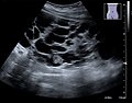
Renal cyst of the left kidney (hyperintense area) as shown on MRI. | |
Specialty | Urology ![]() |
A renal cyst is a fluid collection in or on the kidney. There are several types based on the Bosniak classification. The majority are benign, simple cysts that can be monitored and not intervened upon. However, some are cancerous or are suspicious for cancer and are commonly removed in a surgical procedure called nephrectomy.
Numerous renal cysts are seen in the cystic kidney diseases, which include polycystic kidney disease and medullary sponge kidney.
Renal cysts are classified by malignant risk using the Bosniak classification system. The system was created by Morton Bosniak (1929–2016), a faculty member at the New York University Langone Medical Center in New York City. [1]
The Bosniak classification categorizes renal cysts into five groups. [2]
This category includes renal cysts with multiple thin septa, a septum thicker than hairline, slightly thick wall, or with calcification, which may be thick. It also includes intrarenal cysts larger than 3 centimetres (1.2 inches) if:
Category IIF cysts have a 5–10% risk of being kidney cancer, and therefore follow-up is recommended. However, there is no consensus recommendation on the appropriate interval of follow up. [7]
The presence of measurable contrast enhancement of the lesion is the most important characteristic in distinguishing between high-risk cysts (classifications III and IV) from the typically benign, low-risk Bosniak I, II, and IIF cysts. [8] Such contrast enhancement should be at least 10 to 15 Hounsfield units higher when compared with unenhanced images.
Bosniak category | |||||
---|---|---|---|---|---|
I | II | IIF | III | IV | |
Attenuation [3] | 0–20 HU | 60–70 Hounsfield units | |||
Walls [3] | Thin and smooth | Small and fine calcifications | Nodular or irregular calcifications | Thick, heterogeneous. Gross calcifications with enhancement | |
Solid components [3] | No | Yes | |||
The complex cyst can be further evaluated with doppler ultrasonography, and for Bosniak classification and follow-up of complex cysts, either contrast-enhanced ultrasound (CEUS) or contrast CT is used. [12]
This system is more directly focused on the most appropriate management. These alternatives are broadly to ignore the cyst, schedule follow-up or perform a surgical excision of it. When a cyst shows discrepancy in severity across categories, it is the most worrisome feature that is used in deciding about management. There is no established rule regarding the follow-up frequency, but one possibility is after 6 months, which can later be doubled if unchanged. [5]
Recommended management [5] | |||
---|---|---|---|
Ignore | Follow | Excise | |
Calcification |
| Thick, nodular | |
If radiodensity > 20 HU without radiocontrast |
|
|
|
Septations | Thin and smooth | Slightly greater than hairline |
|
Enhancement (increase with radiocontrast) | < 10 HU | 10–15 HU | > 15 HU |
Multilocular | If infection | All others | |
Nodularity [5] | Very small and nonenhancing | All others | |
Wall thickening [5] | If infection | All others | |
Parapelvic cysts originate from around the kidney at the adjacent renal parenchyma, and plunge into the renal sinus. Peripelvic cysts are contained entirely within the renal sinus, possibly related to dilated lymphatic channels. When viewed on CT in absence of contrast, they can mimic hydronephrosis. [14] If symptomatic, they can be laparoscopically decorticated - removal of the outer layer or cortex. [15]
Up to 27 percent of individuals older than 50 years may have simple renal cysts that cause no symptoms. [16]
{{cite book}}
: |website=
ignored (help){{cite web}}
: CS1 maint: multiple names: authors list (link)