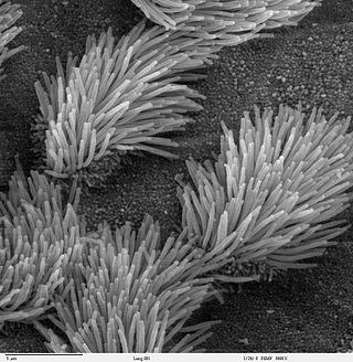
The cilium is a membrane-bound organelle found on most types of eukaryotic cell. Cilia are absent in bacteria and archaea. The cilium has the shape of a slender threadlike projection that extends from the surface of the much larger cell body. Eukaryotic flagella found on sperm cells and many protozoans have a similar structure to motile cilia that enables swimming through liquids; they are longer than cilia and have a different undulating motion.

Gastrulation is the stage in the early embryonic development of most animals, during which the blastula, or in mammals the blastocyst, is reorganized into a two-layered or three-layered embryo known as the gastrula. Before gastrulation, the embryo is a continuous epithelial sheet of cells; by the end of gastrulation, the embryo has begun differentiation to establish distinct cell lineages, set up the basic axes of the body, and internalized one or more cell types including the prospective gut.

Dextrocardia is a rare congenital condition in which the apex of the heart is located on the right side of the body, rather than the more typical placement towards the left. There are two main types of dextrocardia: dextrocardia of embryonic arrest and dextrocardia situs inversus. Dextrocardia situs inversus is further divided.

Situs inversus is a congenital condition in which the major visceral organs are reversed or mirrored from their normal positions. The normal arrangement of internal organs is known as situs solitus. Although cardiac problems are more common, many people with situs inversus have no medical symptoms or complications resulting from the condition, and until the advent of modern medicine, it was usually undiagnosed.
Primary ciliary dyskinesia (PCD) is a rare, autosomal recessive genetic ciliopathy, that causes defects in the action of cilia lining the upper and lower respiratory tract, sinuses, Eustachian tube, middle ear, fallopian tube, and flagella of sperm cells. The alternative name of "immotile ciliary syndrome" is no longer favored as the cilia do have movement, but are merely inefficient or unsynchronized. When accompanied by situs inversus the condition is known as Kartagener syndrome.
Situs ambiguus is a rare congenital defect in which the major visceral organs are distributed abnormally within the chest and abdomen. Clinically heterotaxy spectrum generally refers to any defect of Left-right asymmetry and arrangement of the visceral organs; however, classical heterotaxy requires multiple organs to be affected. This does not include the congenital defect situs inversus, which results when arrangement of all the organs in the abdomen and chest are mirrored, so the positions are opposite the normal placement. Situs inversus is the mirror image of situs solitus, which is normal asymmetric distribution of the abdominothoracic visceral organs. Situs ambiguus can also be subdivided into left-isomerism and right isomerism based on the defects observed in the spleen, lungs and atria of the heart.
Lefty are a class of proteins that are closely related members of the TGF-beta superfamily of growth factors. These proteins are secreted and play a role in left-right asymmetry determination of organ systems during development. Mutations of the genes encoding these proteins have been associated with left-right axis malformations, particularly in the heart and lungs.
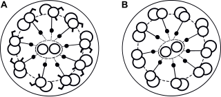
An asymmetric cell division produces two daughter cells with different cellular fates. This is in contrast to symmetric cell divisions which give rise to daughter cells of equivalent fates. Notably, stem cells divide asymmetrically to give rise to two distinct daughter cells: one copy of the original stem cell as well as a second daughter programmed to differentiate into a non-stem cell fate.
The heart is the first functional organ in a vertebrate embryo. There are 5 stages to heart development.

Cerberus is a protein that in humans is encoded by the CER1 gene. Cerberus is a signaling molecule which contributes to the formation of the head, heart and left-right asymmetry of internal organs. This gene varies slightly from species to species but its overall functions seem to be similar.
Levocardia refers to the most common location of the human heart, on the left side of the thoracic cavity. This is opposed to dextrocardia, in which the heart is in the right side. Neither levocardia nor dextrocardia are indicative of the orientation of the visceral organs, which can be in situs solitus, where the remainder of the organs are on normal side as well; or situs inversus, in which the viscera are on the opposite side as normal. The latter condition may or may not be associated with clinically relevant abnormalities.

Paired-like homeodomain transcription factor 2 also known as pituitary homeobox 2 is a protein that in humans is encoded by the PITX2 gene.

Nodal homolog is a secretory protein that in humans is encoded by the NODAL gene which is located on chromosome 10q22.1. It belongs to the transforming growth factor beta superfamily. Like many other members of this superfamily it is involved in cell differentiation in early embryogenesis, playing a key role in signal transfer from the primitive node, in the anterior primitive streak, to lateral plate mesoderm (LPM).

Renal–hepatic–pancreatic dysplasia is an autosomal recessive congenital disorder characterized by pancreatic fibrosis, renal dysplasia and hepatic dysgenesis. An association with NPHP3 has been described. It was characterized in 1959.
Symmetry breaking in biology is the process by which uniformity is broken, or the number of points to view invariance are reduced, to generate a more structured and improbable state. Symmetry breaking is the event where symmetry along a particular axis is lost to establish a polarity. Polarity is a measure for a biological system to distinguish poles along an axis. This measure is important because it is the first step to building complexity. For example, during organismal development, one of the first steps for the embryo is to distinguish its dorsal-ventral axis. The symmetry-breaking event that occurs here will determine which end of this axis will be the ventral side, and which end will be the dorsal side. Once this distinction is made, then all the structures that are located along this axis can develop at the proper location. As an example, during human development, the embryo needs to establish where is ‘back’ and where is ‘front’ before complex structures, such as the spine and lungs, can develop in the right location. This relationship between symmetry breaking and complexity was articulated by P.W. Anderson. He speculated that increasing levels of broken symmetry in many-body systems correlates with increasing complexity and functional specialization. In a biological perspective, the more complex an organism is, the higher number of symmetry-breaking events can be found.
The Nodal signaling pathway is a signal transduction pathway important in regional and cellular differentiation during embryonic development.
Homeobox protein notochord (NOTO) is a transcription factor encoded by the gene notochord homeobox (NOTO) located on the short arm of chromosome 2 (2p13.2) in humans (Homo sapiens). An ortholog of NOTO is found in the house mouse (Mus musculus), among other species, as the gene notochord homeobox (Noto) located on chromosome 6, which encodes the homologous transcription factor homeobox protein notochord (Noto).
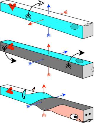
The axial twist theory is a scientific theory put forward to explain a range of unusual aspects of the body plan of vertebrates. It proposes that the rostral part of the head is "turned around" regarding the rest of the body. This end-part consists of the face as well as part of the brain. According to the theory, the vertebrate body has a left-handed chirality.
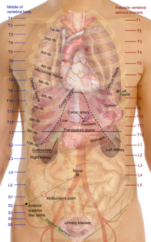
Left-right asymmetry is the process in early embryonic development that breaks the normal symmetry in the bilateral embryo. In vertebrates, left-right asymmetry is established early in development at a structure called the left-right organizer and leads to activation of different signalling pathways on the left and right of the embryo. This in turn cause several organs in adults to develop LR asymmetry, such as the tilt of the heart, the different number lung lobes on each side of the body and the position of the stomach and spleen on the right side of the body. If this process does not occur correctly in humans it can result in the syndromes heterotaxy or situs inversus.