Situs ambiguus

Last updated
Situs ambiguus
Other namesSitus ambiguous, heterotaxy, heterotaxia
Specialty Cardiology

Situs ambiguus is a rare congenital defect in which the major visceral organs are distributed abnormally within the chest and abdomen. Clinically heterotaxy spectrum generally refers to any defect of Left-right asymmetry and arrangement of the visceral organs; however, classical heterotaxy requires multiple organs to be affected. This does not include the congenital defect situs inversus, [1] which results when arrangement of all the organs in the abdomen and chest are mirrored, so the positions are opposite the normal placement. Situs inversus is the mirror image of situs solitus, which is normal asymmetric distribution of the abdominothoracic visceral organs. Situs ambiguus can also be subdivided into left-isomerism and right isomerism based on the defects observed in the spleen, lungs and atria of the heart.

Contents

Individuals with situs inversus or situs solitus do not experience fatal dysfunction of their organ systems, as general anatomy and morphology of the abdominothoracic organ-vessel systems are conserved. Due to abnormal arrangement of organs in situs ambiguus, orientation across the left-right axis of the body is disrupted early in fetal development, resulting in severely flawed cardiac development and function in 50–80% of cases. They also experience complications with systemic and pulmonary blood vessels, significant morbidity, and sometimes death. [2] All patients with situs ambiguus lack lateralization and symmetry of organs in the abdominal and thoracic cavities and are clinically considered to have a form of heterotaxy syndrome.

Heterotaxy syndrome with atrial isomerism occurs in 1 out of every 10,000 live births and is associated with approximately 3% of congenital heart disease cases. [3] Additional estimation of incidence and prevalence of isomerism proves difficult due to failure to diagnose and underestimation of the disease by clinicians. Furthermore, right isomerism is much more easily recognized than left isomerism, contributing to the failure to diagnose. [4]

Situs ambiguus is a growing field of research with findings dating back to 1973. [5]

Signs and symptoms

There are a variety of clinical manifestations of situs ambiguus. Acute symptoms can be due to both cardiac and non-cardiac defects. Cyanosis or blue skin coloration, primarily affecting the lips and fingernails, can indicate a systemic or circulatory issue. Poor feeding, failure to thrive, and rapid shallow breathing may also be observed due to poor circulation. Upon examination, arrhythmia and heart murmur may raise further suspicion of a cardiac abnormality. Non-cardiac symptoms include impairments of the liver and gastrointestinal tract. Biliary atresia, or inflammation and destruction of the bile ducts, may lead to jaundice. Vomiting and swelling of the abdominal region are features that suggest improper positioning of the intestines. Poor positioning of the intestine also makes it more prone to blockage, which can result in numerous chronic health issues. [4] Asplenia and polysplenia are also possible features of heterotaxy syndrome. [6]

Due to abnormal cardiac development, patients with situs ambiguus usually develop right atrial isomerism consisting of two bilaterally paired right atria, or left atrial isomerism consisting of two bilaterally paired left atria. Clinical features and symptoms can vary dependent upon assignment of left versus right atrial isomerism. In either instance, the apex of the heart will be poorly positioned, which should alert a clinician of the likelihood of atrial isomerism. It is estimated that 5–10% of isomeric patients have mesocardia, in which the heart is positioned at the center of the thorax, 25–50% have dextrocardia, in which the apex of the heart is pointed toward the right side of the thorax, and 50–70% have levocardia, in which the apex of the heart is pointed toward the left side of the thorax. [2]

Right atrial appendage isomerism

Right atrial appendage isomerism, also called right atrial isomerism, is a cardiac development defect in which the heart has bilateral right atria and atrial attachments in the muscle wall, as opposed to the normal right atrium and left atrium. In right atrial isomerism, the pulmonary blood oxygen tract is damaged due to right-left shunting of blood. In addition, the atrial septum which distinguishes the 2 atria is absent. These impairments, in addition to congestion in the pulmonary tract, allows deoxygenated blood to mix with oxygenated blood, contributing to cyanosis and possible respiratory distress. Poor systemic circulation also results due to improper positioning of the aorta. [2]

Left atrial appendage isomerism

Left atrial appendage isomerism, also called left atrial isomerism, is a cardiac development defect in which the heart has two bilateral left atria and atrial appendages in the muscle wall. Left atrial isomerism can have varied clinical manifestations, including a later onset of symptoms. Heart failure is often a concern because the inferior vena cava is disrupted due to the inappropriate morphology of the left ventricle to support the vena cava. [2]

Conductive nodes and tissues

Abnormal development of the heart results in impaired doubles of conductive nodes, as well as faulty electrical fibers throughout the ventricles. Individuals with right atrial isomerism develop two sinoatrial nodes, as they have 2 mirrored right atria, whereas those with left atrial isomerism fail to develop a sinus node at all. Thus, patients with left atrial isomerism are more likely to experience atrial fibrillation, or irregular rapid heart beat, and abnormal heart rhythm, known as atrial flutter. Development of the atrioventricular node and bundle of His largely depends on physiological looping of the ventricles. Abnormal looping of the ventricles contributes to arrhythmia and heart block in fetuses. [2]

Bronchial tree and lungs

Isomerism of the bronchial tree is not typically damaging and presents no significant clinical complications. [7] Pulmonary valve stenosis results in issues of blood flow to the lungs.

Abdominal organs

Abdominal organs, including the liver, stomach, intestinal tract, and spleen may be randomly arranged throughout the left-right axis of the body. Distribution of these organs largely dictates treatment, clinical outcomes, and further evaluation.

The liver is typically symmetrical across the left-right axis in patients with situs ambiguus, which is abnormal. A majority of left atrial isomeric patients have defects throughout the biliary tree, which is responsible for bile production, even when the gall bladder is functional and morphologically normal. This biliary atresia can lead to acute problems such as nutrient malabsorption, pale stools, dark urine, and abdominal swelling. If this condition continues without proper treatment, cirrhosis and liver failure become a major concern. Biliary atresia is not usually observed in patients with right atrial isomerism. [2]

Random positioning of the stomach is often one of the first signals of situs ambiguus upon examination. Malrotation of the entire intestinal tract, or improper folding and bulging of the stomach and intestines, results in bowel obstruction. This impairment leads to vomiting, abdominal distension, mucus and blood in the stool. Patients may also experience abdominal pain. Intestinal malrotation is more commonly identified in patients with right atrial isomerism than in those with left atrial isomerism.[ citation needed ]

Isomeric patients often experience disruptions to splenic development during embryogenesis, resulting in an overall lack a spleen (asplenia) or development of many spleens (polysplenia). Asplenia is most often observed in patients with right atrial isomerism. Polysplenia results in 90% of patients with left atrial isomerism. Although they have many spleens, each is usually ineffective resulting in functional asplenia. Rarely, left atrial isomeric patients have a single, normal, functional spleen. Patients lacking a functional spleen are in danger of sepsis and must be monitored. [2]

Causes

Situs ambiguus has been linked to family history of malformations [8] [9] and maternal cocaine use, [10] suggesting both genetic and environmental factors play a role. [11] Several genes in the TGF-beta pathway, which controls left-right patterning of visceral organs across the body axis, have been indicated in sporadic and familial cases of atrial isomerism. There is also overlap between genes associated with situs ambiguus and primary ciliary dyskinesia, likely due to the important role of cilia in establishing left-right asymmetry. [12] The planar cell polarity has an important role in positioning these cilia, and thus genes within this pathway are increasingly associated with situs ambiguus. [13]

Disrupted mitochondria function has also been recently linked to heterotaxy. [14]

Pathophysiology

Molecular and cellular mechanism

Several genes have been identified in normal development of the right-left axis. [15] These genes have been extensively researched. Gene mutations that lead to atrial isomerism is a growing area of research. Mutations in genes that encode proteins in the TGF-beta pathway, including NODAL, NKX2-5, and ZIC3, have been linked to tetralogy of Fallot and hypoplastic left heart syndrome. [16] [17] Mutations in the ZIC3 gene, which encodes a zinc finger family transcription factor, is linked to a 50% risk of atrial isomerism in families. It is also an X-linked disorder, so testing for ZIC3 mutations is highly encouraged in male births. [18]

The most prevalent and best characterized genetic associations of heterotaxy include: [2]

Type OMIM GeneLocus
HTX1 306955 ZIC3 Xq26.2
HTX2 605376 CFC1 2q21.1
HTX3 606325 PA26 6q21
HTX4 613751 ACVR2B 992
HTX5 270100 NODAL 10q22.1
HTX6 614779 CCDC11 18q21.1

Classical pathology

Bronchial tree and lungs

Pathophysiology in the bronchial tree can be observed by radiography. Under normal development, the bronchial tree consists of two main bronchi that are anatomically different:

In situs ambiguus, there is a duplication of either the hyparterial or eparterial bronchus. These features are not associated with any significant clinical complications. [19] Mechanisms leading to bronchial duplication are not thoroughly understood.

In pulmonary valve stenosis, there is a reduction in blood flow to the lungs due to an obstruction of the heart at the pulmonic valve. This contributes to cyanosis and pulmonary hypertension. [20]

Diagnosis

For proper diagnosis of situs ambiguus, cardiac and non-cardiac features must be evaluated. Diagnostic criteria for atrial isomerism includes observation of symmetry of thoracic visceral organs upon echocardiogram, arrhythmia upon electrocardiogram, and chest x-ray for confirmation of the heart's location across the left-right axis. Additional radiographic and cross-sectional imaging may be obtained to evaluate both cardiac and non-cardiac manifestations of situs ambiguus. In addition, a series of gastrointestinal tests can be conducted for observation of intestinal malrotation, as well as a scan of the liver and spleen for biliary function. [2]

Diagnostic techniques for cardiac causes

Diagnostic techniques for non-cardiac causes

Other diagnostic features

Management

Each of the symptoms of situs ambiguus must be managed with appropriate treatment dependent upon the organ system involved. Intestinal malrotation is treated surgically using the Ladd procedure. This procedure widens a fold in the peritoneum so that the intestines can be placed in non-rotated formation. It is not possible to return the bowel to a normal morphology [21] However, 89% of patients that undergo the Ladd surgery experience a complete resolution of symptoms.

Following cholangiogram, a Kasai procedure is usually performed in cases of biliary atresia. In this surgery, a Y-shaped shunt is used to passage bile from the liver directly to the intestine. If this is unsuccessful, liver transplantation can be considered based on the overall health of the patient. The Kasai procedure is successful in approximately 80% of patients. [22] Following the operation, patients are advised to take fat-soluble vitamins, choleretics, and anti-inflammatory medications.

Functionally asplenic patients have an elevated lifetime risk of sepsis, as they have no functional spleen for fighting infection. For this reason, asplenic patients are under constant observation for any signs of fever or infection. In the case of infection, patients are placed on controlled empiric antibiotic therapy to avoid development of antibiotic resistance. [23] This therapy battles infection by both gram-positive and gram-negative bacteria.

Right-atrial and left-atrial isomerism and associated pulmonary issues are treated in a series of steps based on the severity of symptoms. Isomeric patients are first treated by inserting a shunt that will move incoming blood through the pulmonary circuit. The Fontan procedure routes blood through the patient's single ventricle, to the lungs, and into systemic circulation. This process is favorable in patients aged 2 to 5 years old. About 20–30% of patients will require a heart transplant. [24] Left-atrial isomeric patients have less severe complications, as they typically have 2 functional ventricles. In this case, they can undergo biventricular repair to form 2 separate ventricles and functional associated valves.

Prognosis

Prognosis for patients with situs ambiguus is quite varied, owed to the spectrum of clinical manifestations. [25] Infants who experience severe cyanosis at birth may die within hours of delivery if medical intervention is not immediate. Alternatively, longevity of neonates with mild cardiac lesions is unaffected. [26] Ten percent of patients born with right atrial isomerism die by the age of 5 without intervention. Improvements in therapies has increased the 5-year survival to 30–74% for right atrial isomeric patients and 65–84% for left atrial isomeric patients based on the cause of their disease.

Research

There have been vast amounts of research on the clinical features, racial disparities, and physiological mechanisms of heterotaxy syndrome dating back to 1973.

Mishra et al. published a review in November 2015 describing current knowledge of cardiac and non-cardiac abnormalities associated with situs ambiguus. The author stresses the importance of genetic testing prior to deciding a prognosis for affected patients. [27] She also proposes prenatal screening and evaluation in cases at risk for development of situs ambiguus.

Recent studies have shown higher rates of heterotaxy syndrome among Hispanic infants of Mexican descent, as well as female infants of non-Hispanic black and white mothers. Additional studies must be done to clarify the mechanisms behind racial disparities in heterotaxy syndrome. [28] Individuals of Asian descent show a higher prevalence of heterotaxy syndrome in general than members of the Western world. [29]

The National Birth Defects Prevention study (October 2014) attempted to link clinical presentations of situs ambiguus to demographics in an epidemiological study. [30] This proved a difficult task due to the vast differences in presentation of this disorder. However, the authors are hopeful that finding a link can help inform clinical decision-making, predictive analyses, and future outcomes.

See also

Related Research Articles

<span class="mw-page-title-main">Tetralogy of Fallot</span> Type of congenital heart defect

Tetralogy of Fallot (TOF), formerly known as Steno-Fallot tetralogy, is a congenital heart defect characterized by four specific cardiac defects. Classically, the four defects are:

<span class="mw-page-title-main">Dextrocardia</span> Medical condition

Dextrocardia is a rare congenital condition in which the apex of the heart is located on the right side of the body, rather than the more typical placement towards the left. There are two main types of dextrocardia: dextrocardia of embryonic arrest and dextrocardia situs inversus. Dextrocardia situs inversus is further divided.

<span class="mw-page-title-main">Situs inversus</span> Condition in which organs are reversed

Situs inversus is a congenital condition in which the major visceral organs are reversed or mirrored from their normal positions. The normal arrangement of internal organs is known as situs solitus. Although cardiac problems are more common, many people with situs inversus have no medical symptoms or complications resulting from the condition, and until the advent of modern medicine, it was usually undiagnosed.

<span class="mw-page-title-main">Primary ciliary dyskinesia</span> Medical condition

Primary ciliary dyskinesia (PCD) is a rare, autosomal recessive genetic ciliopathy, that causes defects in the action of cilia lining the upper and lower respiratory tract, sinuses, Eustachian tube, middle ear, Fallopian tube, and flagella of sperm cells. The alternative name of "immotile ciliary syndrome" is no longer favored as the cilia do have movement, but are merely inefficient or unsynchronized. When accompanied by situs inversus the condition is known as Kartagener syndrome.

<span class="mw-page-title-main">Atrial septal defect</span> Human heart defect present at birth

Atrial septal defect (ASD) is a congenital heart defect in which blood flows between the atria of the heart. Some flow is a normal condition both pre-birth and immediately post-birth via the foramen ovale; however, when this does not naturally close after birth it is referred to as a patent (open) foramen ovale (PFO). It is common in patients with a congenital atrial septal aneurysm (ASA).

<span class="mw-page-title-main">Jugular venous pressure</span>

The jugular venous pressure is the indirectly observed pressure over the venous system via visualization of the internal jugular vein. It can be useful in the differentiation of different forms of heart and lung disease. Classically three upward deflections and two downward deflections have been described.

<span class="mw-page-title-main">Ventricular septal defect</span> Medical condition

A ventricular septal defect (VSD) is a defect in the ventricular septum, the wall dividing the left and right ventricles of the heart. The extent of the opening may vary from pin size to complete absence of the ventricular septum, creating one common ventricle. The ventricular septum consists of an inferior muscular and superior membranous portion and is extensively innervated with conducting cardiomyocytes.

<span class="mw-page-title-main">Fontan procedure</span>

The Fontan procedure or Fontan–Kreutzer procedure is a palliative surgical procedure used in children with univentricular hearts. It involves diverting the venous blood from the inferior vena cava (IVC) and superior vena cava (SVC) to the pulmonary arteries without passing through the morphologic right ventricle; i.e., the systemic and pulmonary circulations are placed in series with the functional single ventricle. The procedure was initially performed in 1968 by Francis Fontan and Eugene Baudet from Bordeaux, France, published in 1971, simultaneously described in 1971 by Guillermo Kreutzer from Buenos Aires, Argentina, and finally published in 1973.

<span class="mw-page-title-main">Eisenmenger syndrome</span> Medical condition

Eisenmenger syndrome or Eisenmenger's syndrome is defined as the process in which a long-standing left-to-right cardiac shunt caused by a congenital heart defect causes pulmonary hypertension and eventual reversal of the shunt into a cyanotic right-to-left shunt. Because of the advent of fetal screening with echocardiography early in life, the incidence of heart defects progressing to Eisenmenger syndrome has decreased.

<span class="mw-page-title-main">Atrium (heart)</span> Part of the human heart

The atrium is one of two upper chambers in the heart that receives blood from the circulatory system. The blood in the atria is pumped into the heart ventricles through the atrioventricular valves.

<span class="mw-page-title-main">Transposition of the great vessels</span> Group of congenital heart defects

Transposition of the great vessels (TGV) is a group of congenital heart defects involving an abnormal spatial arrangement of any of the great vessels: superior and/or inferior venae cavae, pulmonary artery, pulmonary veins, and aorta. Congenital heart diseases involving only the primary arteries belong to a sub-group called transposition of the great arteries (TGA), which is considered the most common congenital heart lesion that presents in neonates.

<span class="mw-page-title-main">Atrioventricular septal defect</span> Medical condition

Atrioventricular septal defect (AVSD) or atrioventricular canal defect (AVCD), also known as "common atrioventricular canal" (CAVC) or "endocardial cushion defect" (ECD), is characterized by a deficiency of the atrioventricular septum of the heart that creates connections between all four of its chambers. It is caused by an abnormal or inadequate fusion of the superior and inferior endocardial cushions with the mid portion of the atrial septum and the muscular portion of the ventricular septum.

<span class="mw-page-title-main">Norwood procedure</span>

The Norwood procedure is the first of three surgeries intended to create a new functional systemic circuit in patients with hypoplastic left heart syndrome and other complex heart defects with single ventricle physiology. The first successful Norwood procedure involving the use of a cardiopulmonary bypass was reported by Dr. William Imon Norwood, Jr. and colleagues in 1981.

Cor triatriatum is a congenital heart defect where the left atrium or right atrium is subdivided by a thin membrane, resulting in three atrial chambers.

<span class="mw-page-title-main">Situs solitus</span>

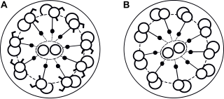
Situs solitus is the medical term referring to the normal position of thoracic and abdominal organs. Anatomically, this means that the heart is on the left with the pulmonary atrium on the right and the systemic atrium on the left along with the cardiac apex. Right-sided organs are the liver, the gall bladder and a trilobed lung as well as the inferior vena cava, while left-sided organs are the stomach, single spleen, a bilobed lung, and the aorta.

<span class="mw-page-title-main">Asplenia with cardiovascular anomalies</span> Medical condition

Asplenia with cardiovascular anomalies, also known as Ivemark syndrome and right atrial isomerism, is an example of a heterotaxy syndrome. These uncommon congenital disorders are characterized by defects in the heart, spleen and paired organs such as the lungs and kidneys. Another name is "asplenia-cardiovascular defect-heterotaxy".

<span class="mw-page-title-main">Lutembacher's syndrome</span> Medical condition

Lutembacher's syndrome is a very rare form of congenital heart disease that affects one of the chambers of the heart as well as a valve. It is commonly known as both congenital atrial septal defect (ASD) and acquired mitral stenosis (MS). Congenital atrial septal defect refers to a hole being in the septum or wall that separates the two atria; this condition is usually seen in fetuses and infants. Mitral stenosis refers to mitral valve leaflets sticking to each other making the opening for blood to pass from the atrium to the ventricles very small. With the valve being so small, blood has difficulty passing from the left atrium into the left ventricle. Septal defects that may occur with Lutembacher's syndrome include: Ostium primum atrial septal defect or ostium secundum which is more prevalent.

Polysplenia is a congenital disease manifested by multiple small accessory spleens, rather than a single, full-sized, normal spleen. Polysplenia sometimes occurs alone, but it is often accompanied by other developmental abnormalities. Conditions associated with polysplenia include gastrointestinal abnormalities, such as intestinal malrotation or biliary atresia, as well as cardiac abnormalities, such as dextrocardia.

<span class="mw-page-title-main">Arrhythmia</span> Group of medical conditions characterized by irregular heartbeat

Arrhythmias, also known as cardiac arrhythmias, heart arrhythmias, or dysrhythmias, are irregularities in the heartbeat, including when it is too fast or too slow. A resting heart rate that is too fast – above 100 beats per minute in adults – is called tachycardia, and a resting heart rate that is too slow – below 60 beats per minute – is called bradycardia. Some types of arrhythmias have no symptoms. Symptoms, when present, may include palpitations or feeling a pause between heartbeats. In more serious cases, there may be lightheadedness, passing out, shortness of breath or chest pain. While most cases of arrhythmia are not serious, some predispose a person to complications such as stroke or heart failure. Others may result in sudden death.

Isolated levocardia is a rare type of organs' situs inversus in which the heart is still in normal position but other abdominal viscera are transposed. Isolated levocardia may occur with heart defects and patients without having operations have low life expectancy: only about 5% to 13% of patients survive more than 5 years. Therefore, even though the risk of cardiac surgeries is high, once patients are diagnosed, operations are suggested to be held as soon as possible. Isolated levocardia is congenital. So far, there is not sufficient evidence to prove that chromosome abnormalities will result in isolated levocardia, and the cause of isolated levocardia is still unknown.

References

  1. "heterotaxy syndrome". United States National Library of Medicine . Retrieved 23 May 2016.
  2. 1 2 3 4 5 6 7 8 9 Lowenthal, A.; et al. (September 26, 2015). "Anatomy, clinical manifestations and diagnosis of heterotaxy (isomerism of the atrial appendages)". Up To Date. Retrieved November 4, 2015.
  3. Zhu, Lirong; Belmont, John W.; Ware, Stephanie M. (2005-10-26). "Genetics of human heterotaxias". European Journal of Human Genetics. 14 (1): 17–25. doi: 10.1038/sj.ejhg.5201506 . ISSN   1018-4813. PMID   16251896.
  4. 1 2 Kim, Soo-Jin (2011). "Heterotaxy Syndrome". Korean Circulation Journal. 41 (5): 227–32. doi:10.4070/kcj.2011.41.5.227. PMC   3116098 . PMID   21731561.
  5. Freedom, Robert M.; Treves, S. (1973-05-01). "Splenic Scintigraphy and Radionuclide Venography in the Heterotaxy Syndrome". Radiology. 107 (2): 381–386. doi:10.1148/107.2.381. ISSN   0033-8419. PMID   4695908.
  6. Van Praagh, R; et al. (December 15, 1990). "Atrial isomerism in the heterotaxy syndromes with asplenia, or polysplenia, or normally formed spleen: an erroneous concept". Am J Cardiol. 66 (20): 1504–6. doi:10.1016/0002-9149(90)90543-a. PMID   2252000.
  7. Cohen, Meryl S.; Anderson, Robert H.; Cohen, Mitchell I.; Atz, Andrew M.; Fogel, Mark; Gruber, Peter J.; Lopez, Leo; Rome, Jonathan J.; Weinberg, Paul M. (2007). "Controversies, genetics, diagnostic assessment, and outcomes relating to the heterotaxy syndrome". Cardiology in the Young. 17 (S2): 29–43. doi:10.1017/s104795110700114x. PMID   18039397. S2CID   206304327.
  8. Martínez-Frías ML (March 2001). "Heterotaxia as an outcome of maternal diabetes: an epidemiological study". American Journal of Medical Genetics. 99 (2): 142–6. doi:10.1002/1096-8628(2000)9999:999<00::AID-AJMG1139>3.0.CO;2-Z. PMID   11241474.
  9. Maeyama K, Kosaki R, Yoshihashi H, Casey B, Kosaki K (March 2001). "Mutation analysis of left-right axis determining genes in NOD and ICR, strains susceptible to maternal diabetes". Teratology. 63 (3): 119–26. doi:10.1002/tera.1022. PMID   11283968.
  10. Kuehl KS, Loffredo C (November 2002). "Risk factors for heart disease associated with abnormal sidedness". Teratology. 66 (5): 242–8. doi:10.1002/tera.10099. PMID   12397632.
  11. Kuehl KS, Loffredo CA (March 2003). "Population-based study of l-transposition of the great arteries: possible associations with environmental factors". Birth Defects Research. Part A, Clinical and Molecular Teratology. 67 (3): 162–7. doi:10.1002/bdra.10015. PMID   12797457.
  12. Sutherland, Mardi J.; Ware, Stephanie M. (15 November 2009). "Disorders of left-right asymmetry: Heterotaxy and situs inversus". American Journal of Medical Genetics Part C: Seminars in Medical Genetics. 151C (4): 307–317. doi:10.1002/ajmg.c.30228. PMID   19876930. S2CID   23490441.
  13. Axelrod, Jeffrey D. (1 February 2020). "Planar cell polarity signaling in the development of left–right asymmetry". Current Opinion in Cell Biology. 62: 61–69. doi:10.1016/j.ceb.2019.09.002. PMC   9258637 . PMID   31654871. S2CID   204918577.
  14. Burkhalter MD, Sridhar A, Sampaio P, Jacinto R, Burczyk MS, Donow C, Angenendt M, Competence Network for Congenital Heart Defects Investigators, Hempel M, Walther P, Pennekamp P, Omran H, Lopes SS, Ware SM, Philipp M (2019). "Imbalanced mitochondrial function provokes heterotaxy via aberrant ciliogenesis". J Clin Invest. 129 (7): 2841–2855. doi:10.1172/JCI98890. PMC   6597216 . PMID   31094706.{{cite journal}}: CS1 maint: multiple names: authors list (link)
  15. Chen, CM; et al. (April 2010). "Transcriptional Control of Left–Right Patterning in Cardiac Development". Pediatric Cardiology. 31 (3): 371–377. doi:10.1007/s00246-009-9610-3. PMID   20054532. S2CID   300003.
  16. Kosaki, K; Bassi, M T; Kosaki, R; Lewin, M; Belmont, J; Schauer, G; Casey, B (1999-03-01). "Characterization and mutation analysis of human LEFTY A and LEFTY B, homologues of murine genes implicated in left-right axis development". American Journal of Human Genetics. 64 (3): 712–721. doi:10.1086/302289. ISSN   0002-9297. PMC   1377788 . PMID   10053005.
  17. Mohapatra, Bhagyalaxmi; Casey, Brett; Li, Hua; Ho-Dawson, Trang; Smith, Liana; Fernbach, Susan D.; Molinari, Laura; Niesh, Stephen R.; Jefferies, John Lynn (2009-03-01). "Identification and functional characterization of NODAL rare variants in heterotaxy and isolated cardiovascular malformations". Human Molecular Genetics. 18 (5): 861–871. doi:10.1093/hmg/ddn411. ISSN   1460-2083. PMC   2722226 . PMID   19064609.
  18. Gebbia, Marinella; Ferrero, Giovanni B.; Pilia, Giuseppe; Bassi, Maria T.; Aylsworth, Arthur S.; Penman-Splitt, Miranda; Bird, Lynne M.; Bamforth, John S.; Burn, John (1997-11-01). "X-linked situs abnormalities result from mutations in ZIC3". Nature Genetics. 17 (3): 305–308. doi:10.1038/ng1197-305. PMID   9354794. S2CID   22916101.
  19. Applegate, Kimberly E.; Goske, Marilyn J.; Pierce, Gregory; Murphy, Daniel (1999-07-01). "Situs Revisited: Imaging of the Heterotaxy Syndrome". RadioGraphics. 19 (4): 837–852. doi: 10.1148/radiographics.19.4.g99jl31837 . ISSN   0271-5333. PMID   10464794.
  20. Lowenthal, Alexander; et al. (October 27, 2015). "Management and outcome of heterotaxy (isomerism of the atrial appendages)". UpToDate. Retrieved December 8, 2015.
  21. Brandt, Mary; et al. (October 24, 2015). "Intestinal Malrotation". UpToDate. Retrieved December 10, 2015.
  22. Erlichman, Jessi; et al. (August 3, 2015). "Biliary Atresia". UpToDate. Retrieved December 9, 2015.
  23. Pasternack, Mark S; et al. (December 11, 2014). "Clinical features and management of sepsis in the asplenic patient". UpToDate. Retrieved December 10, 2015.
  24. Anderson PA, Sleeper LA, Mahony L, et al. (2008). ""Contemporary outcomes after the Fontan procedure: A Pediatric Heart Network multicenter study."". Journal of the American College of Cardiology. 52 (2): 85–93. doi:10.1016/j.jacc.2008.01.074. PMC   4385517 . PMID   18598886.
  25. Shahrouki, Puja; Lee, Edward Wolfgang; Ruehm, Stefan (February 2022). "Polysplenia syndrome and sickle cell trait: extensive deep venous thrombosis from venous stasis and hypercoagulability". Clinical Imaging. 82: 127–131. doi:10.1016/j.clinimag.2021.11.017. ISSN   0899-7071. PMID   34813990. S2CID   244443675.
  26. Hashmi, Aijaz; Abu-Sulaiman, Riyadh; McCrindle, Brian W; Smallhorn, Jeffrey F; Williams, William G; Freedom, Robert M (1998-04-01). "Management and Outcomes of Right Atrial Isomerism: A 26-Year Experience". Journal of the American College of Cardiology. 31 (5): 1120–1126. doi: 10.1016/S0735-1097(98)00062-X . PMID   9562017.
  27. Mishra, Smita (2015-11-26). "Cardiac and Non-Cardiac Abnormalities in Heterotaxy Syndrome". The Indian Journal of Pediatrics. 82 (12): 1135–1146. doi:10.1007/s12098-015-1925-x. ISSN   0019-5456. PMID   26612104. S2CID   207388492.
  28. Lopez, Keila N.; Marengo, Lisa K.; Canfield, Mark A.; Belmont, John W.; Dickerson, Heather A. (2015-09-01). "Racial disparities in heterotaxy syndrome". Birth Defects Research Part A: Clinical and Molecular Teratology. 103 (11): 941–950. doi:10.1002/bdra.23416. ISSN   1542-0760. PMID   26333177.
  29. Kim, Soo-Jin; Kim, Woong-Han; Lim, Hong-Gook; Lee, Jae-Young (July 2008). "Outcome of 200 patients after an extracardiac Fontan procedure". The Journal of Thoracic and Cardiovascular Surgery. 136 (1): 108–116. doi:10.1016/j.jtcvs.2007.12.032. hdl: 10371/60103 . PMID   18603062.
  30. Lin, Angela E.; Krikov, Sergey; Riehle-Colarusso, Tiffany; Frías, Jaime L.; Belmont, John; Anderka, Marlene; Geva, Tal; Getz, Kelly D.; Botto, Lorenzo D. (2014-10-01). "Laterality defects in the national birth defects prevention study (1998–2007): Birth prevalence and descriptive epidemiology". American Journal of Medical Genetics Part A. 164 (10): 2581–2591. doi:10.1002/ajmg.a.36695. ISSN   1552-4833. PMC   4462240 . PMID   25099286.