Rasmussen syndrome is a condition characterized by multiple trichoepitheliomas.
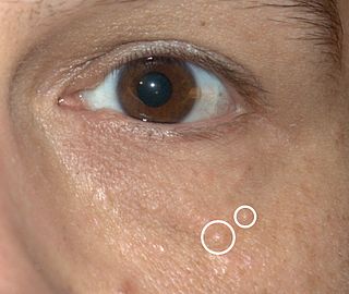
Syringomas are harmless eccrine sweat duct tumors, typically found clustered on eyelids, although they may also be found in the armpits, abdomen, chest, neck, scalp or groin area including genitals in a symmetric pattern. They are skin-colored or yellowish firm, rounded bumps, 1–3 mm in diameter, and may be confused with xanthoma, milia, hidrocystoma, trichoepithelioma, and xanthelasma. They are more common in women and are most commonly found in middle-aged Asian women. While they can present at any time in life, they typically present during adolescence. They are usually not associated with any other symptoms although can sometimes cause itchiness or irritation.

Blue nevus is a type of melanocytic nevus. The blue colour is caused by the pigment being deeper in the skin than in ordinary nevi. In principle they are harmless but they can sometimes be mimicked by malignant lesions, i.e. some melanomas can look like a blue nevus.

Rombo syndrome is a very rare genetic disorder characterized mainly by atrophoderma vermiculatum of the face, multiple milia, telangiectases, acral erythema, peripheral vasodilation with cyanosis and a propensity to develop basal cell carcinomas. The lesions become visible in late childhood, began at ages 7 to 10 years and are most pronounced on the face, At that time a pronounced, somewhat cyanotic redness of the lips and hands was evident as well as moderate follicular atrophy of the skin on the cheeks. In adulthood, whitish-yellow, milia-like papules and telangiectatic vessels developed. The papules were present particularly on the cheeks and forehead, gradually becoming very conspicuous and dominating the clinical picture. Trichoepitheliomas were found in 1 case. In adults, the eyelashes and eyebrows were either missing or irregularly distributed with defective and maldirected growth. Basal cell carcinomas were a frequent complication. The skin atrophy was referred to as vermiculate atrophoderma. Basal cell carcinomas may develop around the age of 35. Histological observations during the early stage include irregularly distributed and atrophic hair follicles, milia, dilated dermal vessels, lack of elastin or elastin in clumps. After light irradiation a tendency to increased repair activity was observed both in epidermis and in the dermal fibroblasts. Histologic sections showed the dermis to be almost devoid of elastin in most areas with clumping of elastic material in other areas. The disorder had been transmitted through at least 4 generations with instances of male-to-male transmission.
Generalized trichoepitheliomas are characterized histologically by replacement of the hair follicles by trichoepithelioma-like epithelial proliferations associated with hyperplastic sebaceous glands.

Cutaneous lymphoid hyperplasia refers to a groups of benign cutaneous disorders characterized by collections of lymphocytes, macrophages, and dendritic cells in the skin. Conditions included in this groups are:
CD30+ cutaneous T-cell lymphoma, also known as primary cutaneous anaplastic large cell lymphoma, is a cutaneous (skin) condition characterized by solitary or localized skin lesions that have a tendency to ulcerate.
Secondary cutaneous CD30+ large-cell lymphoma is a cutaneous condition that may arise in cases of mycosis fungoides, and in patients with lymphomatoid papulosis.
Non-mycosis fungoides CD30− cutaneous large T-cell lymphoma is a cutaneous condition that usually presents as solitary or generalized plaques, nodules, or tumors of short duration.

Spiradenoma, also spiroma or eccrine spiradenoma, is a cutaneous condition that is typically characterized, clinically, as a solitary, deep-seated dermal nodule of approximately one centimeter, occurring on the ventral surface of the body. Spiradenoma lesions are benign sudoriferous tumors, and have also been described as cystic epitheliomas of the sweat glands.
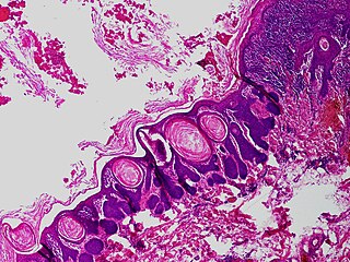
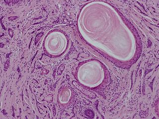
Trichofolliculoma is a cutaneous condition characterized by a benign, highly structured tumor of the pilosebaceous unit.
Multiple familial trichoepithelioma is a cutaneous condition characterized by multiple cystic and solid nodules appearing on the face.
A giant solitary trichoepithelioma is a cutaneous condition characterized by a skin lesion that may be up to several centimeters in diameter.
A desmoplastic trichoepithelioma is a cutaneous condition characterized by a solitary, firm skin lesion on the face.

Trichoblastomas are a cutaneous condition characterized by benign neoplasms of follicular germinative cells. Trichoblastic fibroma is a designation used to characterize small nodular trichoblastomas with conspicuous fibrocytic stroma, sometimes constituting over 50% of the lesion.
Brooke–Fordyce syndrome is a condition characterized by multiple trichoepitheliomas.
Sarcoidosis involves the skin in about 25% of patients. The most common lesions are erythema nodosum, plaques, maculopapular eruptions, subcutaneous nodules, and lupus pernio. Treatment is not required, since the lesions usually resolve spontaneously in two to four weeks. Although it may be disfiguring, cutaneous sarcoidosis rarely causes major problems.


