Immunoperoxidase is a type of immunostain used in molecular biology, medical research, and clinical diagnostics. In particular, immunoperoxidase reactions refer to a sub-class of immunohistochemical or immunocytochemical procedures in which the antibodies are visualized via a peroxidase-catalyzed reaction.

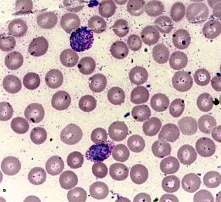
Romanowsky staining, also known as Romanowsky–Giemsa staining, is a prototypical staining technique that was the forerunner of several distinct but similar stains widely used in hematology and cytopathology. Romanowsky-type stains are used to differentiate cells for microscopic examination in pathological specimens, especially blood and bone marrow films, and to detect parasites such as malaria within the blood. Stains that are related to or derived from the Romanowsky-type stains include Giemsa, Jenner, Wright, Field, May–Grünwald and Leishman stains. The staining technique is named after the Russian physician Dmitri Leonidovich Romanowsky (1861–1921), who was one of the first to recognize its potential for use as a blood stain.
Methylene blue, also known as methylthioninium chloride, is a medication and dye. As a medication, it is mainly used to treat methemoglobinemia. Specifically, it is used to treat methemoglobin levels that are greater than 30% or in which there are symptoms despite oxygen therapy. It has previously been used for cyanide poisoning and urinary tract infections, but this use is no longer recommended. It is typically given by injection into a vein.

An automated analyser is a medical laboratory instrument designed to measure different chemicals and other characteristics in a number of biological samples quickly, with minimal human assistance. These measured properties of blood and other fluids may be useful in the diagnosis of disease.

Staining is a technique used to enhance contrast in samples, generally at the microscopic level. Stains and dyes are frequently used in histology and in the medical fields of histopathology, hematology, and cytopathology that focus on the study and diagnoses of disease at a microscopic level. Stains may be used to define biological tissues, cell populations, or organelles within individual cells.

A blood film—or peripheral blood smear—is a thin layer of blood smeared on a glass microscope slide and then stained in such a way as to allow the various blood cells to be examined microscopically. Blood films are examined in the investigation of hematological (blood) disorders and are routinely employed to look for blood parasites, such as those of malaria and filariasis.

In biochemistry, immunostaining is any use of an antibody-based method to detect a specific protein in a sample. The term "immunostaining" was originally used to refer to the immunohistochemical staining of tissue sections, as first described by Albert Coons in 1941. However, immunostaining now encompasses a broad range of techniques used in histology, cell biology, and molecular biology that use antibody-based staining methods.

A fluorophore is a fluorescent chemical compound that can re-emit light upon light excitation. Fluorophores typically contain several combined aromatic groups, or planar or cyclic molecules with several π bonds.
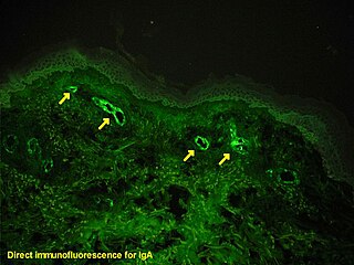
Immunofluorescence is a technique used for light microscopy with a fluorescence microscope and is used primarily on microbiological samples. This technique uses the specificity of antibodies to their antigen to target fluorescent dyes to specific biomolecule targets within a cell, and therefore allows visualization of the distribution of the target molecule through the sample. The specific region an antibody recognizes on an antigen is called an epitope. There have been efforts in epitope mapping since many antibodies can bind the same epitope and levels of binding between antibodies that recognize the same epitope can vary. Additionally, the binding of the fluorophore to the antibody itself cannot interfere with the immunological specificity of the antibody or the binding capacity of its antigen. Immunofluorescence is a widely used example of immunostaining and is a specific example of immunohistochemistry. This technique primarily makes use of fluorophores to visualise the location of the antibodies.

A fluorescence microscope is an optical microscope that uses fluorescence instead of, or in addition to, scattering, reflection, and attenuation or absorption, to study the properties of organic or inorganic substances. "Fluorescence microscope" refers to any microscope that uses fluorescence to generate an image, whether it is a more simple set up like an epifluorescence microscope or a more complicated design such as a confocal microscope, which uses optical sectioning to get better resolution of the fluorescence image.

Crystal violet or gentian violet, also known as methyl violet 10B or hexamethyl pararosaniline chloride, is a triarylmethane dye used as a histological stain and in Gram's method of classifying bacteria. Crystal violet has antibacterial, antifungal, and anthelmintic properties and was formerly important as a topical antiseptic. The medical use of the dye has been largely superseded by more modern drugs, although it is still listed by the World Health Organization.
A vital stain in a casual usage may mean a stain that can be applied on living cells without killing them. Vital stains have been useful for diagnostic and surgical techniques in a variety of medical specialties. In supravital staining, living cells have been removed from an organism, whereas intravital staining is done by injecting or otherwise introducing the stain into the body. The term vital stain is used by some authors to refer to an intravital stain, and by others interchangeably with a supravital stain, the core concept being that the cell being examined is still alive. In a more strict sense, the term vital staining has a meaning contrasting with supravital staining. While in supravital staining the living cells take up the stain, in "vital staining" – the most accepted but apparently paradoxical meaning of this term, the living cells exclude the stain i.e. stain negatively and only the dead cells stain positively and thus viability can be assessed by counting the percentage of total cells that stain negatively. Very bulky or highly charged stains that don't cross live plasma membrane are used as vital stains and supravital stains are those that are either small or are pumped actively into live cells. Since supravital and intravital nature of the staining depends on the dye, a combination of supravital and vital dyes can also be used in a sophisticated way to better classify cells into distinct subsets.
Leishman stain, also known as Leishman's stain, is used in microscopy for staining blood smears. It is generally used to differentiate between and identify white blood cells, malaria parasites, and trypanosomas. It is based on a methanolic mixture of "polychromed" methylene blue and eosin. The methanolic stock solution is stable and also serves the purpose of directly fixing the smear eliminating a prefixing step. If a working solution is made by dilution with an aqueous buffer, the resulting mixture is very unstable and cannot be used for long. Leishman stain is named after its inventor, the Scottish pathologist William Boog Leishman. It is a version of the Romanowsky stain, and is thus similar to and partially replaceable by Giemsa stain, Jenner's stain, and Wright's stain.
New methylene blue is an organic compound of the thiazine class of heterocycles. It is used as a stain and as an antimicrobial agent. It is classified as an azine dye, and the chromophore is a cation, the anion is often unspecified.

Immunocytochemistry (ICC) is a common laboratory technique that is used to anatomically visualize the localization of a specific protein or antigen in cells by use of a specific primary antibody that binds to it. The primary antibody allows visualization of the protein under a fluorescence microscope when it is bound by a secondary antibody that has a conjugated fluorophore. ICC allows researchers to evaluate whether or not cells in a particular sample express the antigen in question. In cases where an immunopositive signal is found, ICC also allows researchers to determine which sub-cellular compartments are expressing the antigen.

Toxic granulation refers to dark coarse granules found in granulocytes, particularly neutrophils, in patients with inflammatory conditions.

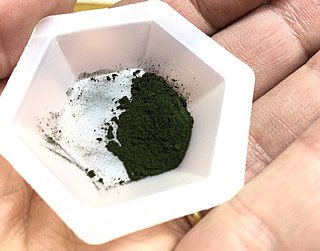
Brilliant cresyl blue is a supravital stain used for counting reticulocytes. It is classified as an oxazine dye. N95 dust masks, eye shields, and gloves must all be worn when handling the chemical.
Cresyl violet is an organic compound with the chemical formula C19H18ClN3O. It is a basic dye and is used as a common stain in histology.

Chromoendoscopy is a medical procedure wherein dyes are instilled into the gastrointestinal tract at the time of visualization with fibre-optic endoscopy. The purposes of chromoendoscopy is chiefly enhance the characterization of tissues, although dyes may be used for other functional purposes. The detail achieved with chromoendoscopy can often allow for identification of the tissue type or pathology based upon the pattern uncovered.

A white blood cell differential is a medical laboratory test that provides information about the types and amounts of white blood cells in a person's blood. The test, which is usually ordered as part of a complete blood count (CBC), measures the amounts of the five normal white blood cell types – neutrophils, lymphocytes, monocytes, eosinophils and basophils – as well as abnormal cell types if they are present. These results are reported as percentages and absolute values, and compared against reference ranges to determine whether the values are normal, low, or high. Changes in the amounts of white blood cells can aid in the diagnosis of many health conditions, including viral, bacterial, and parasitic infections and blood disorders such as leukemia.
