Laryngeal cancer

Last updated
Laryngeal cancer
Other namesCancer of the larynx, laryngeal carcinoma
Tumor Laryngis-01.jpg
Larynx cancer – endoscopic view
Specialty Oncology
Deaths94,800 (2018) [1]

Laryngeal cancer or throat cancer is a kind of cancer that can develop in any part of the larynx. It is typically a squamous-cell carcinoma, reflecting its origin from the epithelium of the larynx.

Contents

The prognosis is affected by the location of the tumour. For the purposes of staging, the larynx is divided into three anatomical regions: the glottis (true vocal cords, anterior and posterior commissures); the supraglottis (epiglottis, arytenoids and aryepiglottic folds, and false cords); and the subglottis. Most laryngeal cancers originate in the glottis, with supraglottic and subglottic tumours being less frequent.

Laryngeal cancer may spread by: direct extension to adjacent structures, metastasis to regional cervical lymph nodes, or via the blood stream. The most common site of distant metastases is the lung. Laryngeal cancer occurred in 177,000 people in 2018, and resulted in 94,800 deaths (an increase from 76,000 deaths in 1990). [1] [2] Five-year survival rates in the United States are 60.3%. [3]

Signs and symptoms

The symptoms of laryngeal cancer depend on the size and location of the tumour. Symptoms may include the following: [4] [5]

Adverse effects of treatment can include changes in appearance, difficulty eating, dry mouth, or loss of voice that may require learning alternate methods of speaking. [6]

Risk factors

The most important risk factor for laryngeal cancer is tobacco smoking. Death from laryngeal cancer is 20 times more likely for the heaviest smokers than for their non-smoking peers. [7] Heavy chronic consumption of alcohol, particularly alcoholic spirits, is also a significant risk factor. When present in combination, the usages of alcohol and tobacco appear to have a synergistic effect. Other reported risk factors include being of low socioeconomic status, male sex, or age greater than 55 years.[ citation needed ]

Occupational exposure to environmental factors such as wood dust, paint fumes, and certain chemicals used in the metalworking, petroleum, plastics, and textile industries [8] is also believed to be a risk factor for laryngeal cancers. Infections by some strains of Papillomaviridae carry some risk of laryngeal carcinoma. [9]

People with a history of head and neck cancer are known to be at higher risk (about 25%) of developing a second, separate cancer of the head, neck, or lung. This is likely due to chronic exposure to the carcinogenic effects of alcohol and tobacco. In this situation, a field change effect may occur, where the epithelial tissues start to become diffusely dysplastic with a reduced threshold for malignant change. This risk may be reduced by quitting alcohol and tobacco.[ citation needed ]

Diagnosis

Larynx and nearby structures
Cavitas nasi: Nasal cavity
Cavis orum: oral cavity
Glottis: Larynx
Plica vocalis: Vocal cords
Trachea
Oesophagus: Esophagus Kehlkopf Schema.png
Larynx and nearby structures
Cavitas nasi: Nasal cavity
Cavis orum: oral cavity
Glottis: Larynx
Plica vocalis: Vocal cords
Trachea
Oesophagus: Esophagus

Diagnosis is made by the doctor on the basis of a medical history, physical examination, and special investigations which may include a chest x-ray, CT or MRI scans, and tissue biopsy. The examination of the larynx requires some expertise, which may require specialist referral.[ citation needed ]

The physical exam includes a systematic examination of the whole patient to assess general health and to look for signs of associated conditions and metastatic disease. The neck and supraclavicular fossa are palpated to feel for cervical adenopathy, other masses, and laryngeal crepitus. The oral cavity and oropharynx are examined under direct vision. The larynx may be examined by indirect laryngoscopy using a small angled mirror with a long handle (akin to a dentist's mirror) and a strong light. Indirect laryngoscopy can be highly effective, but requires skill and practice for consistent results. For this reason, many specialist clinics now use fibre-optic nasal endoscopy where a thin and flexible endoscope, inserted through the nostril, is used to clearly visualise the entire pharynx and larynx. Nasal endoscopy is a quick and easy procedure performed in clinic. Local anaesthetic spray may be used.[ citation needed ]

If there is a suspicion of cancer, biopsy is performed, usually under general anaesthetic. This provides histological proof of cancer type and grade. If the lesion appears to be small and well localised, the surgeon may undertake excision biopsy, where an attempt is made to completely remove the tumour at the time of first biopsy. In this situation, the pathologist will not only be able to confirm the diagnosis, but can also comment on the completeness of excision, i.e., whether the tumour has been completely removed. A full endoscopic examination of the larynx, trachea, and esophagus is often performed at the time of biopsy.[ citation needed ]

For small glottic tumours further imaging may be unnecessary. In most cases, tumour staging is completed by scanning the head and neck region to assess the local extent of the tumour and any pathologically enlarged cervical lymph nodes.

The final management plan will depend on the site, stage (tumour size, nodal spread, distant metastasis), and histological type. The overall health and wishes of the patient must also be taken into account. A prognostic multigene classifier has been shown to be potentially useful for the distinction of laryngeal cancer of low or high risk of recurrence and might influence the treatment choice in future. [10]

Staging

Laryngeal tumours are classified according to the guidelines set by academic organisations such as the National Comprehensive Cancer Network (NCCN) . [11] Overall classification, also known as "staging", can help predict treatment options for patients. [12] Staging consists of three separate evaluations. The first is of the tumour/cancer itself ("T"). [12] The second is the extent to which adjacent lymph nodes are involved in the tumour/cancer's spread ("N"). [12] The third is the presence or absence of any distant metastases ("M). [12] The specific “staging” criteria for laryngeal cancer, as utilised in the NCCN’s 2019 Guidelines for Head and Neck Cancers, [13] are:

T

TX: Unable to assess

Tis: Carcinoma in situ

Supraglottis

T1: Tumour present in only one subsite of the supraglottis. Vocal cords have normal mobility.

T2: Tumour invades mucosa. There is no fixation of the larynx.

T3: Tumour causes fixation of the vocal cords, with or without invasion of neighbouring areas.

T4:

Glottis

T1: Tumour only involves the vocal cords. Vocal cords have normal mobility.

  • T1A – One vocal cord
  • T1B – Both vocal cords

T2: Tumour meets at least one of the following criteria:

  • extends to supra- or sub-glottis
  • impairs vocal cord mobility

T3: Tumour meets at least one of the following criteria:

T4: Same as “Supraglottis”

Subglottis

T1: Tumour is only in the subglottis

T2: Tumour involves both subglottis and vocal cords (regardless of cord mobility)

T3: Same as “Glottis

T4: Same as “Supraglottis

N

If Using Clinical (Non-Pathological) Diagnosis

NX: Unable to assess

N0: No involvement of neighbouring lymph nodes

N1: Tumour meets ALL of the following criteria:

N2: Tumour meets ANY of the following criteria

N3: Tumour meets ANY of the following criteria:

If Using Pathological Diagnosis

NX: Same as “Clinical Diagnosis – NX”

N0:  Same as “Clinical Diagnosis – N0”

N1:  Same as “Clinical Diagnosis – N1”

N2: Tumour meets ANY of the following criteria

N3: Tumour meets ANY of the following criteria:

M

M0: No evidence of distant metastasis

M1: Evidence of distant metastasis

Treatment

Larynx, removed
At right: Fingertip,
At the bottom: Holder Kehlkopf.png
Larynx, removed
At right: Fingertip,
At the bottom: Holder

Specific treatment depends on the location, type, and stage of the tumour. [14] Treatment may involve surgery, radiotherapy, or chemotherapy, alone or in combination. [14]

Surgical Treatment

Surgical treatment may involve partial or full removal of the tumour. [15] Neighbouring tissues and structures may or may not be removed, depending on their involvement in the tumour’s structure and spread. [16] Full removal of the larynx may be necessary in some cases.

Adjunct Treatment

Adjunct treatment, most commonly the administration of chemotherapy or radiotherapy, may be necessary. [16] Chemotherapy or radiotherapy may be necessary singly, in combination with each other, or in combination with surgery. [13] Adjunct treatment may be necessary prior to surgical treatment, alongside surgical treatment, or after surgical treatment. Clinical decision-making can be difficult in circumstances where the patient is unable to access necessary adjunct treatment.

Multi-Disciplinary Treatment

Often, successful treatment of and recovery from laryngeal cancer will involve expertise outside of the realms of surgery or oncology. Physical therapists, occupational therapists, speech therapists, psychiatrists, psychologists, oral/maxillofacial surgeons,   dentists, neurologists, neurosurgeons, and endocrinologists may all become involved in the care of patients with laryngeal cancer.

Epidemiology

Incidence is five in 100,000 (12,500 new cases per year) in the US. [17] The American Cancer Society estimated that 9,510 men and women (7,700 men and 1,810 women) would be diagnosed with and 3,740 men and women would die of laryngeal cancer in 2006.[ citation needed ]

According to the GLOBOCAN 2018 estimates of cancer incidence and mortality produced by the International Agency for Research on Cancer, there were 177,422 new cases of laryngeal cancer worldwide in 2018 (1.0% of the global total.) Among worldwide cancer deaths, 94,771 (1.0%) were due to laryngeal cancer. [18]

In 2019, it is estimated that there will be 12,410 new laryngeal cancer cases in the United States, (3.0 per 100,000). [19] The number of new cases decreases every year at a rate of 2.4%, [19] and this is believed to be related to decreased cigarette smoking in the general population. [20]

Laryngeal cancer is listed as a "rare disease" by the Office of Rare Diseases (ORD) of the National Institutes of Health (NIH). This means that laryngeal cancer affects fewer than 200,000 people in the US. [21]

See also

Related Research Articles

<span class="mw-page-title-main">Larynx</span> Voice box, an organ in the neck of amphibians, reptiles, and mammals

The larynx, commonly called the voice box, is an organ in the top of the neck involved in breathing, producing sound and protecting the trachea against food aspiration. The opening of larynx into pharynx known as the laryngeal inlet is about 4–5 centimeters in diameter. The larynx houses the vocal cords, and manipulates pitch and volume, which is essential for phonation. It is situated just below where the tract of the pharynx splits into the trachea and the esophagus. The word 'larynx' comes from the Ancient Greek word lárunx ʻlarynx, gullet, throat.ʼ

<span class="mw-page-title-main">Kidney cancer</span> Medical condition

Kidney cancer, also known as renal cancer, is a group of cancers that starts in the kidney. Symptoms may include blood in the urine, a lump in the abdomen, or back pain. Fever, weight loss, and tiredness may also occur. Complications can include spread to the lungs or brain.

<span class="mw-page-title-main">Oral cancer</span> Cancer of the lining of the lips, mouth, or upper throat

Oral cancer, also known as mouth cancer, is a cancer of the lining of the lips, mouth, or upper throat. In the mouth, it most commonly starts as a painless white patch, that thickens, develops red patches, an ulcer, and continues to grow. When on the lips, it commonly looks like a persistent crusting ulcer that does not heal, and slowly grows. Other symptoms may include difficult or painful swallowing, new lumps or bumps in the neck, a swelling in the mouth, or a feeling of numbness in the mouth or lips.

Cancer staging is the process of determining the extent to which a cancer has grown and spread. A number from I to IV is assigned, with I being an isolated cancer and IV being a cancer that has metastasized and spread from its origin. The stage generally takes into account the size of a tumor, whether it has invaded adjacent organs, how many regional (nearby) lymph nodes it has spread to, and whether it has appeared in more distant locations (metastasized).

<span class="mw-page-title-main">Invasive carcinoma of no special type</span> Medical condition

Invasive carcinoma of no special type, invasive breast carcinoma of no special type (IBC-NST), invasive ductal carcinoma (IDC), infiltrating ductal carcinoma (IDC) or invasive ductal carcinoma, not otherwise specified (NOS) is a disease. For international audiences this article will use "invasive carcinoma NST" because it is the preferred term of the World Health Organization (WHO).

<span class="mw-page-title-main">Sentinel lymph node</span> First lymph node to receive drainage from a primary tumor

The sentinel lymph node is the hypothetical first lymph node or group of nodes draining a cancer. In case of established cancerous dissemination it is postulated that the sentinel lymph nodes are the target organs primarily reached by metastasizing cancer cells from the tumor.

Prostate cancer staging is the process by which physicians categorize the risk of cancer having spread beyond the prostate, or equivalently, the probability of being cured with local therapies such as surgery or radiation. Once patients are placed in prognostic categories, this information can contribute to the selection of an optimal approach to treatment. Prostate cancer stage can be assessed by either clinical or pathological staging methods. Clinical staging usually occurs before the first treatment and tumour presence is determined through imaging and rectal examination, while pathological staging is done after treatment once a biopsy is performed or the prostate is removed by looking at the cell types within the sample.

<span class="mw-page-title-main">Anaplastic thyroid cancer</span> Medical condition

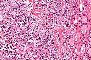
Anaplastic thyroid cancer (ATC), also known as anaplastic thyroid carcinoma, is an aggressive form of thyroid cancer characterized by uncontrolled growth of cells in the thyroid gland. This form of cancer generally carries a very poor prognosis due to its aggressive behavior and resistance to cancer treatments. The cells of anaplastic thyroid cancer are highly abnormal and usually no longer resemble the original thyroid cells and have poor differentiation.

<span class="mw-page-title-main">Papillary thyroid cancer</span> Medical condition

Papillary thyroid cancer is the most common type of thyroid cancer, representing 75 percent to 85 percent of all thyroid cancer cases. It occurs more frequently in women and presents in the 20–55 year age group. It is also the predominant cancer type in children with thyroid cancer, and in patients with thyroid cancer who have had previous radiation to the head and neck. It is often well-differentiated, slow-growing, and localized, although it can metastasize.

<span class="mw-page-title-main">Cervical lymph nodes</span> Lymph nodes found in the neck

Cervical lymph nodes are lymph nodes found in the neck. Of the 800 lymph nodes in the human body, 300 are in the neck. Cervical lymph nodes are subject to a number of different pathological conditions including tumours, infection and inflammation.

<span class="mw-page-title-main">Medullary breast carcinoma</span> Rare type of breast cancer

Medullary breast carcinoma is a rare type of breast cancer that is characterized as a relatively circumscribed tumor with pushing, rather than infiltrating, margins. It is histologically characterized as poorly differentiated cells with abundant cytoplasm and pleomorphic high grade vesicular nuclei. It involves lymphocytic infiltration in and around the tumor and can appear to be brown in appearance with necrosis and hemorrhage. Prognosis is measured through staging but can often be treated successfully and has a better prognosis than other infiltrating breast carcinomas.

<span class="mw-page-title-main">Medullary thyroid cancer</span> Malignant thyroid neoplasm originating from C-cells

Medullary thyroid cancer is a form of thyroid carcinoma which originates from the parafollicular cells, which produce the hormone calcitonin. Medullary tumors are the third most common of all thyroid cancers and together make up about 3% of all thyroid cancer cases. MTC was first characterized in 1959.

Lung cancer staging is the assessment of the extent to which a lung cancer has spread from its original source. As with most cancers, staging is an important determinant of treatment and prognosis. In general, more advanced stages of cancer are less amenable to treatment and have a worse prognosis.

<span class="mw-page-title-main">Hypopharyngeal cancer</span> Medical condition

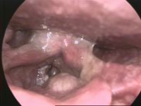
Hypopharyngeal cancer is a disease in which malignant cells grow in the hypopharynx the area where the larynx and esophagus meet.

<span class="mw-page-title-main">Lobular carcinoma in situ</span> Medical condition

Lobular carcinoma in situ (LCIS) is an incidental microscopic finding with characteristic cellular morphology and multifocal tissue patterns. The condition is a laboratory diagnosis and refers to unusual cells in the lobules of the breast. The lobules and acini of the terminal duct-lobular unit (TDLU), the basic functional unit of the breast, may become distorted and undergo expansion due to the abnormal proliferation of cells comprising the structure. These changes represent a spectrum of atypical epithelial lesions that are broadly referred to as lobular neoplasia (LN).

Breast cancer classification divides breast cancer into categories according to different schemes criteria and serving a different purpose. The major categories are the histopathological type, the grade of the tumor, the stage of the tumor, and the expression of proteins and genes. As knowledge of cancer cell biology develops these classifications are updated.

<span class="mw-page-title-main">Oropharyngeal cancer</span> Pharynx cancer that is located in the oropharynx

Oropharyngeal cancer, also known as oropharyngeal squamous cell carcinoma and tonsil cancer, is a disease in which abnormal cells with the potential to both grow locally and spread to other parts of the body are found in the oral cavity, in the tissue of the part of the throat (oropharynx) that includes the base of the tongue, the tonsils, the soft palate, and the walls of the pharynx.

<span class="mw-page-title-main">HPV-positive oropharyngeal cancer</span> Cancer of the throat

Human papillomavirus-positive oropharyngeal cancer, is a cancer of the throat caused by the human papillomavirus type 16 virus (HPV16). In the past, cancer of the oropharynx (throat) was associated with the use of alcohol or tobacco or both, but the majority of cases are now associated with the HPV virus, acquired by having oral contact with the genitals of a person who has a genital HPV infection. Risk factors include having a large number of sexual partners, a history of oral-genital sex or anal–oral sex, having a female partner with a history of either an abnormal Pap smear or cervical dysplasia, having chronic periodontitis, and, among men, younger age at first intercourse and a history of genital warts. HPV-positive OPC is considered a separate disease from HPV-negative oropharyngeal cancer.

Cervical cancer staging is the assessment of cervical cancer to determine the extent of the disease. This is important for determining disease prognosis and treatment. Cancer staging generally runs from stage 0, which is pre-cancerous or non-invasive, to stage IV, in which the cancer has spread throughout a significant part of the body.

In CT scan of the thyroid, focal and diffuse thyroid abnormalities are commonly encountered. These findings can often lead to a diagnostic dilemma, as the CT reflects nonspecific appearances. Ultrasound (US) examination has a superior spatial resolution and is considered the modality of choice for thyroid evaluation. Nevertheless, CT detects incidental thyroid nodules (ITNs) and plays an important role in the evaluation of thyroid cancer.

References

  1. 1 2 "Larynx Cancer Factsheet" (PDF). Global Cancer Observatory. Archived (PDF) from the original on 8 November 2019. Retrieved 8 November 2019.
  2. Naghavi M, Wang H, Lozano R, Davis A, Liang X, Zhou M, et al. (GBD 2013 Mortality and Causes of Death Collaborators) (January 2015). "Global, regional, and national age-sex specific all-cause and cause-specific mortality for 240 causes of death, 1990–2013: a systematic analysis for the Global Burden of Disease Study 2013". Lancet. 385 (9963): 117–171. doi:10.1016/S0140-6736(14)61682-2. PMC   4340604 . PMID   25530442.
  3. "SEER Stat Fact Sheets: Larynx Cancer". NCI. Archived from the original on 17 October 2019. Retrieved 22 January 2020.
  4. Laryngeal cancer Archived 2009-04-15 at the Wayback Machine at Mount Sinai Hospital
  5. DeVita VT, Lawrence TS, Rosenberg SA (2011). Devita, Hellman, and Rosenberg's cancer : principles & practice of oncology (10th ed.). Philadelphia. ISBN   978-1-4511-9294-0.{{cite book}}: CS1 maint: location missing publisher (link)
  6. "Cancer of the Larynx - Causes, Symptoms, Treatment, Diagnosis - MedBroadcast.com". Archived from the original on 2015-10-18. Retrieved 2018-01-25.
  7. Ridge JA, Glisson BS, Lango MN, Feigenberg S, Horwitz EM (2008). "Head and neck tumors.". In Pazdur R, Wagman LD, Camphausen KA, Hoskins W (eds.). Cancer management: a multidisciplinary approach (PDF). Vol. 11. p. 369. Archived (PDF) from the original on 2022-10-02. Retrieved 2021-11-09.
  8. "Laryngeal Cancer". Archived from the original on December 9, 2022. Retrieved April 7, 2019.
  9. Torrente MC, Rodrigo JP, Haigentz M, Dikkers FG, Rinaldo A, Takes RP, et al. (April 2011). "Human papillomavirus infections in laryngeal cancer". Head & Neck. Head Neck. 33 (4): 581–586. doi:10.1002/hed.21421. PMID   20848441. S2CID   30274997.
  10. Mirisola V, Mora R, Esposito AI, Guastini L, Tabacchiera F, Paleari L, et al. (August 2011). "A prognostic multigene classifier for squamous cell carcinomas of the larynx". Cancer Letters. 307 (1): 37–46. doi:10.1016/j.canlet.2011.03.013. PMID   21481529.
  11. "National Comprehensive Cancer Network – Home". NCCN. Archived from the original on 2023-02-13. Retrieved 2020-11-24.
  12. 1 2 3 4 Amin M, Edge S, Greene F, et al. (2017). AJCC Cancer Staging Manual. New York: Springer.
  13. 1 2 Pfister DG, Spencer S, Adelstein D, Adkins D, Anzai Y, Brizel DM, et al. (July 2020). "Head and Neck Cancers, Version 2.2020, NCCN Clinical Practice Guidelines in Oncology". Journal of the National Comprehensive Cancer Network. 18 (7): 873–898. doi: 10.6004/jnccn.2020.0031 . PMID   32634781. S2CID   220405484.
  14. 1 2 National Comprehensive Cancer Network, "Evidence Blocks for Head and Neck Cancers," 2019.
  15. Nibu KI, Hayashi R, Asakage T, Ojiri H, Kimata Y, Kodaira T, et al. (August 2017). "Japanese Clinical Practice Guideline for Head and Neck Cancer". Auris, Nasus, Larynx. 44 (4): 375–380. doi:10.1016/j.anl.2017.02.004. PMID   28325607.
  16. 1 2 Grégoire V, Lefebvre JL, Licitra L, Felip E (May 2010). "Squamous cell carcinoma of the head and neck: EHNS-ESMO-ESTRO Clinical Practice Guidelines for diagnosis, treatment and follow-up". Annals of Oncology. 21 (Suppl 5): v184–86. doi: 10.1093/annonc/mdq185 . PMID   20555077.
  17. Beenken SW. "Laryngeal Cancer (Cancer of the larynx)". Armenian Health Network, Health.am. Archived from the original on 2020-11-27. Retrieved 2007-03-22.
  18. Bray F, Ferlay J, Soerjomataram I, Siegel RL, Torre LA, Jemal A (November 2018). "Global cancer statistics 2018: GLOBOCAN estimates of incidence and mortality worldwide for 36 cancers in 185 countries". CA: A Cancer Journal for Clinicians. Wiley. 68 (6): 394–424. doi: 10.3322/caac.21492 . PMID   30207593. S2CID   52188256.
  19. 1 2 "Cancer Stat Facts: Larynx Cancer". Archived from the original on 2022-11-22. Retrieved 2019-08-09.
  20. "Throat Cancer Statistics | Cases of Throat Cancer Per Year". www.cancer.org. Archived from the original on 2019-07-27. Retrieved 2020-12-04.
  21. "Annual Report on the Rare Diseases and Conditions Research". National Institutes of Health. Archived from the original on 2010-12-03. Retrieved 2007-03-22.