Adenocarcinoma of the lung

Last updated
Adenocarcinoma of the lung
Other namesPulmonary adenocarcinoma
Adenocarcinoma of the lung.jpg
A gross pathological specimen of a pulmonary adenocarcinoma, removed in a lobectomy.
Specialty Oncology

Adenocarcinoma of the lung is the most common type of lung cancer, and like other forms of lung cancer, it is characterized by distinct cellular and molecular features. [1] It is classified as one of several non-small cell lung cancers (NSCLC), to distinguish it from small cell lung cancer which has a different behavior and prognosis. Lung adenocarcinoma is further classified into several subtypes and variants. [2] The signs and symptoms of this specific type of lung cancer are similar to other forms of lung cancer, and patients most commonly complain of persistent cough and shortness of breath.

Contents

Adenocarcinoma is more common in patients with a history of cigarette smoking, and is the most common form of lung cancer in younger women and Asian populations. The pathophysiology of adenocarcinoma is complicated, but generally follows a histologic progression from cells found in healthy lungs to distinctly dysmorphic, or irregular cells. There are several distinct molecular and genetic pathways that contribute to this progression. Like many lung cancers, adenocarcinoma of the lung is often advanced by the time of diagnosis. Once a lesion or tumor is identified with various imaging modalities, such as computed tomography (CT) or X-ray, a biopsy is required to confirm the diagnosis.

Treatment of this lung cancer is based upon the specific subtype and the extent of spread from the primary tumor. Surgical resection, chemotherapy, radiotherapy, targeted therapy and immunotherapy are used in attempt to eradicate the cancerous cells based upon these factors. [3]

Signs and symptoms

The majority of patients who are diagnosed with lung cancer usually present with locally advanced or metastatic disease. Only about one third of patients have stage I disease when diagnosed. [4] The symptoms that the patient exhibits usually reflect the extent of the cancer's spread. Lung cancers that are discovered early may cause symptoms localized to the respiratory system. However, lung cancer that is advanced will cause patients to experience additional signs and symptoms secondary to the cancer spreading to other organ systems. [5] In order of highest frequency, the most common signs of lung cancer include: [6]

Clinicians should have a high level of suspicion for lung cancer, especially in patients with a smoking history. Patients with recurring or unresolving lung infections (e.g. bronchitis and pneumonia) that are unresponsive to antibiotics should also be further evaluated for lung cancer. In nonsmokers, women and East Asians are more likely to present with symptoms of an underlying lung cancer at younger ages. [5] Importantly, many of these signs are commonly due to other causes that are not cancer. [6] A detailed medical history should be obtained from each patient to determine the relevance of further diagnostic workup and management.[ citation needed ]

Extrapulmonary manifestations

Adenocarcinoma, like other forms of lung cancer, is usually advanced or metastatic at time of diagnosis. Patients may complain of signs or symptoms outside of the respiratory tract that represent a hematologic or metabolic complication of the malignancy without, however, resulting necessarily from obstruction or metastasis. These go under the name of paraneoplastic syndromes, which often indicate advanced disease and worse prognosis[ citation needed ]. The most common paraneoplastic syndromes associated with adenocarcinoma of the lung are described below:

Causes

Risk factors

According to the Nurses' Health Study, the risk of pulmonary adenocarcinoma increases substantially after a long duration of tobacco smoking: smokers with a previous smoking duration of 30–40 years are more than twice as likely to develop lung adenocarcinoma compared to never-smokers (relative risk of approximately 2.4); a duration of more than 40 years increases relative risk to 5. [8]

This cancer usually is seen peripherally in the lungs, as opposed to small cell lung cancer and squamous cell lung cancer, which both tend to be more centrally located, [9] [10] although it may also occur as central lesions. [10] For unknown reasons, it often arises in relation to peripheral lung scars. The current theory is that the scar probably occurred secondary to the tumor, rather than causing the tumor. [10] The adenocarcinoma has an increased incidence in smokers, and is the most common type of lung cancer seen in non-smokers and women. [10] Deeper inhalation of cigarette smoke results in peripheral lesions that are often the case in adenocarcinomas of the lung. Generally, adenocarcinoma grows more slowly and forms smaller masses than the other subtypes. [10] However, it tends to metastasize at an early stage. [10]

Mechanism

Pathogenesis

Micrograph showing an ALK positive adenocarcinoma of the lung. ALK immunostain. ALK positive lung adenocarcinoma - ALK IHC -- high mag.jpg
Micrograph showing an ALK positive adenocarcinoma of the lung. ALK immunostain.
Micrograph showing a ROS1 positive adenocarcinoma of the lung. ROS1 immunostain. Adenocarcinoma - ROS1 positive - ROS1 -- high mag.jpg
Micrograph showing a ROS1 positive adenocarcinoma of the lung. ROS1 immunostain.

Large scale studies such as The Cancer Genome Atlas (TCGA) have systematically characterized recurrent somatic alterations likely driving lung adenocarcinoma initiation and development. [11]

Gene mutations and copy number alterations

Since smoking is a strong mutagenic factor, lung adenocarcinoma is one of the tumor types with the highest number of mutations. [12] Common somatic mutations in lung adenocarcinoma affect many oncogenes and tumor suppressor genes, including TP53 (mutated in 46% of cases), EGFR (27%), KRAS (32%), KEAP1, STK11 and NF1. [3] EGFR and KRAS mutations tend to appear in a mutually exclusive fashion. KRAS mutations are associated with smoking habits, whereas EGFR mutations occur more frequently in females, people of Asian ethnicity and never-smokers. [2]

Copy number amplifications in oncogenes such as TERT, MDM2, EGFR, MET, and MYC have been reported, as well as deletions of tumor suppressor genes such as CDKN2A. [11]

Frequent alterations occur in genes belonging to the receptor tyrosine kinase pathway, of which EGFR is the most prominent example. This pathway is involved in cell proliferation and survival and it is often deregulated in cancer. As a consequence, targeted therapies have been developed to inhibit mutant pathway components. [11] [13]

Chromosomal rearrangements

Three membrane associated tyrosine kinase receptors are recurrently involved in fusions or rearrangements in adenocarcinomas: ALK, ROS1, and RET, and more than eighty other translocations have also been reported in adenocarcinomas of the lung. [14]

In ALK rearrangements, the most common partner gene is EML4. [3] EML4-ALK fusions tend to occur in tumors that do not carry EGFR or KRAS mutations and have also a lower frequency of TP53 mutations. ALK and ROS fusions offer opportunities for targeted therapies with tyrosine kinase inhibitors[ citation needed ].

Pathophysiology

The respiratory tract can be divided into two main components: the conducting airways and the gas exchange airways. The gas exchange airways are made of alveoli, small microscopic air sacs that are responsible for the exchange of oxygen and carbon dioxide during normal breathing. Alveoli are composed of two cell types, type I and type II pneumocytes. Type I pneumocytes cover 95% of alveolar surfaces, and are not able to regenerate. Type II pneumocytes are more common, making up 60% of the cells within alveolar epithelium, but constitute only 3% of the alveolar surface. [15]

There are several factors that contribute to the transformation of normal alveolar epithelium into dysplastic, or pre-cancerous, lesions. Adenocarcinoma of the lung develops in a step-wise progression as type II pneumocytes undergo consecutive molecular changes that disrupt normal cell regulation and turnover. Atypical adenomatous hyperplasia (AAH) is considered a pre-cancerous lesion, and is thought to further progress to adenocarcinoma in situ and invasive adenocarcinoma of the lung. The lesions of AAH are <5 mm, can be single or multiple, and have a ground glass appearance on CT imaging. As more genetic mutations and dysregulation of normal cell signaling pathways accumulate, AAH can progress to adenocarcinoma in situ (AIS). AIS lesions are classified as small tumors <3 cm with abnormal type II pneumocyte cell growth that is limited to the alveolar spaces i.e. without invasion into the stroma, pleura, or vasculature. This type of growth is termed "lepidic" and is characteristic of adenocarcinoma of the lung in its earliest stages. [15]

Diagnosis

A diagnosis of lung cancer may be suspected on the basis of typical symptoms, particularly in a person with smoking history. Symptoms such as coughing up blood and unintentional weight loss may prompt further investigation, such as medical imaging.

Classification

CT scan- adenocarcinoma of the left lung Adenocarcinoma - CT scan (5499628365).jpg
CT scan- adenocarcinoma of the left lung

The majority of lung cancers can be characterized as either small cell lung cancer (SCLC) or non-small cell lung cancer (NSCLC). Lung adenocarcinoma is one of the three major subtypes of NSCLC, which also include squamous carcinoma and large cell carcinoma. [15]

Historically, there has been much debate in the most accurate method of describing adenocarcinoma of the lung and several revisions of classification systems have been published. Most recently, the International Multidisciplinary Classification of Lung Adenocarcinoma was published in 2011 and represents the consensus of several organizations to more accurately describe this specific type of lung cancer. [2] The current classification system aims to more reliably predict prognosis and determination of therapeutic management. [4]

The tumor size, pattern of cell growth, and depth of cell invasion into normal lung tissue are considered in determining classification. The following names represent a step-wise pathologic progression in the natural course of adenocarcinoma development; Adenocarcinoma in situ (AIS), Minimally invasive adenocarcinoma (MIA), and Invasive adenocarcinoma. [15] Invasive adenocarcinoma of the lung includes a heterogenous mixture of subtypes and variants.

The 2011 consensus describes five subtypes of invasive adenocarcinomas based on the cell pattern that is most predominant. These subtypes are described below:

Histopathology of lepidic predominant adenocarcinoma. Histopathology of lepidic predominant adenocarcinoma.jpg
Histopathology of lepidic predominant adenocarcinoma.
Acinar pattern. Histopathology of lung adenocarcinoma with acinar pattern.png
Acinar pattern.
Solid pattern. Histopathology of lung adenocarcinoma with solid pattern.jpg
Solid pattern.

Cell patterns identifying subtypes are associated with prognosis, ranging from favorable (lepidic) to intermediate (acinar and papillary) to poor (micropapillary and solid). [2]

Four discrete variants of invasive adenocarcinomas not assignable to these five subtypes are also included in the current classification:

Imaging

A chest x-ray (radiograph) is often the first imaging test performed when a person presents with cough or chest pain, particularly in the primary care setting. A chest radiograph may detect a lung nodule/mass that is suggestive of cancer, although sensitivity and specificity are limited.[ citation needed ]

CT imaging provides better evaluation of the lungs, with higher sensitivity and specificity for lung cancer compared to chest radiograph (although still significant false positive rate [27] ). Computed tomography (CT) that is specifically aimed at evaluating lung cancer includes the chest and the upper abdomen. This allows for evaluation of other relevant anatomic structures such as nearby lymph nodes, adrenal glands, liver, and bones which may show evidence of metastatic spread of disease. [4] Indeed, the US Preventative Services Task Force recommends annual screening with low-dose CT in adults aged 55 to 80 years who have a 30 pack-year smoking history and currently smoke or have quit within the past 15 years, with certain caveats (see Lung cancer screening). [28]

Nuclear medicine imaging, such as PET/CT and bone scan, may also be helpful to diagnose and detect metastatic disease elsewhere in the body. [7] PET/CT uses a metabolically active tracer that allows clinicians to identify areas of the body that are hypermetabolic. Increased uptake of the tracer occurs in malignant cells and areas of inflammation or infection. Integrating the imaging reflective of metabolic activity with normal CT imaging allows for higher sensitivity and specificity compared to PET alone. [4]

MRI is reserved for patients with advanced disease where intracranial, or brain, involvement is likely. It is also helpful for evaluating the extent of chest wall, diaphragmatic, brachial plexus (such as in the case of superior sulcus tumors), or spine involvement. [4]

Histopathology

If possible, a biopsy of any suspected lung tumor is performed in order to make a microscopic evaluation of the cells involved and is ultimately required to confirm diagnosis. [7] Biopsy should be attempted in distant lesions first to establish a histologic diagnosis and to simultaneously confirm metastatic staging. The biopsy material is also used to analyze whether the tumor express any specific mutations suitable for tageted therapy (e.g. EGFR mutation or ALK mutation). Biopsy can be accomplished via bronchoscopy, transthoracic needle biopsy, and video-assisted thorascopic surgery (VATS). [4]

While sputum cytology has been shown to have limited utility, thoracentesis, or aspiration of pleural fluid with an ultrasound-guided needle, should be performed when pleural effusion is present. When malignant cells are identified in the pleural aspirate of patients highly suspect for lung cancer, a definitive diagnosis and staging (stage IV adenocarcinoma of the lung) is established. [4]

Adenocarcinoma of the lung tends to stain mucin positive as it is derived from the mucus-producing glands of the lungs. Similar to other adenocarcinoma, if this tumor is well differentiated (low grade) it will resemble the normal glandular structure. Poorly differentiated adenocarcinoma will not resemble the normal glands (high grade) and will be detected by seeing that they stain positive for mucin (which the glands produce). Adenocarcinoma can also be distinguished by staining for TTF-1, a cell marker for adenocarcinoma. [29]

As discussed previously, the category of adenocarcinoma includes are range of subtypes, and any one tumor tends to be heterogeneous in composition. Several major subtypes are currently recognized by the World Health Organization (WHO) [1] and the International Association for the Study of Lung Cancer (IASLC) / American Thoracic Society (ATS) / European Respiratory Society (ERS): [30] [31] [32] lepidic predominant adenocarcinoma, acinar predominant adenocarcinoma, papillary predominant adenocarcinoma, micropapillary predominant adenocarcinoma, solid predominant adenocarcinoma, and solid predominant with mucin production. In as many as 80% of these tumors, components of more than one subtype will be recognized. Surgically resected tumors should be classified by comprehensive histological subtyping, describing patterns of involvement in increments of 5%. The predominant histologic subtype is then used to classify the tumor overall. [2] The predominant subtype is prognostic for survival after complete resection. [33]

To reveal the adenocarcinomatous lineage of the solid variant, demonstration of intracellular mucin production may be performed. Foci of squamous metaplasia and dysplasia may be present in the epithelium proximal to adenocarcinomas, but these are not the precursor lesions for this tumor. Rather, the precursor of peripheral adenocarcinomas has been termed atypical adenomatous hyperplasia (AAH). [10] Microscopically, AAH is a well-demarcated focus of epithelial proliferation, containing cuboidal to low-columnar cells resembling club cells or type II pneumocytes. [10] These demonstrate various degrees of cytologic atypia, including hyperchromasia, pleomorphism, prominent nucleoli. [10] However, the atypia is not to the extent as seen in frank adenocarcinomas. [10] Lesions of AAH are monoclonal, and they share many of the molecular aberrations (like KRAS mutations) that are associated with adenocarcinomas. [10]

Signet ring and clear cell adenocarcinoma are no longer histological subtypes, but rather cytological features that can occur in tumour cells of multiple histological subtypes, most often solid adenocarcinoma. [30]

Treatment

The treatment of adenocarcinoma of the lung depends on several factors including stage, resectability, performance status, histology and genomic alterations acquired by the individual tumor. [34] As in most cancer types, treatment approaches can be broadly divided into 5 categories: surgery, chemotherapy, radiotherapy, targeted therapy and immunotherapy.[ citation needed ]

Surgery

Early stage (I, II and IIIA) lung adenocarcinomas are typically treated surgically to remove the tumor with pneumonectomy or lobectomy, if it is found to be resectable with imaging studies and biopsies and if the patient is considered able to tolerate surgery. [10] Video-assisted thorascopic surgery (VATS) is often adopted, which consists in the insertion of a thorascope inside a small incision made in the chest; a lobe can be removed via the scope through this small incision. [34]

Incidence of adenocarcinoma of the lung (in yellow) as compared to other lung cancer types, with fractions of non-smokers versus smokers shown for each type. Pie chart of lung cancers.svg
Incidence of adenocarcinoma of the lung (in yellow) as compared to other lung cancer types, with fractions of non-smokers versus smokers shown for each type.

Chemotherapy

For advanced (stage IV) and unresectable lung tumors, the first-line therapy is platinum-based doublet chemotherapy, combining cisplatin or carboplatin with another cytotoxic agent. [3] Regimens strongly depend on each patient performance status and response, and when the risk of adverse events could worsen quality of life significantly, basic supportive care is more recommended. Chemotherapy is also used as an adjuvant therapy following surgery to kill remaining cancer cells in patients with stage IIA, IIB and IIIA NSCLC. [34]

Radiotherapy

Adenocarcinoma is a non-small cell lung carcinoma, and it is not as responsive to radiation therapy compared to small cell lung carcinoma. [10] However, radiotherapy may be used as an adjuvant therapy for patients who have undergone a resection surgery to reduce the risk of lung cancer relapse. It may also benefit inoperable tumors that are localized to the chest and be part of palliative care to improve quality of life in patients not responding to surgery or chemotherapy. [34]

Targeted therapy

Targeted therapy is available for lung adenocarcinomas with certain molecular characteristics. Tyrosine kinase inhibitors (TKIs) have been developed to target mutant components of the receptor tyrosine kinase pathway such as EGFR, ALK [36] and ROS1, which show frequent alterations in lung adenocarcinomas.

First-generation EGFR TKIs, including gefitinib and erlotinib, have been shown to be more effective in treating EGFR-mutated patients with respect to cytotoxic chemotherapy. Second-generation inhibitors such as afatinib and dacomitinib provided a broader scope of application as they are able to target not only the protein EGFR itself but also other members of the EGFR family, such as HER2 and HER4 (also known as ERBB2 and ERBB4), and they have shown improved progression-free survival compared to gefitinib. As the most common cause of acquired resistance to first-generation TKIs is a second EGFR mutation on codon 790, a third-generation EGFR TKI, osimertinib, has been developed to target this new mutation as well. [3] MET amplification is another known mechanism of acquired resistance. [2]

ALK inhibitors such as crizotinib showed to be effective against tumors harboring ALK fusions. Most patients previously treated with crizotinib benefited from second-generation ALK inhibitors including ceritinib, alectinib and brigatinib. Resistance to ALK inhibitors can occur with novel acquired ALK mutations or amplifications. [3]

Also ROS1-positive tumors have shown high sensitivity to ALK inhibitors due to the high homology between the kinase domains of ROS1 and ALK. [3]

Immunotherapy

Immune response can be prevented via activation of immune checkpoints, which consist in the binding of a ligand protein (e.g. PD-L1) to a receptor (e.g. PD-1) on the immune cell surface. As a consequence, cancer cells expressing PD-L1 can inactivate T cells thus fostering tumor growth. Immune checkpoint inhibitors have been developed to restore T cell-mediated antitumor immunity by blocking either the ligand or the receptor.[ citation needed ]

Immune checkpoint inhibitors have been approved for NSCLC, including anti-PD-1 nivolumab and pembrolizumab. Anti-PD-1 agents are used for patients with advanced NSCLC whose tumors progress after first-line cytotoxic chemotherapy. Pembrolizumab was established as a new standard of care for patients with advanced or metastatic NSCLC with high PD-L1 expression levels, and responses are even more pronounced for tumor with a high mutational burden (i.e. having an elevated number of mutations). [3]

Therapeutic approaches combining multiple immune checkpoint inhibitors or one immune checkpoint inhibitors and a cytotoxic agent are undergoing clinical trials as of 2018. [3] [37] Howerver, the KEYNOTE-598 phase III trial has reported in 2021 that adding ipilimumab to pembrolizumab for NSCLC patients with PD-L1 tumor proportion score ≥50% does not confer any efficacy benefit, but may introduce greater toxicity. [38] The potential role of anti-PD-1 agents as neoadjuvant therapy in resectable NSCLCs is also being investigated. [39]

Epidemiology

As for other lung cancer subtypes, lung adenocarcinoma incidence is strongly associated with smoking.

Incidence of pulmonary adenocarcinoma has been increasing in many developed Western nations in the past few decades, with a share reaching 43.3% of all lung cancers in the US as of 2012, [40] thus replacing squamous cell lung carcinoma as the most common type of lung cancer. This can be largely attributed to the decreasing smoking rates, which favors the adenocarcinoma histology. Indeed, although smoking is still its strongest risk factor, lung adenocarcinoma is by far the most common among lifelong non-smokers (<100 cigarettes in a lifetime). [41]

Related Research Articles

<span class="mw-page-title-main">Lung cancer</span> Malignant tumor characterized by uncontrolled cell growth in lung tissue

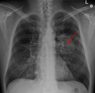
Lung cancer, also known as lung carcinoma, is a malignant tumor that begins in the lung. Lung cancer is caused by genetic damage to the DNA of cells in the airways, often caused by cigarette smoking or inhaling damaging chemicals. Damaged airway cells gain the ability to multiply unchecked, causing the growth of a tumor. Without treatment, tumors spread throughout the lung, damaging lung function. Eventually lung tumors metastasize, spreading to other parts of the body.

<span class="mw-page-title-main">Gefitinib</span> Drug used in fighting breast, lung, and other cancers

Gefitinib, sold under the brand name Iressa, is a medication used for certain breast, lung and other cancers. Gefitinib is an EGFR inhibitor, like erlotinib, which interrupts signaling through the epidermal growth factor receptor (EGFR) in target cells. Therefore, it is only effective in cancers with mutated and overactive EGFR, but resistances to gefitinib can arise through other mutations. It is marketed by AstraZeneca and Teva.

<span class="mw-page-title-main">Erlotinib</span> EGFR inhibitor for treatment of non-small-cell lung cancer

Erlotinib, sold under the brand name Tarceva among others, is a medication used to treat non-small cell lung cancer (NSCLC) and pancreatic cancer. Specifically it is used for NSCLC with mutations in the epidermal growth factor receptor (EGFR) — either an exon 19 deletion (del19) or exon 21 (L858R) substitution mutation — which has spread to other parts of the body. It is taken by mouth.

<span class="mw-page-title-main">Non-small-cell lung cancer</span> Any type of epithelial lung cancer other than small-cell lung carcinoma

Non-small-cell lung cancer (NSCLC), or non-small-cell lung carcinoma, is any type of epithelial lung cancer other than small-cell lung cancer (SCLC). NSCLC accounts for about 85% of all lung cancers. As a class, NSCLCs are relatively insensitive to chemotherapy, compared to small-cell carcinoma. When possible, they are primarily treated by surgical resection with curative intent, although chemotherapy has been used increasingly both preoperatively and postoperatively.

<span class="mw-page-title-main">KRAS</span> Protein-coding gene in humans

KRAS is a gene that provides instructions for making a protein called K-Ras, a part of the RAS/MAPK pathway. The protein relays signals from outside the cell to the cell's nucleus. These signals instruct the cell to grow and divide (proliferate) or to mature and take on specialized functions (differentiate). It is called KRAS because it was first identified as a viral oncogene in the KirstenRAt Sarcoma virus. The oncogene identified was derived from a cellular genome, so KRAS, when found in a cellular genome, is called a proto-oncogene.

<span class="mw-page-title-main">Adenocarcinoma in situ of the lung</span> Medical condition

Adenocarcinoma in situ (AIS) of the lung —previously included in the category of "bronchioloalveolar carcinoma" (BAC)—is a subtype of lung adenocarcinoma. It tends to arise in the distal bronchioles or alveoli and is defined by a non-invasive growth pattern. This small solitary tumor exhibits pure alveolar distribution and lacks any invasion of the surrounding normal lung. If completely removed by surgery, the prognosis is excellent with up to 100% 5-year survival.

<span class="mw-page-title-main">ROS1</span> Protein-coding gene in the species Homo sapiens

Proto-oncogene tyrosine-protein kinase ROS is an enzyme that in humans is encoded by the ROS1 gene.

Large-cell lung carcinoma (LCLC), or large-cell carcinoma (LCC) in short, is a heterogeneous group of undifferentiated malignant neoplasms that lack the cytologic and architectural features of small cell carcinoma and glandular or squamous differentiation. LCC is categorized as a type of NSCLC which originates from epithelial cells of the lung. LCLC is histologically characterized by the presence of large, undifferentiated cells that lack distinctive features of either squamous cell carcinoma or adenocarcinoma. Typically seen in LCLC tumor cells are abundant pale staining cytoplasm and prominent nucleoli.

<span class="mw-page-title-main">Combined small-cell lung carcinoma</span> Medical condition

Combined small cell lung carcinoma is a form of multiphasic lung cancer that is diagnosed by a pathologist when a malignant tumor, arising from transformed cells originating in lung tissue, contains a component of;small cell lung carcinoma (SCLC), admixed with one components of any histological variant of non-small cell lung carcinoma (NSCLC) in any relative proportion.

Treatment of lung cancer refers to the use of medical therapies, such as surgery, radiation, chemotherapy, immunotherapy, percutaneous ablation, and palliative care, alone or in combination, in an attempt to cure or lessen the adverse impact of malignant neoplasms originating in lung tissue.

Targeted therapy of lung cancer refers to using agents specifically designed to selectively target molecular pathways responsible for, or that substantially drive, the malignant phenotype of lung cancer cells, and as a consequence of this (relative) selectivity, cause fewer toxic effects on normal cells.

HOHMS is the medical acronym for "Higher-Order HistoMolecular Stratification", a term and concept which was first applied to lung cancer research and treatment theory.

Mucinous cystadenocarcinoma of the lung (MCACL) is a very rare malignant mucus-producing neoplasm arising from the uncontrolled growth of transformed epithelial cells originating in lung tissue.

Sarcomatoid carcinoma of the lung is a term that encompasses five distinct histological subtypes of lung cancer, including (1) pleomorphic carcinoma, (2) spindle cell carcinoma, (3) giant cell carcinoma, (4) carcinosarcoma, or (5) pulmonary blastoma.

<span class="mw-page-title-main">Crizotinib</span> ALK inhibitor for treatment of non-small-cell lung cancer

Crizotinib, sold under the brand name Xalkori among others, is an anti-cancer medication used for the treatment of non-small cell lung carcinoma (NSCLC). It acts as an ALK and ROS1 inhibitor.

<span class="mw-page-title-main">ALK inhibitor</span>

ALK inhibitors are anti-cancer drugs that act on tumours with variations of anaplastic lymphoma kinase (ALK) such as an EML4-ALK translocation. They fall under the category of tyrosine kinase inhibitors, which work by inhibiting proteins involved in the abnormal growth of tumour cells. All the current approved ALK inhibitors function by binding to the ATP pocket of the abnormal ALK protein, blocking its access to energy and deactivating it. A majority of ALK-rearranged NSCLC harbour the EML4-ALK fusion, although as of 2020, over 92 fusion partners have been discovered in ALK+ NSCLC. For each fusion partner, there can be several fusion variants depending on the position the two genes were fused at, and this may have implications on the response of the tumour and prognosis of the patient.

<span class="mw-page-title-main">ALK positive lung cancer</span> Medical condition

ALK positive lung cancer is a primary malignant lung tumor whose cells contain a characteristic abnormal configuration of DNA wherein, most frequently, the echinoderm microtubule-associated protein-like 4 (EML4) gene is fused to the anaplastic lymphoma kinase (ALK) gene. Less frequently, there will be novel translocation partners for the ALK gene, in place of EML4. This abnormal gene fusion leads to the production of a protein that appears, in many cases, to promote and maintain the malignant behavior of the cancer cells.

<span class="mw-page-title-main">Brigatinib</span> ALK inhibitor for treatment of non-small-cell lung cancer

Brigatinib, sold under the brand name Alunbrig among others, is a small-molecule targeted cancer therapy being developed by Ariad Pharmaceuticals, Inc. Brigatinib acts as both an anaplastic lymphoma kinase (ALK) and epidermal growth factor receptor (EGFR) inhibitor.

<span class="mw-page-title-main">Squamous-cell carcinoma of the lung</span> Medical condition

Squamous-cell carcinoma (SCC) of the lung is a histologic type of non-small-cell lung carcinoma (NSCLC). It is the second most prevalent type of lung cancer after lung adenocarcinoma and it originates in the bronchi. Its tumor cells are characterized by a squamous appearance, similar to the one observed in epidermal cells. Squamous-cell carcinoma of the lung is strongly associated with tobacco smoking, more than any other forms of NSCLC.

<span class="mw-page-title-main">Atezolizumab</span> Monoclonal anti-PD-L1 antibody

Atezolizumab, sold under the brand name Tecentriq, is a monoclonal antibody medication used to treat urothelial carcinoma, non-small cell lung cancer (NSCLC), small cell lung cancer (SCLC), hepatocellular carcinoma and alveolar soft part sarcoma, but discontinued for use in triple-negative breast cancer (TNBC). It is a fully humanized, engineered monoclonal antibody of IgG1 isotype against the protein programmed cell death-ligand 1 (PD-L1).

References

  1. 1 2 Travis WD, Brambilla E, Müller-Hermelink HK, Harris CC, eds. (2004). Pathology and Genetics of Tumours of the Lung, Pleura, Thymus and Heart (PDF). World Health Organization Classification of Tumours. Lyon: IARC Press. ISBN   978-92-832-2418-1. Archived from the original (PDF) on 2009-08-23. Retrieved 27 March 2010.
  2. 1 2 3 4 5 6 Travis WD, Brambilla E, Noguchi M, Nicholson AG, Geisinger KR, Yatabe Y, et al. (February 2011). "International association for the study of lung cancer/american thoracic society/european respiratory society international multidisciplinary classification of lung adenocarcinoma". Journal of Thoracic Oncology. 6 (2): 244–285. doi:10.1097/JTO.0b013e318206a221. PMC   4513953 . PMID   21252716.
  3. 1 2 3 4 5 6 7 8 9 Herbst RS, Morgensztern D, Boshoff C (January 2018). "The biology and management of non-small cell lung cancer". Nature. 553 (7689): 446–454. Bibcode:2018Natur.553..446H. doi:10.1038/nature25183. PMID   29364287. S2CID   4463109.
  4. 1 2 3 4 5 6 7 8 9 10 Fishman JA, Kotloff R, Grippi MA, Pack AI, Senior RM, Elias JA (2015-04-14). Grippi MA (ed.). Fishman's pulmonary diseases and disorders. McGraw-Hill Education. ISBN   9780071807289. OCLC   898053564.
  5. 1 2 Horn L, Pao W, Johnson DH (2012). "Chapter 89". In Longo DL, Kasper DL, Jameson JL, Fauci AS, Hauser SL, Loscalzo J (eds.). Harrison's Principles of Internal Medicine (18th ed.). McGraw-Hill. ISBN   978-0-07-174889-6.
  6. 1 2 "Non-Small Cell Lung Cancer Signs and Symptoms". Cancer.org. American Cancer Society. May 16, 2016. Retrieved March 11, 2018.
  7. 1 2 3 "Tests for Non-Small Cell Lung Cancer". American Cancer Society. June 23, 2017. Retrieved March 11, 2018.
  8. Kenfield SA, Wei EK, Stampfer MJ, Rosner BA, Colditz GA (June 2008). "Comparison of aspects of smoking among the four histological types of lung cancer". Tobacco Control. 17 (3): 198–204. doi:10.1136/tc.2007.022582. PMC   3044470 . PMID   18390646.
  9. Travis WD, Travis LB, Devesa SS (January 1995). "Lung cancer". Cancer. 75 (1 Suppl): 191–202. doi:10.1002/1097-0142(19950101)75:1+<191::AID-CNCR2820751307>3.0.CO;2-Y. PMID   8000996. S2CID   221577784.
  10. 1 2 3 4 5 6 7 8 9 10 11 12 13 Mitchell RS, Kumar V, Abbas AK, Fausto N (2007). "Chapter 13, box on morphology of adenocarcinoma". Robbins Basic Pathology (8th ed.). Philadelphia: Saunders. ISBN   978-1-4160-2973-1.
  11. 1 2 3 Cancer Genome Atlas Research Network; et al. (July 2014). "Comprehensive molecular profiling of lung adenocarcinoma". Nature. 511 (7511): 543–550. Bibcode:2014Natur.511..543T. doi:10.1038/nature13385. PMC   4231481 . PMID   25079552.
  12. Ellrott K, Bailey MH, Saksena G, Covington KR, Kandoth C, Stewart C, et al. (March 2018). "Scalable Open Science Approach for Mutation Calling of Tumor Exomes Using Multiple Genomic Pipelines". Cell Systems. 6 (3): 271–281.e7. doi:10.1016/j.cels.2018.03.002. PMC   6075717 . PMID   29596782.
  13. "Lung cancer". Genetics Home Reference. Retrieved 2019-05-06.
  14. "Lung: Translocations in Adenocarcinoma". Atlas of Genetics and Cytogenetics in Oncology and Haematology.
  15. 1 2 3 4 Brunicardi (2014). Schwartz's Principles of Surgery (10 th ed.). McGraw-Hill. OCLC   941117341.
  16. Chen CL, Chen CC, Yu WH, Chen SH, Chang YC, Hsu TI; et al. (2021). "An annotation-free whole-slide training approach to pathological classification of lung cancer types using deep learning". Nat Commun. 12 (1): 1193. doi:10.1038/s41467-021-21467-y. PMC   7896045 . PMID   33608558.{{cite journal}}: CS1 maint: multiple names: authors list (link)
    - "This article is licensed under a Creative Commons Attribution 4.0 International License"
  17. Li, Meng; Wu, Ning; Zhang, Li; Sun, Wei; Wang, Jianwei; Lv, Lv; Ren, Jiansong; Lin, Dongmei (2017). "Pathologic N0 Status in Clinical T1N0M0 Lung Adenocarcinoma is Predictable by the Solid Component Proportion with Quantitative CT Number Analysis". Scientific Reports. 7 (1). doi:10.1038/s41598-017-16701-x. ISSN   2045-2322.
    - Creative Commons Attribution 4.0 International License
  18. Weerakkody, Yuranga. "Lepidic-predominant adenocarcinoma of the lung | Radiology Reference Article | Radiopaedia.org". Radiopaedia. Retrieved 21 January 2022.
  19. Weerakkody, Yuranga. "Acinar-predominant adenocarcinoma of the lung | Radiology Reference Article | Radiopaedia.org". Radiopaedia. Retrieved 21 January 2022.
  20. Weerakkody, Yuranga. "Papillary-predominant adenocarcinoma of the lung | Radiology Reference Article | Radiopaedia.org". Radiopaedia. Retrieved 21 January 2022.
  21. Muzio, Bruno Di. "Micropapillary-predominant adenocarcinoma of the lung | Radiology Reference Article | Radiopaedia.org". Radiopaedia. Retrieved 21 January 2022.
  22. Muzio, Bruno Di. "Solid-predominant adenocarcinoma of the lung with mucin production | Radiology Reference Article | Radiopaedia.org". Radiopaedia. Retrieved 21 January 2022.
  23. Weerakkody, Yuranga. "Invasive mucinous adenocarcinoma of the lung | Radiology Reference Article | Radiopaedia.org". Radiopaedia. Retrieved 21 January 2022.
  24. Weerakkody, Yuranga. "Colloid adenocarcinoma of the lung | Radiology Reference Article | Radiopaedia.org". Radiopaedia. Retrieved 21 January 2022.
  25. Weerakkody, Yuranga. "Fetal adenocarcinoma of the lung | Radiology Reference Article | Radiopaedia.org". Radiopaedia. Retrieved 21 January 2022.
  26. Weerakkody, Yuranga. "Primary pulmonary enteric adenocarcinoma | Radiology Reference Article | Radiopaedia.org". Radiopaedia. Retrieved 21 January 2022.
  27. Gossner J (April 2014). "Lung cancer screening-don't forget the chest radiograph". World Journal of Radiology. 6 (4): 116–118. doi: 10.4329/wjr.v6.i4.116 . PMC   4000607 . PMID   24778773.
  28. U.S. Preventive Services Task Force (December 2016). "Final Recommendation Statement: Lung Cancer: Screening" . Retrieved 2018-03-10.
  29. World Cancer Report 2014. World Health Organization. 2014. pp. Chapter 5.1. ISBN   978-9283204299.
  30. 1 2 Van Schil PE, Asamura H, Rusch VW, Mitsudomi T, Tsuboi M, Brambilla E, Travis WD (February 2012). "Surgical implications of the new IASLC/ATS/ERS adenocarcinoma classification". The European Respiratory Journal. 39 (2): 478–486. doi: 10.1183/09031936.00027511 . PMID   21828029.
  31. Travis WD, Brambilla E, Van Schil P, Scagliotti GV, Huber RM, Sculier JP, et al. (August 2011). "Paradigm shifts in lung cancer as defined in the new IASLC/ATS/ERS lung adenocarcinoma classification". The European Respiratory Journal. 38 (2): 239–243. doi: 10.1183/09031936.00026711 . PMID   21804158.
  32. Vazquez M, Carter D, Brambilla E, Gazdar A, Noguchi M, Travis WD, et al. (International Early Lung Cancer Action Program Investigators) (May 2009). "Solitary and multiple resected adenocarcinomas after CT screening for lung cancer: histopathologic features and their prognostic implications". Lung Cancer. 64 (2): 148–154. doi:10.1016/j.lungcan.2008.08.009. PMC   2849638 . PMID   18951650.
  33. Russell PA, Wainer Z, Wright GM, Daniels M, Conron M, Williams RA (September 2011). "Does lung adenocarcinoma subtype predict patient survival?: A clinicopathologic study based on the new International Association for the Study of Lung Cancer/American Thoracic Society/European Respiratory Society international multidisciplinary lung adenocarcinoma classification". Journal of Thoracic Oncology. 6 (9): 1496–1504. doi: 10.1097/JTO.0b013e318221f701 . PMID   21642859. S2CID   26574271.
  34. 1 2 3 4 Zappa C, Mousa SA (June 2016). "Non-small cell lung cancer: current treatment and future advances". Translational Lung Cancer Research. 5 (3): 288–300. doi: 10.21037/tlcr.2016.06.07 . PMC   4931124 . PMID   27413711.
  35. Smokers defined as current or former smoker of more than 1 year of duration. See image page in Commons for percentages in numbers. Reference:
  36. Arbour KC, Riely GJ (February 2017). "Diagnosis and Treatment of Anaplastic Lymphoma Kinase-Positive Non-Small Cell Lung Cancer". Hematology/Oncology Clinics of North America. 31 (1): 101–111. doi:10.1016/j.hoc.2016.08.012. PMC   5154547 . PMID   27912826.
  37. Hellmann MD, Ciuleanu TE, Pluzanski A, Lee JS, Otterson GA, Audigier-Valette C, et al. (May 2018). "Nivolumab plus Ipilimumab in Lung Cancer with a High Tumor Mutational Burden". The New England Journal of Medicine. 378 (22): 2093–2104. doi: 10.1056/NEJMoa1801946 . PMC   7193684 . PMID   29658845.
  38. Boyer M, Şendur M, Rodríguez-Abreu D, Park K, Lee DH, Çiçin I, Yumuk PF, Orlandi FJ, Leal TA, Molinier O, Soparattanapaisarn N, Langleben A, Califano R, Medgyasszay B, Hsia TC, Otterson GA, Xu L, Piperdi B, Samkari A, Reck M (Jul 2021). "Pembrolizumab Plus Ipilimumab or Placebo for Metastatic Non-Small-Cell Lung Cancer With PD-L1 Tumor Proportion Score ≥ 50%: Randomized, Double-Blind Phase III KEYNOTE-598 Study". J Clin Oncol. 39 (20): 2327–2338. doi: 10.1200/jco.20.03579 . PMID   33513313. S2CID   231754357.
  39. Forde PM, Chaft JE, Smith KN, Anagnostou V, Cottrell TR, Hellmann MD, et al. (May 2018). "Neoadjuvant PD-1 Blockade in Resectable Lung Cancer". The New England Journal of Medicine. 378 (21): 1976–1986. doi:10.1056/NEJMoa1716078. PMC   6223617 . PMID   29658848.
  40. "Browse the Tables and Figures - SEER Cancer Statistics Review (CSR) 1975-2012". SEER. Retrieved 2019-02-22.
  41. Gandara DR, Hammerman PS, Sos ML, Lara PN, Hirsch FR (May 2015). "Squamous cell lung cancer: from tumor genomics to cancer therapeutics". Clinical Cancer Research. 21 (10): 2236–2243. doi:10.1158/1078-0432.CCR-14-3039. PMC   4862209 . PMID   25979930.