Related Research Articles

The striatum, or corpus striatum, is a nucleus in the subcortical basal ganglia of the forebrain. The striatum is a critical component of the motor and reward systems; receives glutamatergic and dopaminergic inputs from different sources; and serves as the primary input to the rest of the basal ganglia.

The cerebral cortex, also known as the cerebral mantle, is the outer layer of neural tissue of the cerebrum of the brain in humans and other mammals. The cerebral cortex mostly consists of the six-layered neocortex, with just 10% consisting of allocortex. It is separated into two cortices, by the longitudinal fissure that divides the cerebrum into the left and right cerebral hemispheres. The two hemispheres are joined beneath the cortex by the corpus callosum. The cerebral cortex is the largest site of neural integration in the central nervous system. It plays a key role in attention, perception, awareness, thought, memory, language, and consciousness. The cerebral cortex is part of the brain responsible for cognition.
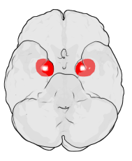
The amygdala is one of two almond-shaped clusters of nuclei located deep and medially within the temporal lobes of the brain's cerebrum in complex vertebrates, including humans. Shown to perform a primary role in the processing of memory, decision making, and emotional responses, the amygdalae are considered part of the limbic system. The term "amygdala" was first introduced by Karl Friedrich Burdach in 1822.

The sensory nervous system is a part of the nervous system responsible for processing sensory information. A sensory system consists of sensory neurons, neural pathways, and parts of the brain involved in sensory perception and interoception. Commonly recognized sensory systems are those for vision, hearing, touch, taste, smell, balance and visceral sensation. Sense organs are transducers that convert data from the outer physical world to the realm of the mind where people interpret the information, creating their perception of the world around them.
The mesolimbic pathway, sometimes referred to as the reward pathway, is a dopaminergic pathway in the brain. The pathway connects the ventral tegmental area in the midbrain to the ventral striatum of the basal ganglia in the forebrain. The ventral striatum includes the nucleus accumbens and the olfactory tubercle.

The olfactory system, or sense of smell, is the sensory system used for smelling (olfaction). Olfaction is one of the special senses, that have directly associated specific organs. Most mammals and reptiles have a main olfactory system and an accessory olfactory system. The main olfactory system detects airborne substances, while the accessory system senses fluid-phase stimuli.

Dopaminergic pathways in the human brain are involved in both physiological and behavioral processes including movement, cognition, executive functions, reward, motivation, and neuroendocrine control. Each pathway is a set of projection neurons, consisting of individual dopaminergic neurons.

The ventral tegmental area (VTA), also known as the ventral tegmental area of Tsai, or simply ventral tegmentum, is a group of neurons located close to the midline on the floor of the midbrain. The VTA is the origin of the dopaminergic cell bodies of the mesocorticolimbic dopamine system and other dopamine pathways; it is widely implicated in the drug and natural reward circuitry of the brain. The VTA plays an important role in a number of processes, including reward cognition and orgasm, among others, as well as several psychiatric disorders. Neurons in the VTA project to numerous areas of the brain, ranging from the prefrontal cortex to the caudal brainstem and several regions in between.
Cortical maps are collections (areas) of minicolumns in the brain cortex that have been identified as performing a specific information processing function.

The auditory cortex is the part of the temporal lobe that processes auditory information in humans and many other vertebrates. It is a part of the auditory system, performing basic and higher functions in hearing, such as possible relations to language switching. It is located bilaterally, roughly at the upper sides of the temporal lobes – in humans, curving down and onto the medial surface, on the superior temporal plane, within the lateral sulcus and comprising parts of the transverse temporal gyri, and the superior temporal gyrus, including the planum polare and planum temporale.

The insular cortex is a portion of the cerebral cortex folded deep within the lateral sulcus within each hemisphere of the mammalian brain.

Affective neuroscience is the study of how the brain processes emotions. This field combines neuroscience with the psychological study of personality, emotion, and mood. The basis of emotions and what emotions are remains an issue of debate within the field of affective neuroscience.
Emotional contagion is a form of social contagion that involves the spontaneous spread of emotions and related behaviors. Such emotional convergence can happen from one person to another, or in a larger group. Emotions can be shared across individuals in many ways, both implicitly or explicitly. For instance, conscious reasoning, analysis, and imagination have all been found to contribute to the phenomenon. The behaviour has been found in humans, other primates, dogs, and chickens.

The olfactory tubercle (OT), also known as the tuberculum olfactorium, is a multi-sensory processing center that is contained within the olfactory cortex and ventral striatum and plays a role in reward cognition. The OT has also been shown to play a role in locomotor and attentional behaviors, particularly in relation to social and sensory responsiveness, and it may be necessary for behavioral flexibility. The OT is interconnected with numerous brain regions, especially the sensory, arousal, and reward centers, thus making it a potentially critical interface between processing of sensory information and the subsequent behavioral responses.
The primary olfactory cortex (POC) is a portion of the cerebral cortex. It is found in the inferior part of the temporal lobe of the brain. It receives input from the olfactory tract. It is involved in the sense of smell (olfaction).

The reward system is a group of neural structures responsible for incentive salience, associative learning, and positively-valenced emotions, particularly ones involving pleasure as a core component. Reward is the attractive and motivational property of a stimulus that induces appetitive behavior, also known as approach behavior, and consummatory behavior. A rewarding stimulus has been described as "any stimulus, object, event, activity, or situation that has the potential to make us approach and consume it is by definition a reward". In operant conditioning, rewarding stimuli function as positive reinforcers; however, the converse statement also holds true: positive reinforcers are rewarding.
Emotional responsivity is the ability to acknowledge an affective stimuli by exhibiting emotion. It is a sharp change of emotion according to a person's emotional state. Increased emotional responsivity refers to demonstrating more response to a stimulus. Reduced emotional responsivity refers to demonstrating less response to a stimulus. Any response exhibited after exposure to the stimulus, whether it is appropriate or not, would be considered as an emotional response. Although emotional responsivity applies to nonclinical populations, it is more typically associated with individuals with schizophrenia and autism.
Visual object recognition refers to the ability to identify the objects in view based on visual input. One important signature of visual object recognition is "object invariance", or the ability to identify objects across changes in the detailed context in which objects are viewed, including changes in illumination, object pose, and background context.
Emotion perception refers to the capacities and abilities of recognizing and identifying emotions in others, in addition to biological and physiological processes involved. Emotions are typically viewed as having three components: subjective experience, physical changes, and cognitive appraisal; emotion perception is the ability to make accurate decisions about another's subjective experience by interpreting their physical changes through sensory systems responsible for converting these observed changes into mental representations. The ability to perceive emotion is believed to be both innate and subject to environmental influence and is also a critical component in social interactions. How emotion is experienced and interpreted depends on how it is perceived. Likewise, how emotion is perceived is dependent on past experiences and interpretations. Emotion can be accurately perceived in humans. Emotions can be perceived visually, audibly, through smell and also through bodily sensations and this process is believed to be different from the perception of non-emotional material.
Social cognitive neuroscience is the scientific study of the biological processes underpinning social cognition. Specifically, it uses the tools of neuroscience to study "the mental mechanisms that create, frame, regulate, and respond to our experience of the social world". Social cognitive neuroscience uses the epistemological foundations of cognitive neuroscience, and is closely related to social neuroscience. Social cognitive neuroscience employs human neuroimaging, typically using functional magnetic resonance imaging (fMRI). Human brain stimulation techniques such as transcranial magnetic stimulation and transcranial direct-current stimulation are also used. In nonhuman animals, direct electrophysiological recordings and electrical stimulation of single cells and neuronal populations are utilized for investigating lower-level social cognitive processes.
References
- ↑ Sah, P.; Faber, E. S. L.; Lopez De Armentia, M.; Power, J. (July 2003). "The amygdaloid complex: anatomy and physiology". Physiological Reviews. 83 (3): 803–834. doi:10.1152/physrev.00002.2003. ISSN 0031-9333. PMID 12843409.
- 1 2 García-Amado, María; Prensa, Lucía (2012-06-13). "Stereological Analysis of Neuron, Glial and Endothelial Cell Numbers in the Human Amygdaloid Complex". PLOS ONE. 7 (6): e38692. doi: 10.1371/journal.pone.0038692 . ISSN 1932-6203. PMC 3374818 . PMID 22719923.
- ↑ Majak, Katarzyna; Pitkänen, Asia (2003). "Projections from the periamygdaloid cortex to the amygdaloid complex, the hippocampal formation, and the parahippocampal region: a PHA-L study in the rat". Hippocampus. 13 (8): 922–942. doi:10.1002/hipo.10134. ISSN 1050-9631. PMID 14750655. S2CID 29706209.
- 1 2 3 4 5 6 Anderson, Sarah Ann R.; Michaelides, Michael; Zarnegar, Parisa; Ren, Yanhua; Fagergren, Pernilla; Thanos, Panayotis K.; Wang, Gene-Jack; Bannon, Michael; Neumaier, John F. (2013-12-02). "Impaired periamygdaloid-cortex prodynorphin is characteristic of opiate addiction and depression". Journal of Clinical Investigation. 123 (12): 5334–5341. doi:10.1172/jci70395. ISSN 0021-9738. PMC 3859405 . PMID 24231353.
- 1 2 Kilts, Clinton D.; Egan, Glenn; Gideon, Deborah A.; Ely, Timothy D.; Hoffman, John M. (2003). "Dissociable Neural Pathways Are Involved in the Recognition of Emotion in Static and Dynamic Facial Expressions". NeuroImage. 18 (1): 156–168. doi:10.1006/nimg.2002.1323. PMID 12507452. S2CID 17223464.
- ↑ Schürmann M, Hesse MD, Stephan KE, et al. (February 2005). "Yearning to yawn: the neural basis of contagious yawning". NeuroImage. 24 (4): 1260–4. doi:10.1016/j.neuroimage.2004.10.022. PMID 15670705. S2CID 6269514.
- ↑ Zelano, C. M. (2007). The role of human primary olfactory cortex in olfactory processing. University of California, Berkeley.
- ↑ Freiherr, Jessica (2017). "Cortical Olfactory Processing". Springer Handbook of Odor. Springer Handbooks. Springer, Cham. pp. 97–98. doi:10.1007/978-3-319-26932-0_38. ISBN 9783319269306.