
In anatomy, the atlas (C1) is the most superior (first) cervical vertebra of the spine and is located in the neck. It is named for Atlas of Greek mythology because, just as Atlas supported the globe, it supports the entire head.

The vertebral arteries are major arteries of the neck. Typically, the vertebral arteries originate from the subclavian arteries. Each vessel courses superiorly along each side of the neck, merging within the skull to form the single, midline basilar artery. As the supplying component of the vertebrobasilar vascular system, the vertebral arteries supply blood to the upper spinal cord, brainstem, cerebellum, and posterior part of brain.

The longus capitis muscle, is broad and thick above, narrow below, and arises by four tendinous slips, from the anterior tubercles of the transverse processes of the third, fourth, fifth, and sixth cervical vertebræ, and ascends, converging toward its fellow of the opposite side, to be inserted into the inferior surface of the basilar part of the occipital bone.
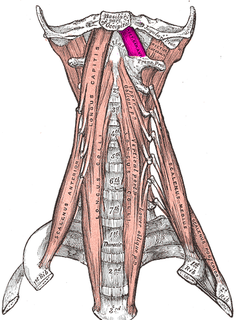
The rectus capitis anterior is a short, flat muscle, situated immediately behind the upper part of the Longus capitis.

The rectus capitis lateralis, a short, flat muscle, arises from the upper surface of the transverse process of the atlas, and is inserted into the under surface of the jugular process of the occipital bone.

The rectus capitis posterior major arises by a pointed tendon from the spinous process of the axis, and, becoming broader as it ascends, is inserted into the lateral part of the inferior nuchal line of the occipital bone and the surface of the bone immediately below the line.

The rectus capitis posterior minor arises by a narrow pointed tendon from the tubercle on the posterior arch of the atlas, and, widening as it ascends, is inserted into the medial part of the inferior nuchal line of the occipital bone and the surface between it and the foramen magnum, and also takes some attachment to the spinal dura mater.

The occipital artery arises from the external carotid artery opposite the facial artery. Its path is below the posterior belly of digastric to the occipital region. This artery supplies blood to the back of the scalp and sternocleidomastoid muscles, and deep muscles in the back and neck.

The nuchal lines are four curved lines on the external surface of the occipital bone:

The basilar part of the occipital bone extends forward and upward from the foramen magnum, and presents in front an area more or less quadrilateral in outline.

The suboccipital triangle is a region of the neck bounded by the following three muscles of the suboccipital group of muscles:
Rectus muscle may refer to:
The prevertebral fascia is a fascia in the neck.
Rectus capitis posterior muscle may refer to:

The following outline is provided as an overview of and topical guide to human anatomy:

The cervical spinal nerve 1 (C1) is a spinal nerve of the cervical segment. C1 carries predominantly motor fibres, but also a small meningeal branch that supplies sensation to parts of the dura around the foramen magnum.

The prevertebral space is a space in the neck.
Rectus capitis may refer to:
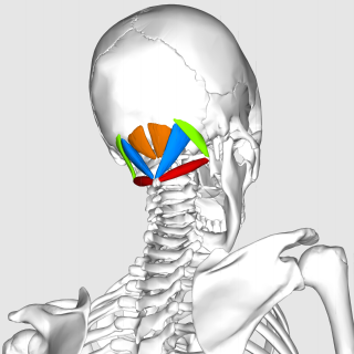
The suboccipital muscles are a group of muscles defined by their location to the occiput. Suboccipital muscles are located below the occipital bone. These are four paired muscles on the underside of the occipital bone; the two straight muscles (rectus) and the two oblique muscles (obliquus).

