Related Research Articles
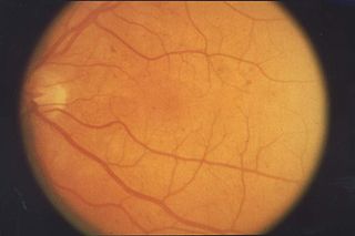
Retinopathy is any damage to the retina of the eyes, which may cause vision impairment. Retinopathy often refers to retinal vascular disease, or damage to the retina caused by abnormal blood flow. Age-related macular degeneration is technically included under the umbrella term retinopathy but is often discussed as a separate entity. Retinopathy, or retinal vascular disease, can be broadly categorized into proliferative and non-proliferative types. Frequently, retinopathy is an ocular manifestation of systemic disease as seen in diabetes or hypertension. Diabetes is the most common cause of retinopathy in the U.S. as of 2008. Diabetic retinopathy is the leading cause of blindness in working-aged people. It accounts for about 5% of blindness worldwide and is designated a priority eye disease by the World Health Organization.
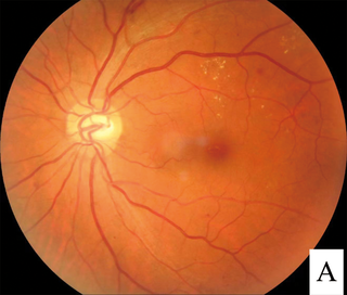
Diabetic retinopathy, is a medical condition in which damage occurs to the retina due to diabetes mellitus. It is a leading cause of blindness in developed countries.

Hypertensive retinopathy is damage to the retina and retinal circulation due to high blood pressure.
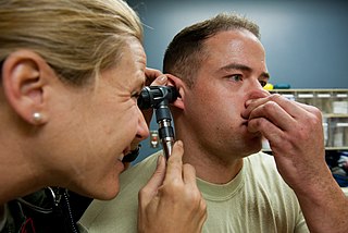
The Valsalva maneuver is performed by a forceful attempt of exhalation against a closed airway, usually done by closing one's mouth and pinching one's nose shut while expelling air out as if blowing up a balloon. Variations of the maneuver can be used either in medical examination as a test of cardiac function and autonomic nervous control of the heart, or to clear the ears and sinuses when ambient pressure changes, as in scuba diving, hyperbaric oxygen therapy, or air travel.

Vitrectomy is a surgery to remove some or all of the vitreous humor from the eye.
Retinopathy of prematurity (ROP), also called retrolental fibroplasia (RLF) and Terry syndrome, is a disease of the eye affecting prematurely born babies generally having received neonatal intensive care, in which oxygen therapy is used due to the premature development of their lungs. It is thought to be caused by disorganized growth of retinal blood vessels which may result in scarring and retinal detachment. ROP can be mild and may resolve spontaneously, but it may lead to blindness in serious cases. Thus, all preterm babies are at risk for ROP, and very low birth-weight is an additional risk factor. Both oxygen toxicity and relative hypoxia can contribute to the development of ROP.

Retinal detachment is a disorder of the eye in which the retina peels away from its underlying layer of support tissue. Initial detachment may be localized, but without rapid treatment the entire retina may detach, leading to vision loss and blindness. It is a surgical emergency.
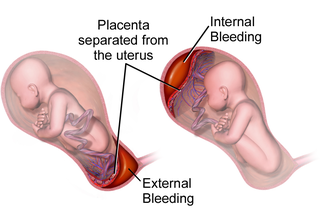
Placental abruption is when the placenta separates early from the uterus, in other words separates before childbirth. It occurs most commonly around 25 weeks of pregnancy. Symptoms may include vaginal bleeding, lower abdominal pain, and dangerously low blood pressure. Complications for the mother can include disseminated intravascular coagulopathy and kidney failure. Complications for the baby can include fetal distress, low birthweight, preterm delivery, and stillbirth.
Retinal hemorrhage is a disorder of the eye in which bleeding occurs in the retina, the light sensitive tissue, located on the back wall of the eye. There are photoreceptor cells in the retina called rods and cones, which transduce light energy into nerve signals that can be processed by the brain to form visual images. Retinal hemorrhage is strongly associated with child abuse in infants and young children and often leaves such abused infants permanently blind. In older children and adults, retinal hemorrhage can be caused by several medical conditions such as hypertension, retinal vein occlusion, anemia, leukemia or diabetes.

Uterine rupture is when the muscular wall of the uterus tears during pregnancy or childbirth. Symptoms, while classically including increased pain, vaginal bleeding, or a change in contractions, are not always present. Disability or death of the mother or baby may result.
Eales disease is a type of obliterative vasculopathy, also known as angiopathia retinae juvenilis, periphlebitis retinae or primary perivasculitis of the retina. It was first described by the British ophthalmologist Henry Eales (1852–1913) in 1880 and is a rare ocular disease characterized by inflammation and possible blockage of retinal blood vessels, abnormal growth of new blood vessels (neovascularization), and recurrent retinal and vitreal hemorrhages.
Hemoperitoneum is the presence of blood in the peritoneal cavity. The blood accumulates in the space between the inner lining of the abdominal wall and the internal abdominal organs. Hemoperitoneum is generally classified as a surgical emergency; in most cases, urgent laparotomy is needed to identify and control the source of the bleeding. In selected cases, careful observation may be permissible. The abdominal cavity is highly distensible and may easily hold greater than five liters of blood, or more than the entire circulating blood volume for an average-sized individual. Therefore, large-scale or rapid blood loss into the abdomen will reliably induce hemorrhagic shock and, if untreated, may rapidly lead to death.
Ocular ischemic syndrome is the constellation of ocular signs and symptoms secondary to severe, chronic arterial hypoperfusion to the eye. Amaurosis fugax is a form of acute vision loss caused by reduced blood flow to the eye; it may be a warning sign of an impending stroke, as both stroke and retinal artery occlusion can be caused by thromboembolism due to atherosclerosis elsewhere in the body. Consequently, those with transient blurring of vision are advised to urgently seek medical attention for a thorough evaluation of the carotid artery. Anterior segment ischemic syndrome is a similar ischemic condition of anterior segment usually seen in post-surgical cases. Retinal artery occlusion leads to rapid death of retinal cells, thereby resulting in severe loss of vision.
Charcot–Bouchard aneurysms are aneurysms of the brain vasculature which occur in small blood vessels. Charcot–Bouchard aneurysms are most often located in the lenticulostriate vessels of the basal ganglia and are associated with chronic hypertension. Charcot–Bouchard aneurysms are a common cause of cerebral hemorrhage.
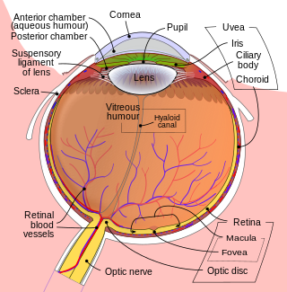
Intraocular hemorrhage is bleeding inside the eye. Bleeding can occur from any structure of the eye where there is vasculature or blood flow, including the anterior chamber, vitreous cavity, retina, choroid, suprachoroidal space, or optic disc.

Laser coagulation or laser photocoagulation surgery is used to treat a number of eye diseases and has become widely used in recent decades. During the procedure, a laser is used to finely cauterize ocular blood vessels to attempt to bring about various therapeutic benefits.
Ovarian apoplexy is a sudden rupture in the ovary, commonly at the site of a cyst, accompanied by hemorrhage in the ovarian tissue and/or intraperitoneal bleeding.

Branch retinal vein occlusion is a common retinal vascular disease of the elderly. It is caused by the occlusion of one of the branches of central retinal vein.
Sickle cell retinopathy can be defined as retinal changes due to blood vessel damage in the eye of a person with a background of sickle cell disease. It can likely progress to loss of vision in late stages due to vitreous hemorrhage or retinal detachment. Sickle cell disease is a structural red blood cell disorder leading to consequences in multiple systems. It is characterized by chronic red blood cell destruction, vascular injury, and tissue ischemia causing damage to the brain, eyes, heart, lungs, kidneys, spleen, and musculoskeletal system.
Thomas David Duane was an American ophthalmologist better known for studies in the field of retina. He identified that the blackouts experienced by wartime pilots during acceleration is due to reduced blood supply to the retina. In 1972, he first described Valsalva retinopathy a form of retinopathy caused by a sudden increase in intrathoracic or intra-abdominal pressure.
References
- 1 2 3 Rajshri, Hirawat; Krishnappa, Nagesha C.; Sharma, Unnatti; Ganne, Pratyusha (1 March 2021). "Long-standing Valsalva retinopathy". BMJ Case Reports. 14 (3): e240812. doi:10.1136/bcr-2020-240812. ISSN 1757-790X. PMC 7938996 . PMID 33674301. S2CID 232129854.
- ↑ Al-Mujaini, Abdullah S.; Montana, Carolina C. (7 April 2008). "Valsalva retinopathy in pregnancy: a case report". Journal of Medical Case Reports. 2 (1): 101. doi:10.1186/1752-1947-2-101. ISSN 1752-1947. PMC 2311321 . PMID 18394189. S2CID 10134512.
- 1 2 "Valsalva Retinopathy: Vision loss after asthma attack".
- 1 2 3 4 5 "Valsalva Retinopathy - EyeWiki". eyewiki.aao.org. Retrieved 2022-04-24.
- 1 2 Salmon JF (13 December 2019). "Retinal vascular disease". Kanski's clinical ophthalmology : a systematic approach (9th ed.). Elsevier. p. 549. ISBN 978-0-7020-7711-1.
- 1 2 Simakurthy, Sriram; Tripathy, Koushik (2022-02-21). "Valsalva Retinopathy". StatPearls. PMID 31424803.
- ↑ "Ocular Manifestations Of Valsalva Maneuver". Clinical and Refractive Optometry.
- ↑ Ophthalmology (Fifth ed.). Edinburgh: Elsevier. 2019. p. 681. ISBN 978-0-323-52820-7.