Acute myeloblastic leukemia with maturation

Last updated
Acute myeloblastic leukemia with maturation
Acute Myelogenous Leukemia with maturation.jpg
Acute Myelogenous Leukemia with maturation. Bone marrow smear, Giemsa stain, 1000x.
Specialty Hematology, oncology

Acute myeloblastic leukemia with maturation (M2) is a subtype of acute myeloid leukemia (AML). [1]

Contents

Acute myeloid leukemia (AML) is a type of cancer affecting blood cells that eventually develop into non-lymphocyte white blood cells. The disease originates from the bone marrow, the soft inner portion of select bones where blood stem cells develop into either lymphocyte or in this particular condition, myeloid cells. This acute disease prevents bone marrow cells from properly maturing, thus causing an accumulation of immature myeloblast cells in the bone marrow.

Acute myeloid leukemia is more lethal than chronic myeloid leukemia, a disease that affects the same myeloid cells, but at a different pace. Many of the immature blast cells in acute myeloid leukemia have a higher loss of function and thus, a higher inability to carry out normal functions than those more developed immature myeloblast cells in chronic myeloid leukemia (O’Donnell et al. 2012). Acute in acute myeloid leukemia means that the amounts of blast cells are increasing at a very high rate. Myeloid refers to the type of white blood cells that are affected by the condition.

Acute myeloid leukemia is the most common acute leukemia that is affecting the adult population. The 5-year survival rate for the cancer stands at around 26% (ACS, 2016).

M2 acute myeloblastic leukemia with maturation refers to the subtype of acute myeloid leukemia characterized by the maturation stages of the myeloid cell development and the location of the AML1 gene. One of the hallmarks of M2 subtype acute myeloid leukemia is the formation of a fusion protein, AML1-ETO or RUNX1-RUNX1T1, due to a translocation of chromosome 8 to chromosome 21 or t(8;21) (Miyoshi et al., 1991, Andrieu et al., 1996). This cytogenetic abnormality has been found in 90% of M2 acute myeloblastic leukemia; while the other 10% constitutes a mix of M1 and M4 acute myeloid leukemia (GFHC, 1990).

Another translocation between chromosome 6p23 and chromosome 9q34 is also associated with the M2 subtype. The t(6;9) causes the formation of a fusion oncogene made of DEK (6p23) and CAN/NUP214 (9q34). This rare translocation has a poor prognosis compared to the t(8;21) because 70% of t(6;9) acute myeloid leukemia patients have the FLT3-ITD mutation (Schwartz et al., 1983, Kottaridis, 2001). The FLT-ITD mutation is one of the most lethal mutations in acute myeloid leukemia (Chi et al., 2008).

M2 acute myeloblastic leukemia with maturation, as classified by the FAB system, constitutes 25% of adult AML.

Cause

This subtype is characterized by a translocation of a part of chromosome 8 to chromosome 21, written as t(8;21). [2] On both sides of the splice the DNA coded for different proteins, RUNX1 and ETO, These two sequences are then transcribed and translated into a single large protein, "M2 AML" which allows the cell to divide unchecked, leading to cancer.

Genetics

Acute myeloid leukemia is a very heterogeneous disease, composed of a variety of translocations and mutations. However, one tenth of all acute myeloid leukemia cases diagnosed have the AML1-ETO fusion oncoprotein due to the t(8;21) translocation. AML1 or RUNX1 is a DNA-binding transcription factor located at the 21q22. ETO is a protein with transcriptional repressing abilities located at the 8q22.

Less than 1% of acute myeloid leukemia patients have the t(6;9) mutation. The rare translocation causes the formation of fusion oncoprotein DEK-NUP214 (Huret, 2005). DEK functions as a transcriptional repressor by interfering with histone acetyl transferases, regulator for a number of stem cells, and activates gene expression in myeloid cells (Koleva et al., 2012). The NUP214 protein is involved in mRNA export as well as nuclear membrane localization and nuclear pore complex (Koser et al., 2011).

Molecular mechanism

Figure 1. Overview of major interaction with tumor suppressor p14 and downstream effects of fusion protein AML1-ETO in M2 acute myeloid leukemia. Elimination of the tumor suppressor ARF gene is often seen in cancer cells. In adult M2 acute myeloblastic leukemia with maturation, ARF expression is suppressed via chromosome translocations that fuse AML1 or Runx1 to an ETO gene. The AML1 or Runx1 gene is in charge of activating transcription of the ARF gene while the ETO protein is involved in transcriptional repression. The AML1-ETO fusion protein ultimately causes transcriptional repression of the p14 gene, which deregulates the expression levels of Mdm2 and p53. The down regulation of ARF, increases Mdm2 levels due to the lack of regulation by the ARF gene. Unregulated, overexpressed Mdm2 will suppress p53 levels. The suppression of p53 levels is an anti-apoptotic mechanism for cancer cells to survive (Elagib, 2006, Weinberg, 2014). AML1-ETO effects on p53 pathway..png
Figure 1. Overview of major interaction with tumor suppressor p14 and downstream effects of fusion protein AML1-ETO in M2 acute myeloid leukemia. Elimination of the tumor suppressor ARF gene is often seen in cancer cells. In adult M2 acute myeloblastic leukemia with maturation, ARF expression is suppressed via chromosome translocations that fuse AML1 or Runx1 to an ETO gene. The AML1 or Runx1 gene is in charge of activating transcription of the ARF gene while the ETO protein is involved in transcriptional repression. The AML1-ETO fusion protein ultimately causes transcriptional repression of the p14 gene, which deregulates the expression levels of Mdm2 and p53. The down regulation of ARF, increases Mdm2 levels due to the lack of regulation by the ARF gene. Unregulated, overexpressed Mdm2 will suppress p53 levels. The suppression of p53 levels is an anti-apoptotic mechanism for cancer cells to survive (Elagib, 2006, Weinberg, 2014).

The fusion oncoprotein involves the gene AML1 (now known as RUNX1) and ETO (now known as RUNX1T1). AML1, located at the 21q22, normally has the ability to activate transcription of the ARF gene and ETO, located at the 8q22, normally has the ability to repress transcription. The fusion protein AML1-ETO is commonly found in acute myeloid leukemia patients. p14ARF is a well known tumor suppressor that serves as the safety net when p53 tumor suppressor's functions are inhibited. Many cancers recognize the potential of the p14ARF tumor suppressor to block cell growth so it is commonly mutated or inhibited in cancer cells. The AML1-ETO is incapable of p14ARF transcription as the fusion protein took on AML1's involvement with ARF gene expression and ETO's transcription repression. The Akt/PKB signaling is a pathway that is pro-survival and growth. By activating Mdm2, the signal transduction pathway will trigger the anti-apoptotic downstream effects of Mdm2. With no p14ARF to regulate and inhibit Mdm2, there will be an increased level of suppression of p53. Mdm2 is a proto-oncogene that directly antagonizes p53 to ubiquitination (Figure 1). The p53 protein is known as the “guardian of the genome” due to its ability to induce DNA repair enzymes and regulate cell cycle advancements. The down regulation of p53 by Mdm2 would lead to unchecked proliferative growth. The direct consequence of having the fusion protein, AML1-ETO, is the lack of p53 regulation in pre-leukemic cells. Therefore, there are an increased number of immature cells that are unable to carry out normal function, which is essentially cancer (Faderi et al., 2000, Song et al. 2005, Weinberg, 2014).

Autophagy in M2 AML

Autophagy is an innate pathway used for degradation of cellular components (Kobayashi, 2015). In recent studies, scientists recognize the significance of autophagy both as a potential anti-apoptotic response to cancer treatments as well as a potential mechanism for getting rid of undesirable fusion proteins such as AML1-ETO. In a 2013 study, scientists demonstrated that the degradation of fusion oncoprotein AML1-ETO is not mediated by autophagy through a set of drug dosage trials testing the levels of AML1-ETO protein expression. The acute myeloid leukemia Kasumi-1 cell line was selected for the experiment due to its AML1-ETO positive characteristics. These cells were treated with increasing concentrations of each histone deacetylase inhibitors – valproic acid (VPA) (epileptic and bipolar drug) or vorinostat (cutaneous T cell lymphoma drug), which are known to induce autophagy associated with loss of the fusion protein. The two inhibitors were added to the cell line in doses of 0, 0.38 uM, 0.74 uM, and 1.5 uM. The cell lysates were then treated with autophagy inhibitors Baf or CQ, or control. Through immunoblotting, there is no reduction of AML1-ETO observed across the different concentrations of VPA or vorinostat. The results indicate that AML1-ETO degradation is not mediated by autophagy, but there is an observed pro-survival autophagy in the leukemic cells (Torgersen et al., 2013). Thus, an inhibition of autophagy would be a viable treatment method for subtype M2 acute myeloid leukemia.

Diagnosis

The first red flag that indicates M2 acute myeloblastic leukemia with maturation is the skewed ratio of white blood cells to red blood cells. Leukemia is initially diagnosed by a peripheral blood smear, a procedure used to check for cell count and cell shapes. Then a bone marrow aspiration and biopsy would be conducted to collect and view the bone, bone marrow, and blood under a microscope. Cytogenetic assays, such as fluorescence in situ hybridization (FISH) would help evaluate the structure and function of the cell's chromosomes.

The criteria for an acute myeloid leukemia case to fall under the M2 subtype is the following: 20%+ nonerythroid cells in peripheral blood or bone marrow are myeloblasts; monocytic precursors are < 20% in bone marrow and granulocytes are 10%+ of cells (Mihova, 2013).

Treatments

Generally, acute myeloid leukemia is treated using chemotherapy consisting of an induction phase and consolidation phase (Dohner et al., 2009). Patients may also consider hematopoietic stem cell transplantation as a second mode of tackling the cancer. The most novel research is being done in tyrosine kinase inhibitors; however M2 acute myeloid leukemia treatment research involves molecules that inhibit the fusion oncoprotein AML1-ETO. Therefore, in terms of M2 subtype acute myeloid leukemia, the most prominent target is the abnormal AML1-ETO fusion protein. Similarly, chronic myeloid leukemia (CML) is comparable to acute myeloid leukemia M2 because it also forms a fusion oncoprotein – BCR-Abl. The developed tyrosine kinase inhibitor, imatinib mesylate, has had a tremendous effect on stopping cancer progression in the majority of chronic myeloid leukemia patients. BCR-Abl is constitutively active due chromosome translocation; therefore it over-phosphorylates the tyrosine kinase. Imatinib mesylate works to block BCR-Abl's activity by blocking the active kinase domain (Fava et al., 2011).

Celastrol is a compound extracted from Tripterygium wilfordii that has anti-cancer properties. It was found to inhibit cell proliferation through the down regulation of AML1-ETO fusion oncoprotein. Celastrol inhibits the fusion oncoprotein by inducing mitochondrial instability and initiating caspase activity. The decrease of AML1-ETO also results in lower levels of C-KIT kinases, Akt/PKB, STAT3, and Erk1/2 – all of which are involved in cell signaling and gene transcription. [3]

Histone deacetylase inhibitors such as valproic acid (VPA), vorinostat, and all-trans retinoic acid (ATRA) are effective in targeting acute myeloid leukemia with the AML1-ETO fusion protein. The HDAC inhibitors are known to induce apoptosis through accumulation of DNA damage, inhibition of DNA repair, and activation of caspases. These inhibitors are extra sensitive to the fusion proteins. Vorinostat has been proven to cause a greater accumulation of DNA damage in fusion protein expressing cells and is directly correlated with the reduction of DNA repair enzymes (Garcia et al., 2008). Romidepsin, a drug in phase two clinical trials, has demonstrated higher efficacy in patients with AML1-ETO fusion protein leukemia (Odenike et al., 2008). Although many clinical evaluations have proven HDAC inhibitors have a promising effect on M2 subtype acute myeloid leukemia, it has not been approved as an official treatment.

In t(6;9) acute myeloid leukemia, FLT3-ITD and the DEK-NUP214 protein are potential targets for treatment. Sorafenib is a kinase inhibitor used as a treatment for kidney and liver cancer. The kinase inhibitor blocks serine-threonine kinase RAF-1 as well as FLT-ITD (Kindler, 2010). The drug has been proven to be effective in reducing FLT3-ITD overexpression (Metzelder et al., 2009). In patients with DEK-NUP214, it was found that the fusion oncoprotein caused an upregulation of mTORC1 (Sanden et al., 2013). Thus, a mTORC inhibitor could be a potential treatment.

Related Research Articles

<span class="mw-page-title-main">Philadelphia chromosome</span> Genetic abnormality in leukemia cancer cells

The Philadelphia chromosome or Philadelphia translocation (Ph) is a specific genetic abnormality in chromosome 22 of leukemia cancer cells. This chromosome is defective and unusually short because of reciprocal translocation, t(9;22)(q34;q11), of genetic material between chromosome 9 and chromosome 22, and contains a fusion gene called BCR-ABL1. This gene is the ABL1 gene of chromosome 9 juxtaposed onto the breakpoint cluster region BCR gene of chromosome 22, coding for a hybrid protein: a tyrosine kinase signaling protein that is "always on", causing the cell to divide uncontrollably by interrupting the stability of the genome and impairing various signaling pathways governing the cell cycle.

<span class="mw-page-title-main">Chronic myelogenous leukemia</span> Medical condition

Chronic myelogenous leukemia (CML), also known as chronic myeloid leukemia, is a cancer of the white blood cells. It is a form of leukemia characterized by the increased and unregulated growth of myeloid cells in the bone marrow and the accumulation of these cells in the blood. CML is a clonal bone marrow stem cell disorder in which a proliferation of mature granulocytes and their precursors is found. It is a type of myeloproliferative neoplasm associated with a characteristic chromosomal translocation called the Philadelphia chromosome.

<span class="mw-page-title-main">Acute promyelocytic leukemia</span> Subtype of acute myeloid leukaemia characterised by accumulation of promyelocytes

Acute promyelocytic leukemia is a subtype of acute myeloid leukemia (AML), a cancer of the white blood cells. In APL, there is an abnormal accumulation of immature granulocytes called promyelocytes. The disease is characterized by a chromosomal translocation involving the retinoic acid receptor alpha (RARA) gene and is distinguished from other forms of AML by its responsiveness to all-trans retinoic acid therapy. Acute promyelocytic leukemia was first characterized in 1957 by French and Norwegian physicians as a hyperacute fatal illness, with a median survival time of less than a week. Today, prognoses have drastically improved; 10-year survival rates are estimated to be approximately 80-90% according to one study.

<span class="mw-page-title-main">Acute myeloid leukemia</span> Cancer of the myeloid line of blood cells

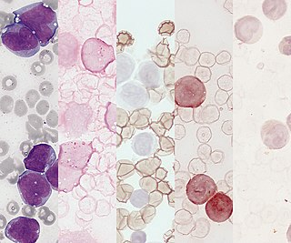
Acute myeloid leukemia (AML) is a cancer of the myeloid line of blood cells, characterized by the rapid growth of abnormal cells that build up in the bone marrow and blood and interfere with normal blood cell production. Symptoms may include feeling tired, shortness of breath, easy bruising and bleeding, and increased risk of infection. Occasionally, spread may occur to the brain, skin, or gums. As an acute leukemia, AML progresses rapidly, and is typically fatal within weeks or months if left untreated.

<span class="mw-page-title-main">Acute monocytic leukemia</span> Medical condition

Acute monocytic leukemia is a type of acute myeloid leukemia. In AML-M5 >80% of the leukemic cells are of monocytic lineage. This cancer is characterized by a dominance of monocytes in the bone marrow. There is an overproduction of monocytes that the body does not need in the periphery. These overproduced monocytes interfere with normal immune cell production which causes many health complications for the affected individual.

<span class="mw-page-title-main">CD135</span> Protein-coding gene in the species Homo sapiens

Cluster of differentiation antigen 135 (CD135) also known as fms like tyrosine kinase 3, receptor-type tyrosine-protein kinase FLT3, or fetal liver kinase-2 (Flk2) is a protein that in humans is encoded by the FLT3 gene. FLT3 is a cytokine receptor which belongs to the receptor tyrosine kinase class III. CD135 is the receptor for the cytokine Flt3 ligand (FLT3L).

<span class="mw-page-title-main">ETV6</span> Protein-coding gene in the species Homo sapiens

ETV6 protein is a transcription factor that in humans is encoded by the ETV6 gene. The ETV6 protein regulates the development and growth of diverse cell types, particularly those of hematological tissues. However, its gene, ETV6 frequently suffers various mutations that lead to an array of potentially lethal cancers, i.e., ETV6 is a clinically significant proto-oncogene in that it can fuse with other genes to drive the development and/or progression of certain cancers. However, ETV6 is also an anti-oncogene or tumor suppressor gene in that mutations in it that encode for a truncated and therefore inactive protein are also associated with certain types of cancers.

<span class="mw-page-title-main">RUNX1</span> Protein-coding gene in humans

Runt-related transcription factor 1 (RUNX1) also known as acute myeloid leukemia 1 protein (AML1) or core-binding factor subunit alpha-2 (CBFA2) is a protein that in humans is encoded by the RUNX1 gene.

<span class="mw-page-title-main">MN1 (gene)</span> Protein-coding gene in the species Homo sapiens

MN1 is a gene found on human chromosome 22, with gene map locus 22q12.3-qter. Its official full name is meningioma 1 because it is disrupted by a balanced translocation (4;22) in a meningioma.

<span class="mw-page-title-main">Zinc finger and BTB domain-containing protein 16</span> Protein found in humans

Zinc finger and BTB domain-containing protein 16 is a protein that in humans is encoded by the ZBTB16 gene.

<span class="mw-page-title-main">HOXA9</span> Protein-coding gene in humans

Homeobox protein Hox-A9 is a protein that in humans is encoded by the HOXA9 gene.

<span class="mw-page-title-main">CBFB</span> Protein-coding gene in the species Homo sapiens

Core-binding factor subunit beta is a protein that in humans is encoded by the CBFB gene.

<span class="mw-page-title-main">RUNX1T1</span> Protein-coding gene in humans

Protein CBFA2T1 is a protein that in humans is encoded by the RUNX1T1 gene.

<span class="mw-page-title-main">MECOM</span> Protein-coding gene in the species Homo sapiens

MDS1 and EVI1 complex locus protein EVI1 (MECOM) also known as ecotropic virus integration site 1 protein homolog (EVI-1) or positive regulatory domain zinc finger protein 3 (PRDM3) is a protein that in humans is encoded by the MECOM gene. EVI1 was first identified as a common retroviral integration site in AKXD murine myeloid tumors. It has since been identified in a plethora of other organisms, and seems to play a relatively conserved developmental role in embryogenesis. EVI1 is a nuclear transcription factor involved in many signaling pathways for both coexpression and coactivation of cell cycle genes.

Acute myelomonocytic leukemia (AMML) is a form of acute myeloid leukemia that involves a proliferation of CFU-GM myeloblasts and monoblasts. AMML occurs with a rapid increase amount in white blood cell count and is defined by more than 20% of myeloblast in the bone marrow. It is classified under "M4" in the French-American-British classification (FAB). It is classified under "AML, not otherwise classified" in the WHO classification.

<span class="mw-page-title-main">Crenolanib</span> Chemical compound

Crenolanib besylate is an investigational inhibitor being developed by AROG Pharmaceuticals, LLC. The compound is currently being evaluated for safety and efficacy in clinical trials for various types of cancer, including acute myeloid leukemia (AML), gastrointestinal stromal tumor (GIST), and glioma. Crenolanib is an orally bioavailable benzimidazole that selectively and potently inhibits signaling of wild-type and mutant isoforms of class III receptor tyrosine kinases (RTK) FLT3, PDGFR α, and PDGFR β. Unlike most RTK inhibitors, crenolanib is a type I mutant-specific inhibitor that preferentially binds to phosphorylated active kinases with the ‘DFG in’ conformation motif.

<span class="mw-page-title-main">Midostaurin</span> Chemical compound

Midostaurin, sold under the brand name Rydapt & Tauritmo both by Novartis, is a multi-targeted protein kinase inhibitor that has been investigated for the treatment of acute myeloid leukemia (AML), myelodysplastic syndrome (MDS) and advanced systemic mastocytosis. It is a semi-synthetic derivative of staurosporine, an alkaloid from the bacterium Streptomyces staurosporeus.

AI-10-49 is a small molecule inhibitor of leukemic oncoprotein CBFβ-SMHHC developed by the laboratory of John Bushweller with efficacy demonstrated by the laboratories of Lucio H. Castilla and Monica Guzman. AI-10-49 allosterically binds to CBFβ-SMMHC and disrupts protein-protein interaction between CBFβ-SMMHC and tumor suppressor RUNX1. This inhibitor is under development as an anti-leukemic drug.

Clonal hypereosinophilia, also termed primary hypereosinophilia or clonal eosinophilia, is a grouping of hematological disorders all of which are characterized by the development and growth of a pre-malignant or malignant population of eosinophils, a type of white blood cell that occupies the bone marrow, blood, and other tissues. This population consists of a clone of eosinophils, i.e. a group of genetically identical eosinophils derived from a sufficiently mutated ancestor cell.

<span class="mw-page-title-main">Gilteritinib</span> Chemical compound

Gilteritinib, sold under the brand name Xospata, is an anti-cancer drug. It acts as an inhibitor of FLT3, hence it is a tyrosine kinase inhibitor.

References

  1. "Acute Myeloid Leukemia - Signs and Symptoms".
  2. Taj AS, Ross FM, Vickers M, et al. (1995). "t(8;21) myelodysplasia, an early presentation of M2 AML". Br. J. Haematol. 89 (4): 890–2. doi:10.1111/j.1365-2141.1995.tb08429.x. PMID   7772527. S2CID   35020709.
  3. Yu, Xianjun; Ruan, Xuzhi; Zhang, Jingxuan; Zhao, Qun (30 April 2016). "Celastrol Induces Cell Apoptosis and Inhibits the Expression of the AML1-ETO/C-KIT Oncoprotein in t(8;21) Leukemia". Molecules. 21 (5): 574. doi: 10.3390/molecules21050574 . PMC   6274014 . PMID   27144550.