Dermatofibrosarcoma protuberans

Last updated
Dermatofibrosarcoma protuberans
Other namesDFSP [1]
Dermatofibrosarcoma protuberans (1) recurrence.JPG
Histopathological image of dermatofibrosarcoma protuberans. Local recurrence long after the first excision. H&E stain
Specialty Oncology   OOjs UI icon edit-ltr-progressive.svg

Dermatofibrosarcoma protuberans (DFSP) [2] is a rare locally aggressive malignant cutaneous soft-tissue sarcoma. DFSP develops in the connective tissue cells in the middle layer of the skin (dermis). [3] Estimates of the overall occurrence of DFSP in the United States are 0.8 to 4.5 cases per million persons per year. [4] [5] In the United States, DFSP accounts for between 1 and 6 percent of all soft-tissue sarcomas [6] and 18 percent of all cutaneous soft-tissue sarcomas. In the Surveillance, Epidemiology and End Results (SEER) tumor registry from 1992 through 2004, DFSP was second only to Kaposi sarcoma.

Contents

Presentation

Dermatofibrosarcoma protuberans begins as a minor firm area of skin most commonly about to 1 to 5 cm in diameter. It can resemble a bruise, birthmark, or pimple. It is a slow-growing tumor and is usually found on the torso but can occur anywhere on the body. [7] About 90% of DFSPs are low-grade sarcomas. About 10% are mixed, containing a high-grade sarcomatous component (DFSP-FS); therefore, they are considered to be intermediate-grade sarcomas. DFSPs rarely lead to a metastasis (fewer than 5% metastasize), but DFSPs can recur locally. DFSPs most often arise in patients who are in their thirties but this may be due to diagnostic delay.

Location

Commonly located on the chest and shoulders, the following is the site distribution of DFPS as was observed in Surveillance, Epidemiology, and End Results (SEER) database between 2000 and 2010. [5] [8]

Variants

The World Health Organization in 2020 classified the fibro sarcomatous DFSP (DFSP-FS) variant (also termed dermatofibrosarcoma protuberans, fibro sarcomatous) of the dermatofibrosarcoma protuberans as a specific form of the intermediate (rarely metastasizing) fibroblastic and myofibroblastic tumors and other variants of this disorder as a specific form of the intermediate (locally aggressive) fibroblastic and myofibroblastic tumors. [9]

Bednar tumors

Bednar, or pigmented DFSP, is distinguished by the dispersal of melanin-rich dendritic cells of the skin. It represents 1–5 percent [10] of all DFSP occurring in people rich in melanin pigments. Bednar is characterized by a dermal spindle cell proliferation like DFSP but distinguished by the additional presence of melanocytic dendritic cells. It occurs at the same rate as DFSP on fairer skin and should be considered to have the same chances of metastasis. [11]

Myxoid DFSP

Myxoid DFSP has areas of myxoid degeneration in the stroma. [12] [13]

Giant cell fibroblastoma

Giant cell fibroblastoma [2] contains giant cells, and is also known as juvenile DFSP. [14] Giant cell fibroblastomas are skin and soft-tissue tumors that usually arise in childhood. They are sometimes seen in association with dermatofibrosarcoma protuberans (DFSP, hybrid lesions) or may transform or recur as DFSP. [15] [13]

Atrophic DFSP

Atrophic DFSP resemble other benign lesions such as morphea, idiopathic atrophoderma, atrophic scar, anetoderma or lipoatrophy. It behaves like classic DFSP. It commonly favours young to middle-aged adults. It has a slow infiltrative growth and a high rate of local recurrence if not completely excised. [13] [16]

Sclerosing DFSP

Sclerosing DFSP is a variant in which the cellularity is low, and the tumor consists of uniform bundles of collagen interspersed with more typical DFSP cells. [13]

Granular cell variant is a rare type in which spindle cells are mingled with richly granular cells, the granules being lysosomal, with prominent nucleoli. [13]

Fibrosarcomatous DFSP (DFSP-FS)

Fibrosarcomatous DFSP is a rare variant of DFSP involving greater aggression, high rates of local occurrences, and higher metastatic potential. [17] DFSP-FS are considered to be intermediate-grade sarcomas, [18] although they rarely metastasize (fewer than 5 percent of cases).

Pathophysiology

More than 90% of DFSP tumors have the chromosomal translocation t(17;22). The translocation fuses the collagen gene (COL1A1) with the platelet-derived growth factor (PDGF) gene. The fibroblast, the cell of origin of this tumor, expresses the fusion gene in the belief that it codes for collagen. However, the resulting fusion protein is processed into a mature platelet-derived growth factor which is a potent growth factor. Fibroblasts contain the receptor for this growth factor. Thus the cell "thinks" it is producing a structural protein, but it creates a self-stimulatory growth signal. The cell divides rapidly and tumor forms.

The tissue is often positive for CD34. [19] [20]

Diagnosis

The pattern of tumor cells radiating from a center is described as "cartwheel pattern". Histopathology of cartwheel pattern in dermatofibrosarcoma protuberans, annotated.jpg
The pattern of tumor cells radiating from a center is described as "cartwheel pattern".

DFSP is a malignant tumor diagnosed with a biopsy, when a portion of the tumor is removed for examination. In order to ensure that enough tissue is removed to make an accurate diagnosis, the initial biopsy of a suspected DFSP is usually done with a core needle or a surgical incision. [21]

Clinical palpation is not entirely reliable for ascertaining the depth of a DFSP infiltration. Magnetic resonance imaging (MRI) is more sensitive addressing the depth of the invasion on some types of DFSP, particularly large or recurring tumors, [22] [23] though MRI is less accurate for identifying infiltration to head and neck tumors.

Diagnostic delay and misdiagnosis

Due to the rarity, initial presentation of flat plaque (skin hardening) and the slow-growing nature of DFSP, it may be months to years without a protuberance (bump). The dissonance between the name of the neoplasm and its clinical presentations may cause a majority of patients to experience a diagnostic delay. A 2019 research study found out of 214 patients a range between less than a year to 42 years before diagnosis (median, four years) from patients noticing a symptom to diagnosis. [24]

Currently, a majority of patients (53%) receive a misdiagnosis by health care providers. The most frequent prebiopsy clinical suspicion included cyst (101 [47.2%]), lipoma (30 [14.0%]), and scar (17 [7.9%]). [24]

It has been suggested an alternative term for DFSP should be dermatofibrosarcoma, often protuberant. [24]

Pregnancy

It is suggested that DFSPs may enlarge more rapidly during pregnancy. Immunohistochemical stains for CD34, S-100 protein, factor XIIIa, and estrogen and progesterone receptors were performed on biopsy specimens. The tumors showed the expression of the progesterone receptor. As with many other stromal neoplasms, DFSPs appear to express low levels of hormone receptors, which may be one factor that accounts for their accelerated growth during pregnancy. [25]

Treatment

Dermatofibrosarcoma protuberans of the left axilla. CT, coronal reconstruction Dermatofibrosarcoma protuberans CT cor.jpg
Dermatofibrosarcoma protuberans of the left axilla. CT, coronal reconstruction

Treatment is primarily surgical, with chemotherapy and radiation therapy used if clear resection margins are not acquired. [26]

Surgical treatment

The type of surgical treatment chosen is dependent on the location of the DFSP occurrence and possible size.

Mohs surgery

Mohs micrographic surgery (MMS) has a high cure rate and lowers the recurrence reduction of DFSP [27] if negative resection margins are achieved.

Wide local excision

Wide local excision (WLE) was the gold standard for treating DFSP but is currently under reevaluation. Presently in the United States, WLE may be suggested after the recurrence of MMS. Larger resection margins are suggested for WLE than MMS. Recurrence rate with WLE is about 8.5% with a lower recurrence rate related to wider excision. [28]

Resection margin

DFSP characteristic features are its capacity to invade surrounding tissues, to a considerable distance from the central focus of the tumor in a "tentacle-like" fashion. This fact, coupled with diagnostic delay, may lead to inadequate initial resection. Inadequate initial treatment results in larger, deeper recurrent lesions, but these can be managed by appropriate wide excision. [29]

Radiation therapy

DFSP is a radioresponsive tumor; radiation therapy (RT) is not used as the first choice for treatment. Conservative resection through MMS or WLE is attempted first. If clear margins are not achieved RT, or chemotherapy is recommended. [30]

Chemotherapy

DFSP was previously regarded and nonresponsive to standard chemotherapy. [31] In 2006 the US FDA approved (imatinib mesylate) for the treatment of DFSP. [32] As is true for all medicinal drugs with name ending in "ib," imatinib is a small molecular pathway inhibitor; imatinib inhibits tyrosine kinase. It may be able to induce tumor regression in patients with recurrent DFSP, unresectable DFSP, or metastatic DFSP. [33] There is clinical evidence that imatinib, which inhibits PDGF-receptors, may be effective for tumors positive for the t(17;22) translocation. It is suggested that imatinib may be a treatment for challenging, locally advanced disease and in the rare metastatic cases. It was approved for use by adult patients with unresectable, recurrent and/or metastatic dermatofibrosarcoma protuberans (DFSP). [34]

Metastatic disease

Distant hematogenous metastases are extremely rare. [35] Metastases to regional lymph nodes are rarer and are most likely in patients who have had multiple local recurrences after inadequate surgical resection. [36] Repeatedly recurring tumors have an increased risk for transformation into a more malignant form (DFSP-FS). The lungs are most frequently affected, but metastases to the brain, [37] bone, [38] and other soft tissues are reported.

Studies

DFSP is not extensively studied due to its rarity and low mortality. The majority of studies are small size case studies or meta-analysis.

The most extensive research study to date was Perspectives of Patients With Dermatofibrosarcoma Protuberans on Diagnostic Delays, Surgical Outcomes, and Nonprotuberance. [24] The lead researcher, Jerad Gardner, spoke at a TED Talk in February 2020 on the topic. [39]

History

R. W. Taylor, in 1890, [40] first identified DFSP as a keloid sarcoma. Later in 1924, Ferdinand-Jean Darier and Ferrand identified it as a progressive recurrent dermatofibroma. In 1925, E. Hoffmann [41] coined the term dermatofibrosarcoma protuberans. Bednar tumor was first described by Bednar in 1957. [42] [43]

ICD coding

The following are the ICD-10 medical codes:

Additional images

See also

Related Research Articles

<span class="mw-page-title-main">Sarcoma</span> Type of malignant tumor (cancer)

A sarcoma is a malignant tumor, a type of cancer that arises from cells of mesenchymal origin. Connective tissue is a broad term that includes bone, cartilage, fat, vascular, or other structural tissues, and sarcomas can arise in any of these types of tissues. As a result, there are many subtypes of sarcoma, which are classified based on the specific tissue and type of cell from which the tumor originates. Sarcomas are primary connective tissue tumors, meaning that they arise in connective tissues. This is in contrast to secondary connective tissue tumors, which occur when a cancer from elsewhere in the body spreads to the connective tissue. Sarcomas are one of five different types of cancer, classified by the cell type from which they originate. The word sarcoma is derived from the Greek σάρκωμα sarkōma 'fleshy excrescence or substance', itself from σάρξsarx meaning 'flesh'.

<span class="mw-page-title-main">Imatinib</span> Chemical compound

Imatinib, sold under the brand names Gleevec and Glivec (both marketed worldwide by Novartis) among others, is an oral targeted therapy medication used to treat cancer. Imatinib is a small molecule inhibitor targeting multiple tyrosine kinases such as CSF1R, ABL, c-KIT, FLT3, and PDGFR-β. Specifically, it is used for chronic myelogenous leukemia (CML) and acute lymphocytic leukemia (ALL) that are Philadelphia chromosome–positive (Ph+), certain types of gastrointestinal stromal tumors (GIST), hypereosinophilic syndrome (HES), chronic eosinophilic leukemia (CEL), systemic mastocytosis, and myelodysplastic syndrome.

<span class="mw-page-title-main">Gastrointestinal stromal tumor</span> Human disease (cancer)

Gastrointestinal stromal tumors (GISTs) are the most common mesenchymal neoplasms of the gastrointestinal tract. GISTs arise in the smooth muscle pacemaker interstitial cell of Cajal, or similar cells. They are defined as tumors whose behavior is driven by mutations in the KIT gene (85%), PDGFRA gene (10%), or BRAF kinase (rare). 95% of GISTs stain positively for KIT (CD117). Most (66%) occur in the stomach and gastric GISTs have a lower malignant potential than tumors found elsewhere in the GI tract.

<span class="mw-page-title-main">Fibrosarcoma</span> Medical condition

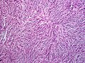
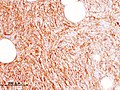
Fibrosarcoma is a malignant mesenchymal tumour derived from fibrous connective tissue and characterized by the presence of immature proliferating fibroblasts or undifferentiated anaplastic spindle cells in a storiform pattern. Fibrosarcomas mainly arise in people between the ages of 25 and 79. It originates in fibrous tissues of the bone and invades long or flat bones such as the femur, tibia, and mandible. It also involves the periosteum and overlying muscle.

<span class="mw-page-title-main">Phyllodes tumor</span> Medical condition

Phyllodes tumors, are a rare type of biphasic fibroepithelial mass that form from the periductal stromal and epithelial cells of the breast. They account for less than 1% of all breast neoplasms. They were previously termed cystosarcoma phyllodes, coined by Johannes Müller in 1838, before being renamed to phyllodes tumor by the World Health Organization in 2003. Phullon, which means 'leaf' in Greek, describes the unique papillary projections characteristic of phyllodes tumors on histology. Diagnosis is made via a core-needle biopsy and treatment is typically surgical resection with wide margins (>1 cm), due to their propensity to recur.

<span class="mw-page-title-main">Aggressive fibromatosis</span> Medical condition

Aggressive fibromatosis or desmoid tumor is a rare condition. Desmoid tumors are a type of fibromatosis and related to sarcoma, though without the ability to spread throughout the body (metastasize). The tumors arise from cells called fibroblasts, which are found throughout the body and provide structural support, protection to the vital organs, and play a critical role in wound healing. These tumors tend to occur in women in their thirties, but can occur in anyone at any age. They can be either relatively slow-growing or malignant. However, aggressive fibromatosis is locally aggressive and can cause life-threatening problems or even death when the tumors compress vital organs such as intestines, kidneys, lungs, blood vessels, or nerves. The condition is rarely fatal. Most cases are sporadic, but some are associated with familial adenomatous polyposis (FAP). Approximately 10% of individuals with Gardner's syndrome, a type of FAP with extracolonic features, have desmoid tumors.

<span class="mw-page-title-main">Undifferentiated pleomorphic sarcoma</span> Medical condition

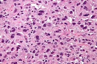
Undifferentiated pleomorphic sarcoma (UPS), also termed pleomorphic myofibrosarcoma, high-grade myofibroblastic sarcoma, and high-grade myofibrosarcoma, is characterized by the World Health Organization (WHO), 2020, as a rare, poorly differentiated neoplasm, i.e. an abnormal growth of cells that have an unclear identity and/or cell of origin. WHO classified it as one of the undifferentiated/unclassified sarcomas in the category of tumors of uncertain differentiation. Sarcomas are cancers known or thought to derive from mesenchymal stem cells that typically develop in bone, muscle, fat, blood vessels, lymphatic vessels, tendons, and ligaments. More than 70 sarcoma subtypes have been described. The UPS subtype of these sarcomas consists of tumor cells that are poorly differentiated and may appear as spindle-shaped cells, histiocytes, and giant cells. UPS is considered a diagnosis that defies formal sub-classification after thorough histologic, immunohistochemical, and ultrastructural examinations fail to identify the type of cells involved.

<span class="mw-page-title-main">Resection margin</span>

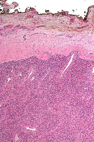
A resection margin or surgical margin is the margin of apparently non-tumorous tissue around a tumor that has been surgically removed, called "resected", in surgical oncology. The resection is an attempt to remove a cancer tumor so that no portion of the malignant growth extends past the edges or margin of the removed tumor and surrounding tissue. These are retained after the surgery and examined microscopically by a pathologist to see if the margin is indeed free from tumor cells. If cancerous cells are found at the edges the operation is much less likely to achieve the desired results.

Giant cell fibroblastoma (GCF) is a rare type of soft-tissue tumor marked by painless nodules in the dermis and subcutaneous tissue. These tumors may come back after surgery, but they do not spread to other parts of the body. They occur mostly in boys. GCF tumor tissues consist of bland spindle-shaped or stellate-shaped cells interspersed among multinucleated giant cells.

Collagenous fibroma, also known as desmoplastic fibroblastoma, is a slow-growing, deep-set, benign fibrous tumor, usually located in the deep subcutis, fascia, aponeurosis, or skeletal muscle of the extremities, limb girdles, or head and neck regions. The World Health Organization in 2020 reclassified desmoplastic fibroblastoma/collagenous fibroma as a specific benign tumor type within the broad category of fibroblastic and myofibroblastic tumors.

<span class="mw-page-title-main">Koenen's tumor</span> Medical condition

Koenen's tumor (KT), also commonly termed periungual angiofibroma, is a subtype of the angiofibromas. Angiofibromas are benign papule, nodule, and/or tumor lesions that are separated into various subtypes based primarily on the characteristic locations of their lesions. KTs are angiofibromas that develop in and under the toenails and/or fingernails. KTs were once considered as the same as another subtype of the angiofibromas viz., acral angiofibromas. While the literature may still sometimes regard KTs as acral angiofibromas, acral angiofibromas are characteristically located in areas close to but not in the toenails and fingernails as well as in the soles of the feet and palms of the hands. KTs are here regarded as distinct from acral angiofibromas.

<span class="mw-page-title-main">Spiradenoma</span> Medical condition

Spiradenomas (SA) are rare, benign cutaneous adnexal tumors that may progress to become their malignant counterparts, i.e. spiradenocarcinomas (SAC). Cutaneous adnexal tumors are a group of skin tumors consisting of tissues that have differentiated towards one of the four primary adnexal structures found in normal skin: hair follicles, sebaceous sweat glands, apocrine sweat glands, and eccrine sweat glands. SA and SAC tumors were regarded as eccrine gland tumors and termed eccrine spiradenomas and eccrine spiradenocarcinomas, respectively. However, more recent studies have found them to be hair follicle tumors and commonly term them spiradenomas and spiradenocarcinomas, respectively. Further confusing the situation, SA-like and SAC-like tumors are also 1) manifestations of the inherited disorder, CYLD cutaneous syndrome (CCS), and 2) have repeatedly been confused with an entirely different tumor, adenoid cystic carcinomas of the salivary gland. Here, SA and SAC are strictly defined as sporadic hair follicle tumors that do not include the hereditary CCS spiradenomas and heridtary spiradenocarcinoms of CCS or the adenoid cystic carcinomas.

<span class="mw-page-title-main">Low-grade fibromyxoid sarcoma</span> Medical condition

Low-grade fibromyxoid sarcoma (LGFMS) is a rare type of low-grade sarcoma first described by H. L. Evans in 1987. LGFMS are soft tissue tumors of the mesenchyme-derived connective tissues; on microscopic examination, they are found to be composed of spindle-shaped cells that resemble fibroblasts. These fibroblastic, spindle-shaped cells are neoplastic cells that in most cases of LGFMS express fusion genes, i.e. genes composed of parts of two different genes that form as a result of mutations. The World Health Organization (2020) classified LGFMS as a specific type of tumor in the category of malignant fibroblastic and myofibroblastic tumors.

Andrea A. Hayes Dixon is an American surgeon. She was the first pediatric surgeon to perform a high-risk, life-saving procedure in children with a rare form of cancer and developed the first orthotropic xenograft model of metastatic Ewing's sarcoma. In 2002, she became the first African American female pediatric surgeon board-certified in the United States.

Fibroblastic and myofibroblastic tumors (FMTs) develop from the mesenchymal stem cells which differentiate into fibroblasts and/or the myocytes/myoblasts that differentiate into muscle cells. FMTs are a heterogeneous group of soft tissue neoplasms. The World Health Organization (2020) defined tumors as being FMTs based on their morphology and, more importantly, newly discovered abnormalities in the expression levels of key gene products made by these tumors' neoplastic cells. Histopathologically, FMTs consist of neoplastic connective tissue cells which have differented into cells that have microscopic appearances resembling fibroblasts and/or myofibroblasts. The fibroblastic cells are characterized as spindle-shaped cells with inconspicuous nucleoli that express vimentin, an intracellular protein typically found in mesenchymal cells, and CD34, a cell surface membrane glycoprotein. Myofibroblastic cells are plumper with more abundant cytoplasm and more prominent nucleoli; they express smooth muscle marker proteins such as smooth muscle actins, desmin, and caldesmon. The World Health Organization further classified FMTs into four tumor forms based on their varying levels of aggressiveness: benign, intermediate, intermediate, and malignant.

Lipofibromatosis-like neural tumor (LPF-NT) is an extremely rare soft tissue tumor first described by Agaram et al in 2016. As of mid-2021, at least 39 cases of LPF-NT have been reported in the literature. LPF-NT tumors have several features that resemble lipofibromatosis (LPF) tumors, malignant peripheral nerve sheath tumors, spindle cell sarcomas, low-grade neural tumors, peripheral nerve sheath tumors, and other less clearly defined tumors; Prior to the Agaram at al report, LPF-NTs were likely diagnosed as variants or atypical forms of these tumors. The analyses of Agaram at al and subsequent studies uncovered critical differences between LPF-NT and the other tumor forms which suggest that it is a distinct tumor entity differing not only from lipofibromatosis but also the other tumor forms.

Myxofibrosarcoma (MFS), although a rare type of tumor, is one of the most common soft tissue sarcomas, i.e. cancerous tumors, that develop in the soft tissues of elderly individuals. Initially considered to be a type of histiocytoma termed fibrous histiocytoma or myxoid variant of malignant fibrous histiocytoma, Angervall et al. termed this tumor myxofibrosarcoma in 1977. In 2020, the World Health Organization reclassified MFS as a separate and distinct tumor in the category of malignant fibroblastic and myofibroblastic tumors.

Sclerosing epithelioid fibrosarcoma (SEF) is a very rare malignant tumor of soft tissues that on microscopic examination consists of small round or ovoid neoplastic epithelioid fibroblast-like cells, i.e. cells that have features resembling both epithelioid cells and fibroblasts. In 2020, the World Health Organization classified SEF as a distinct tumor type in the category of malignant fibroblastic and myofibroblastic tumors. However, current studies have reported that low-grade fibromyxoid sarcoma (LGFMS) has many clinically and pathologically important features characteristic of SEF; these studies suggest that LGSFMS may be an early form of, and over time progress to become, a SEF. Since the World Health Organization has classified LGFMS as one of the malignant fibroblastic and myofibroblastic tumors that is distinctly different than SEF, SEF and LGFMS are here regarded as different tumor forms.

Dermatofibrosarcoma protuberans, fibrosarcomatous (DFSP-FS), also termed fibrosarcomatous dermatofibrosarcoma protuberans, is a rare type of tumor located in the dermis. DFSP-FS tumors have been viewed as: 1) a more aggressive form of the dermatofibrosarcoma protuberans (DFSP) tumors because they have areas that resemble and tend to behave like malignant fibrosarcomas or 2) as a distinctly different tumor than DFSP. DFSP-FS tumors are related to DFSP. For example, surgically removed DFSP tumors often recur with newly developed fibrobosarcoma-like areas. Nonetheless, the World Health Organization (WHO), 2020, classified DFSP and DFSP-FS as different tumors with DFSP being in the category of benign and DFSP-FS in the category of rarely metastasizing fibroblastic and myofibroblastic tumors. This article follows the WHO classification: the 5-15% of DFSP tumors that have any areas of fibrosarcomatous microscopic histopathology are here considered DFSP-FS rather than DFSP tumors.

Gardner fibroma (GF) is a benign fibroblastic tumor. GF tumors typically develop in the dermis and adjacent subcutaneous tissue lying just below the dermis. These tumors typically occur on the back, abdomen, and other superficial sites but in rare cases have been diagnoses in internal sites such as the retroperitoneum and around the large blood vessels in the upper thoracic cavity. The World Health Organization, 2020, classified Gardner fibroma as a benign tumor in the category of fibroblastic and myofibroblastic tumors.

References

  1. "Dermatofibrosarcoma Protuberans Synonyms & Antonyms". Synonyms.com.
  2. 1 2 "NCI Dictionary of Cancer Terms". National Cancer Institute. 2011-02-02. Retrieved 2020-06-09.
  3. "Dermatofibrosarcoma protuberans - Overview - Mayo Clinic". www.mayoclinic.org. Retrieved 2020-06-09.
  4. Rouhani, Panta; Fletcher, Christopher D. M.; Devesa, Susan S.; Toro, Jorge R. (2008-08-01). "Cutaneous soft tissue sarcoma incidence patterns in the U.S. : an analysis of 12,114 cases". Cancer. 113 (3): 616–627. doi: 10.1002/cncr.23571 . ISSN   0008-543X. PMID   18618615. S2CID   23746544.
  5. 1 2 Kreicher, Kathryn L.; Kurlander, David E.; Gittleman, Haley R.; Barnholtz-Sloan, Jill S.; Bordeaux, Jeremy S. (January 2016). "Incidence and Survival of Primary Dermatofibrosarcoma Protuberans in the United States". Dermatologic Surgery. 42 (Suppl 1): S24–31. doi:10.1097/DSS.0000000000000300. ISSN   1524-4725. PMID   26730971. S2CID   12966671.
  6. Kransdorf, M. J. (January 1995). "Malignant soft-tissue tumors in a large referral population: distribution of diagnoses by age, sex, and location". American Journal of Roentgenology. 164 (1): 129–134. doi:10.2214/ajr.164.1.7998525. ISSN   0361-803X. PMID   7998525.
  7. "Dermatofibrosarcoma protuberans".
  8. "America's Children and the Environment: Metadata - Surveillance, Epidemiology, and End Results (SEER) Program". United States Environmental Protection Agency . 10 June 2015.
  9. Sbaraglia M, Bellan E, Dei Tos AP (April 2021). "The 2020 WHO Classification of Soft Tissue Tumours: news and perspectives". Pathologica. 113 (2): 70–84. doi:10.32074/1591-951X-213. PMC   8167394 . PMID   33179614.
  10. Liszewski, Walter; Blanchette, Derek; Cunningham, Ashley M.; Miller, Daniel D. (2016-11-01). "Epidemiology of Bednar tumors in the United States". Journal of the American Academy of Dermatology. 75 (5): 1064–1066. doi: 10.1016/j.jaad.2016.06.018 . ISSN   0190-9622. PMID   27745635.
  11. Kaul, Rashmi; Kaur, Navjot; Dogra, Sunder S; Chander, Bal (2015). "Variant of Dermatofibrosarcoma Protuberans: Bednar Tumor". Indian Journal of Dermatology. 60 (1): 107. doi: 10.4103/0019-5154.147885 . ISSN   0019-5154. PMC   4318047 . PMID   25657441.
  12. Reimann, Julie D. R.; Fletcher, Christopher D. M. (September 2007). "Myxoid dermatofibrosarcoma protuberans: a rare variant analyzed in a series of 23 cases". The American Journal of Surgical Pathology. 31 (9): 1371–1377. doi:10.1097/PAS.0b013e31802ff7e7. ISSN   0147-5185. PMID   17721193. S2CID   1268848.
  13. 1 2 3 4 5 "Rare Variants of Dermatofibrosarcoma Protuberans (DFSP)". News-Medical.net. 2017-10-24. Retrieved 2020-06-09.
  14. Buteau, Anna H.; Keeling, Brett H.; Diaz, Lucia Z.; Larralade, Margarita; Luna, Paula; Krishnan, Chandra; Levy, Moise L. (2018-01-16). "Dermatofibrosarcoma protuberans in pediatric patients: A diagnostic and management challenge". JAAD Case Reports. 4 (2): 155–158. doi:10.1016/j.jdcr.2017.09.022. ISSN   2352-5126. PMC   5789519 . PMID   29387771.
  15. "Giant cell fibroblastoma pathology | DermNet NZ". dermnetnz.org. Retrieved 2020-06-09.
  16. Bakry, Ola; Attia, Abdalla (2012-03-27). "Atrophic dermatofibrosarcoma protuberans". Journal of Dermatological Case Reports. 6 (1): 14–17. doi:10.3315/jdcr.2012.1089. ISSN   1898-7249. PMC   3322104 . PMID   22514584.
  17. Abbott, Jared J.; Oliveira, Andre M.; Nascimento, Antonio G. (April 2006). "The prognostic significance of fibrosarcomatous transformation in dermatofibrosarcoma protuberans". The American Journal of Surgical Pathology. 30 (4): 436–443. doi:10.1097/00000478-200604000-00002. ISSN   0147-5185. PMID   16625088. S2CID   23911032.
  18. Bowne, W. B.; Antonescu, C. R.; Leung, D. H.; Katz, S. C.; Hawkins, W. G.; Woodruff, J. M.; Brennan, M. F.; Lewis, J. J. (2000-06-15). "Dermatofibrosarcoma protuberans: A clinicopathologic analysis of patients treated and followed at a single institution". Cancer. 88 (12): 2711–2720. doi: 10.1002/1097-0142(20000615)88:12<2711::AID-CNCR9>3.0.CO;2-M . ISSN   0008-543X. PMID   10870053.
  19. Sirvent N, Maire G, Pedeutour F (May 2003). "Genetics of dermatofibrosarcoma protuberans family of tumors: from ring chromosomes to tyrosine kinase inhibitor treatment". Genes Chromosomes Cancer. 37 (1): 1–19. doi:10.1002/gcc.10202. PMID   12661001. S2CID   36985697.
  20. Patel KU, Szabo SS, Hernandez VS, et al. (February 2008). "Dermatofibrosarcoma protuberans COL1A1-PDGFB fusion is identified in virtually all dermatofibrosarcoma protuberans cases when investigated by newly developed multiplex reverse transcription polymerase chain reaction and fluorescence in situ hybridization assays". Hum. Pathol. 39 (2): 184–93. doi:10.1016/j.humpath.2007.06.009. PMID   17950782.
  21. "What is Dermatofibrosarcoma Protuberans?".
  22. Serra-Guillén, Carlos; Sanmartín, Onofre; Llombart, Beatriz; Nagore, Eduardo; Deltoro, Carlos; Martín, Isabel; Borella-Estrada, Rafael; Requena, Celia; Martorell-Calatayud, Antonio; Cervera, Jose; Guillén, Carlos (November 2011). "Correlation between preoperative magnetic resonance imaging and surgical margins with modified Mohs for dermatofibrosarcoma protuberans". Dermatologic Surgery. 37 (11): 1638–1645. doi:10.1111/j.1524-4725.2011.02077.x. ISSN   1524-4725. PMID   21679274. S2CID   23878054.
  23. Kransdorf, M. J.; Meis-Kindblom, J. M. (August 1994). "Dermatofibrosarcoma protuberans: radiologic appearance". American Journal of Roentgenology. 163 (2): 391–394. doi:10.2214/ajr.163.2.8037038. ISSN   0361-803X. PMID   8037038.
  24. 1 2 3 4 David, Marjorie Parker; Funderburg, Ashley; Selig, James P.; Brown, Rebecca; Caliskan, Pip M.; Cove, Lee; Dicker, Gayle; Hoffman, Lori; Horne, Tammi; Gardner, Jerad M. (2019-08-30). "Perspectives of Patients With Dermatofibrosarcoma Protuberans on Diagnostic Delays, Surgical Outcomes, and Nonprotuberance". JAMA Network Open. 2 (8): e1910413. doi:10.1001/jamanetworkopen.2019.10413. ISSN   2574-3805. PMC   6724159 . PMID   31469398.
  25. Parlette, L. E.; Smith, C. K.; Germain, L. M.; Rolfe, C. A.; Skelton, H. (November 1999). "Accelerated growth of dermatofibrosarcoma protuberans during pregnancy". Journal of the American Academy of Dermatology. 41 (5 Pt 1): 778–783. doi:10.1016/s0190-9622(99)70023-x. ISSN   0190-9622. PMID   10534646.
  26. Allen A, Ahn C, Sangüeza OP (October 2019). "Dermatofibrosarcoma Protuberans". Dermatol Clin. 37 (4): 483–488. doi:10.1016/j.det.2019.05.006. PMID   31466588. S2CID   201672710.
  27. Malan, Malumani; Xuejingzi, Wu; Quan, Song Ji (2019-08-13). "The efficacy of Mohs micrographic surgery over the traditional wide local excision surgery in the cure of dermatofibrosarcoma protuberans". The Pan African Medical Journal. 33: 297. doi:10.11604/pamj.2019.33.297.17692. ISSN   1937-8688. PMC   6815477 . PMID   31692830.
  28. Kim, Byung Jun; Kim, Hyeonwoo; Jin, Ung Sik; Minn, Kyung Won; Chang, Hak (2015). "Wide Local Excision for Dermatofibrosarcoma Protuberans: A Single-Center Series of 90 Patients". BioMed Research International. 2015: 642549. doi: 10.1155/2015/642549 . ISSN   2314-6133. PMC   4673335 . PMID   26688814.
  29. Khatri, Vijay P.; Galante, Joseph M.; Bold, Richard J.; Schneider, Philip D.; Ramsamooj, Rajendra; Goodnight, James E. (November 2003). "Dermatofibrosarcoma protuberans: reappraisal of wide local excision and impact of inadequate initial treatment". Annals of Surgical Oncology. 10 (9): 1118–1122. doi:10.1245/aso.2003.03.581. ISSN   1068-9265. PMID   14597453. S2CID   41310745.
  30. Suit, H.; Spiro, I.; Mankin, H. J.; Efird, J.; Rosenberg, A. E. (August 1996). "Radiation in management of patients with dermatofibrosarcoma protuberans". Journal of Clinical Oncology. 14 (8): 2365–2369. doi:10.1200/JCO.1996.14.8.2365. ISSN   0732-183X. PMID   8708729.
  31. Noujaim, Jonathan; Thway, Khin; Fisher, Cyril; Jones, Robin L. (December 2015). "Dermatofibrosarcoma protuberans: from translocation to targeted therapy". Cancer Biology & Medicine. 12 (4): 375–384. doi:10.7497/j.issn.2095-3941.2015.0067. ISSN   2095-3941. PMC   4706526 . PMID   26779374.
  32. "Gleevec Gains Simultaneous FDA Approval for Five Rare, Life-Threatening Disorders". Cancer Network. Oncology NEWS International Vol 15 No 11. 15 (11). 2006-11-01. Archived from the original on 2020-06-10. Retrieved 2020-06-10.
  33. Rastogi, Sameer; Dhamija, Ekta; Barwad, Adarsh; Aggarwal, Aditi; Sharma, Atul; Panday, Rambha (December 2018). "Advanced Dermatofibrosarcoma Protuberans Treatment With Imatinib: Experience From a Dedicated Sarcoma Medical Oncology Clinic in India". Journal of Global Oncology. 4 (4): 1–7. doi:10.1200/JGO.18.00007. PMC   6223511 . PMID   30085879.
  34. "Lupin in alliance with Natco receives FDA approval for Imatinib Mesylate Tablets". Lupin. 5 March 2019. Retrieved 2020-06-10.
  35. Mavili, M. E.; Gursu, K. G.; Gokoz, A. (April 1994). "Dermatofibrosarcoma with lymph node involvement". Annals of Plastic Surgery. 32 (4): 438–440. doi:10.1097/00000637-199404000-00022. ISSN   0148-7043. PMID   8210168.
  36. Rutgers, E. J.; Kroon, B. B.; Albus-Lutter, C. E.; Gortzak, E. (June 1992). "Dermatofibrosarcoma protuberans: treatment and prognosis". European Journal of Surgical Oncology: The Journal of the European Society of Surgical Oncology and the British Association of Surgical Oncology. 18 (3): 241–248. ISSN   0748-7983. PMID   1607035.
  37. Mahajan, B. B.; Sumir, Kumar; Singla, Monika (July 2015). "Metastatic dermatofibrosarcoma protuberans: A rare case report from North India". Journal of Cancer Research and Therapeutics. 11 (3): 670. doi: 10.4103/0973-1482.146099 . ISSN   1998-4138. PMID   26458720.
  38. Garg, Mandeep Kumar; Yadav, Mukesh Kumar; Gupta, Suruchi; Kumar, Narender; Khandelwal, Niranjan (2009-09-29). "Dermatofibrosarcoma protuberans with contiguous infiltration of the underlying bone". Cancer Imaging. 9 (1): 63–66. doi:10.1102/1470-7330.2009.0011 (inactive 31 January 2024). ISSN   1740-5025. PMC   2792083 . PMID   19933019.{{cite journal}}: CS1 maint: DOI inactive as of January 2024 (link)
  39. Gardner, Jerad (2020-06-08). "Facebook and Rare Cancer Changed My Life". TED Conferences. Retrieved 2023-09-18.
  40. Shen, Kuang-Hsuan; Leu, Yi-Shing (2017-09-01). "Dermatofibrosarcoma protuberans of the cheek". Journal of Cancer Research and Practice. 4 (3): 119–121. doi: 10.1016/j.jcrpr.2017.03.001 . ISSN   2311-3006.
  41. Hoffmann, Erich (1925). "I. Über das knollentreibende Fibrosarkom der Haut (Dermatofibrosarkoma protuberans)". Dermatology. 43 (1–2): 1–28. doi:10.1159/000250699. ISSN   1018-8665.
  42. "Bednar tumour: an infrequent diagnosis | British Journal of Medical Practitioners". www.bjmp.org. Retrieved 2020-06-09.
  43. Bednar, B. (March 1957). "Storiform neurofibromas of the skin, pigmented and nonpigmented". Cancer. 10 (2): 368–376. doi: 10.1002/1097-0142(195703/04)10:2<368::aid-cncr2820100218>3.0.co;2-3 . ISSN   0008-543X. PMID   13426994. S2CID   42490204.
  44. 1 2 3 "Dermatofibrosarcoma protuberans (DFSP)". www.pathologyoutlines.com. Retrieved 2020-06-09.