This article is about embryonic development in all types of animals, including humans. For information specific to human embryonic development, see Human embryonic development. For information specific to plants, see Plant embryonic development.
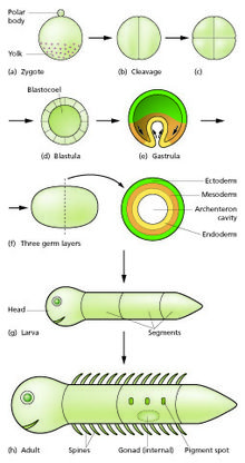
Diagram of stages of embryo development to a larval and adult stage.
The main stages of animal embryonic development are as follows:
The zygote undergoes a series of cell divisions (called cleavage) to form a structure called a morula.
The morula develops into a structure called a blastula through a process called blastulation.
The blastula develops into a structure called a gastrula through a process called gastrulation.
The gastrula then undergoes further development, including the formation of organs (organogenesis).
The embryo then transforms into the next stage of development, the nature of which varies between different animal species (examples of possible next stages include a fetus and a larva).
The egg cell is generally asymmetric, having an animal pole (future ectoderm). It is covered with protective envelopes, with different layers. The first envelope – the one in contact with the membrane of the egg – is made of glycoproteins and is known as the vitelline membrane (zona pellucida in mammals). Different taxa show different cellular and acellular envelopes englobing the vitelline membrane.[2][5]
Fertilization is the fusion of gametes to produce a new organism. In animals, the process involves a sperm fusing with an ovum, which eventually leads to the development of an embryo. Depending on the animal species, the process can occur within the body of the female in internal fertilization, or outside in the case of external fertilization. The fertilized egg cell is known as the zygote.[2][5]
To prevent more than one sperm fertilizing the egg (polyspermy), fast block and slow block to polyspermy are used. Fast block, the membrane potential rapidly depolarizing and then returning to normal, happens immediately after an egg is fertilized by a single sperm. Slow block begins in the first few seconds after fertilization and is when the release of calcium causes the cortical reaction, in which various enzymes are released from cortical granules in the eggs plasma membrane, causing the expansion and hardening of the outside membrane, preventing more sperm from entering.[6][5]
Cell division with no significant growth, producing a cluster of cells that is the same size as the original zygote, is called cleavage. At least four initial cell divisions occur, resulting in a dense ball of at least sixteen cells called the morula. In the early mouse embryo, the sister cells of each division remain connected during interphase by microtubule bridges.[7] The different cells derived from cleavage, up to the blastula stage, are called blastomeres. Depending mostly on the amount of yolk in the egg, the cleavage can be holoblastic (total) or meroblastic (partial).[8][9]
Holoblastic cleavage occurs in animals with little yolk in their eggs,[10] such as humans and other mammals who receive nourishment as embryos from the mother, via the placenta or milk, such as might be secreted from a marsupium. Meroblastic cleavage occurs in animals whose eggs have more yolk (i.e. birds and reptiles). Because cleavage is impeded in the vegetal pole, there is an uneven distribution and size of cells, being more numerous and smaller at the animal pole of the zygote.[8][9]
In holoblastic eggs, the first cleavage always occurs along the vegetal-animal axis of the egg, and the second cleavage is perpendicular to the first. From here the spatial arrangement of blastomeres can follow various patterns, due to different planes of cleavage, in various organisms:
Cleavage patterns followed by holoblastic and meroblastic eggs in animals
In amniotes, the cells of the morula are at first closely aggregated, but soon they become arranged into an outer or peripheral layer, the trophoblast, which does not contribute to the formation of the embryo proper, and an inner cell mass, from which the embryo is developed. Fluid collects between the trophoblast and the greater part of the inner cell-mass, and thus the morula is converted into a vesicle, called the blastodermic vesicle. The inner cell mass remains in contact, however, with the trophoblast at one pole of the ovum; this is named the embryonic pole, since it indicates the location where the future embryo will develop.[18][9]
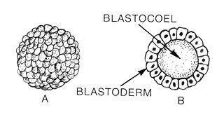
After the seventh cleavage has produced 128 cells, the morula becomes a blastula.[8] The blastula is usually a spherical layer of cells (the blastoderm) surrounding a fluid-filled or yolk-filled cavity the blastocoel.[citation needed]
Mammals at this stage form a structure called the blastocyst, characterized by an inner cell mass that is distinct from the surrounding blastula.[19][20][21] The blastocyst is similar in structure to the blastula but their cells have different fates. In the mouse, primordial germ cells arise from the inner cell mass (the epiblast) as a result of extensive genome-wide reprogramming.[22] Reprogramming involves global DNA demethylation facilitated by the DNA base excision repair pathway as well as chromatin reorganization, and results in cellular totipotency.[23][20]
Before gastrulation, the cells of the trophoblast become differentiated into two layers: The outer layer forms a syncytium (i.e., a layer of protoplasm studded with nuclei, but showing no evidence of subdivision into cells), termed the syncytiotrophoblast, while the inner layer, the cytotrophoblast, consists of well-defined cells. As already stated, the cells of the trophoblast do not contribute to the formation of the embryo proper; they form the ectoderm of the chorion and play an important part in the development of the placenta. On the deep surface of the inner cell mass, a layer of flattened cells, called the endoderm, is differentiated and quickly assumes the form of a small sac, called the yolk sac. Spaces appear between the remaining cells of the mass and, by the enlargement and coalescence of these spaces, a cavity called the amniotic cavity is gradually developed. The floor of this cavity is formed by the embryonic disk, which is composed of a layer of prismatic cells – the embryonic ectoderm, derived from the inner cell mass and lying in apposition with the endoderm.[18][20]
Formation of the germ layers
Comparative vertebrate embryology.
The embryonic disc becomes oval and then pear-shaped, the wider end being directed forward. Towards the narrow, posterior end, an opaque primitive streak, is formed and extends along the middle of the disc for about half of its length; at the anterior end of the streak there is a knob-like thickening termed the primitive node or knot, (known as Hensen's knot in birds). A shallow groove, the primitive groove, appears on the surface of the streak, and the anterior end of this groove communicates by means of an aperture, the blastopore, with the yolk sac. The primitive streak is produced by a thickening of the axial part of the ectoderm, the cells of which multiply, grow downward, and blend with those of the subjacent endoderm. From the sides of the primitive streak a third layer of cells, the mesoderm, extends laterally between the ectoderm and endoderm; the caudal end of the primitive streak forms the cloacal membrane. The blastoderm now consists of three layers, an outer ectoderm, a middle mesoderm, and an inner endoderm; each has distinctive characteristics and gives rise to certain tissues of the body. For many mammals, it is sometime during formation of the germ layers that implantation of the embryo in the uterus of the mother occurs.[18][20]
During gastrulation cells migrate to the interior of the blastula, subsequently forming two (in diploblastic animals) or three (triploblastic) germ layers. The embryo during this process is called a gastrula. The germ layers are referred to as the ectoderm, mesoderm and endoderm. In diploblastic animals only the ectoderm and the endoderm are present.[8]* Among different animals, different combinations of the following processes occur to place the cells in the interior of the embryo:
Epiboly – expansion of one cell sheet over other cells[9]
Ingression – migration of individual cells into the embryo (cells move with pseudopods)[9]
In most animals, a blastopore is formed at the point where cells are migrating inward. Two major groups of animals can be distinguished according to the blastopore's fate. In deuterostomes the anus forms from the blastopore, while in protostomes it develops into the mouth.[9]
Formation of the early nervous system – neural groove, tube and notochord
In front of the primitive streak, two longitudinal ridges, caused by a folding up of the ectoderm, make their appearance, one on either side of the middle line formed by the streak. These are named the neural folds; they commence some little distance behind the anterior end of the embryonic disk, where they are continuous with each other, and from there gradually extend backward, one on either side of the anterior end of the primitive streak. Between these folds is a shallow median groove, the neural groove. The groove gradually deepens as the neural folds become elevated, and ultimately the folds meet and coalesce in the middle line and convert the groove into a closed tube, the neural tube or canal, the ectodermal wall of which forms the rudiment of the nervous system. After the coalescence of the neural folds over the anterior end of the primitive streak, the blastopore no longer opens on the surface but into the closed canal of the neural tube, and thus a transitory communication, the neurenteric canal, is established between the neural tube and the primitive digestive tube. The coalescence of the neural folds occurs first in the region of the hind brain, and from there extends forward and backward; toward the end of the third week, the front opening (anterior neuropore) of the tube finally closes at the anterior end of the future brain, and forms a recess that is in contact, for a time, with the overlying ectoderm; the hinder part of the neural groove presents for a time a rhomboidal shape, and to this expanded portion the term sinus rhomboidalis has been applied. Before the neural groove is closed, a ridge of ectodermal cells appears along the prominent margin of each neural fold; this is termed the neural crest or ganglion ridge, and from it the spinal and cranial nerve ganglia and the ganglia of the sympathetic nervous system are developed.[citation needed] By the upward growth of the mesoderm, the neural tube is ultimately separated from the overlying ectoderm.[24][9]
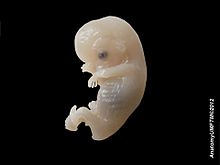
Dissection of human embryo
The cephalic end of the neural groove exhibits several dilatations that, when the tube is closed, assume the form of the three primary brain vesicles, and correspond, respectively, to the future forebrain (prosencephalon), midbrain (mesencephalon), and hindbrain (rhombencephalon) (Fig. 18). The walls of the vesicles are developed into the nervous tissue and neuroglia of the brain, and their cavities are modified to form its ventricles. The remainder of the tube forms the spinal cord (medulla spinalis); from its ectodermal wall the nervous and neuroglial elements of the spinal cord are developed, while the cavity persists as the central canal.[24][9]
Formation of the early septum
The extension of the mesoderm takes place throughout the whole of the embryonic and extra-embryonic areas of the ovum, except in certain regions. One of these is seen immediately in front of the neural tube. Here the mesoderm extends forward in the form of two crescentic masses, which meet in the middle line so as to enclose behind them an area that is devoid of mesoderm. Over this area, the ectoderm and endoderm come into direct contact with each other and constitute a thin membrane, the buccopharyngeal membrane, which forms a septum between the primitive mouth and pharynx.[18][9]
Early formation of the heart and other primitive structures
In front of the buccopharyngeal area, where the lateral crescents of mesoderm fuse in the middle line, the pericardium is afterward developed, and this region is therefore designated the pericardial area. A second region where the mesoderm is absent, at least for a time, is that immediately in front of the pericardial area. This is termed the proamniotic area, and is the region where the proamnion is developed; in humans, however, it appears that a proamnion is never formed. A third region is at the hind end of the embryo, where the ectoderm and endoderm come into apposition and form the cloacal membrane.[18][9]
Somitogenesis is the process by which somites (primitive segments) are produced. These segmented tissue blocks differentiate into skeletal muscle, vertebrae, and dermis of all vertebrates.[25]
Somitogenesis begins with the formation of somitomeres (whorls of concentric mesoderm) marking the future somites in the presomitic mesoderm (unsegmented paraxial). The presomitic mesoderm gives rise to successive pairs of somites, identical in appearance that differentiate into the same cell types but the structures formed by the cells vary depending upon the anteroposterior (e.g., the thoracic vertebrae have ribs, the lumbar vertebrae do not). Somites have unique positional values along this axis and it is thought that these are specified by the Hoxhomeotic genes.[25]
Toward the end of the second week after fertilization, transverse segmentation of the paraxial mesoderm begins, and it is converted into a series of well-defined, more or less cubical masses, also known as the somites, which occupy the entire length of the trunk on either side of the middle line from the occipital region of the head. Each segment contains a central cavity (known as a [myocoel), which, however, is soon filled with angular and spindle-shape cells. The somites lie immediately under the ectoderm on the lateral aspect of the neural tube and notochord, and are connected to the lateral mesoderm by the intermediate cell mass. Those of the trunk may be arranged in the following groups, viz.: cervical 8, thoracic 12, lumbar 5, sacral 5, and coccygeal from 5 to 8. Those of the occipital region of the head are usually described as being four in number. In mammals, somites of the head can be recognized only in the occipital region, but a study of the lower vertebrates leads to the belief that they are present also in the anterior part of the head and that, altogether, nine segments are represented in the cephalic region.[26][25]
At some point after the different germ layers are defined, organogenesis begins. The first stage in vertebrates is called neurulation, where the neural plate folds forming the neural tube (see above).[8] Other common organs or structures that arise at this time include the heart and somites (also above), but from now on embryogenesis follows no common pattern among the different taxa of the animalia.[2]
In most animals organogenesis, along with morphogenesis, results in a larva. The hatching of the larva, which must then undergo metamorphosis, marks the end of embryonic development.[2]
Ontogeny is the origination and development of an organism, usually from the time of fertilization of the egg to adult. The term can also be used to refer to the study of the entirety of an organism's lifespan.
An embryo is an initial stage of development of a multicellular organism. In organisms that reproduce sexually, embryonic development is the part of the life cycle that begins just after fertilization of the female egg cell by the male sperm cell. The resulting fusion of these two cells produces a single-celled zygote that undergoes many cell divisions that produce cells known as blastomeres. The blastomeres are arranged as a solid ball that when reaching a certain size, called a morula, takes in fluid to create a cavity called a blastocoel. The structure is then termed a blastula, or a blastocyst in mammals.
The mesoderm is the middle layer of the three germ layers that develops during gastrulation in the very early development of the embryo of most animals. The outer layer is the ectoderm, and the inner layer is the endoderm.
Blastulation is the stage in early animal embryonic development that produces the blastula. In mammalian development the blastula develops into the blastocyst with a differentiated inner cell mass and an outer trophectoderm. The blastula is a hollow sphere of cells known as blastomeres surrounding an inner fluid-filled cavity called the blastocoel. Embryonic development begins with a sperm fertilizing an egg cell to become a zygote, which undergoes many cleavages to develop into a ball of cells called a morula. Only when the blastocoel is formed does the early embryo become a blastula. The blastula precedes the formation of the gastrula in which the germ layers of the embryo form.
Gastrulation is the stage in the early embryonic development of most animals, during which the blastula, or in mammals the blastocyst, is reorganized into a two-layered or three-layered embryo known as the gastrula. Before gastrulation, the embryo is a continuous epithelial sheet of cells; by the end of gastrulation, the embryo has begun differentiation to establish distinct cell lineages, set up the basic axes of the body, and internalized one or more cell types including the prospective gut.
The ectoderm is one of the three primary germ layers formed in early embryonic development. It is the outermost layer, and is superficial to the mesoderm and endoderm. It emerges and originates from the outer layer of germ cells. The word ectoderm comes from the Greek ektos meaning "outside", and derma meaning "skin".
The blastocyst is a structure formed in the early embryonic development of mammals. It possesses an inner cell mass (ICM) also known as the embryoblast which subsequently forms the embryo, and an outer layer of trophoblast cells called the trophectoderm. This layer surrounds the inner cell mass and a fluid-filled cavity known as the blastocoel. In the late blastocyst, the trophectoderm is known as the trophoblast. The trophoblast gives rise to the chorion and amnion, the two fetal membranes that surround the embryo. The placenta derives from the embryonic chorion and the underlying uterine tissue of the mother.
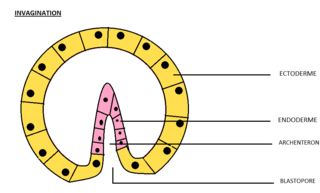
Invagination is the process of a surface folding in on itself to form a cavity, pouch or tube. In developmental biology, invagination is a mechanism that takes place during gastrulation. This mechanism or cell movement happens mostly in the vegetal pole. Invagination consists of the folding of an area of the exterior sheet of cells towards the inside of the blastula. In each organism, the complexity will be different depending on the number of cells. Invagination can be referenced as one of the steps of the establishment of the body plan. The term, originally used in embryology, has been adopted in other disciplines as well.
The blastocoel, also spelled blastocoele and blastocele, and also called cleavage cavity, or segmentation cavity is a fluid-filled or yolk-filled cavity that forms in the blastula during very early embryonic development. At this stage in mammals the blastula develops into the blastocyst containing an inner cell mass, and outer trophectoderm.
The somites are a set of bilaterally paired blocks of paraxial mesoderm that form in the embryonic stage of somitogenesis, along the head-to-tail axis in segmented animals. In vertebrates, somites subdivide into the dermatomes, myotomes, sclerotomes and syndetomes that give rise to the vertebrae of the vertebral column, rib cage, part of the occipital bone, skeletal muscle, cartilage, tendons, and skin.
A germ layer is a primary layer of cells that forms during embryonic development. The three germ layers in vertebrates are particularly pronounced; however, all eumetazoans produce two or three primary germ layers. Some animals, like cnidarians, produce two germ layers making them diploblastic. Other animals such as bilaterians produce a third layer between these two layers, making them triploblastic. Germ layers eventually give rise to all of an animal's tissues and organs through the process of organogenesis.
Organogenesis is the phase of embryonic development that starts at the end of gastrulation and continues until birth. During organogenesis, the three germ layers formed from gastrulation form the internal organs of the organism.
A neurula is a vertebrate embryo at the early stage of development in which neurulation occurs. The neurula stage is preceded by the gastrula stage; consequentially, neurulation is preceded by gastrulation. Neurulation marks the beginning of the process of organogenesis.
The primitive node is the organizer for gastrulation in most amniote embryos. In birds it is known as Hensen's node, and in amphibians it is known as the Spemann-Mangold organizer. It is induced by the Nieuwkoop center in amphibians, or by the posterior marginal zone in amniotes including birds.
In amniote embryonic development, the epiblast is one of two distinct cell layers arising from the inner cell mass in the mammalian blastocyst, or from the blastula in reptiles and birds, the other layer is the hypoblast. It drives the embryo proper through its differentiation into the three primary germ layers, ectoderm, mesoderm and endoderm, during gastrulation. The amniotic ectoderm and extraembryonic mesoderm also originate from the epiblast.
The bilaminar embryonic disc, bilaminar blastoderm or embryonic disc is the distinct two-layered structure of cells formed in an embryo. In the development of the human embryo this takes place by day eight. It is formed when the inner cell mass, also known as the embryoblast, forms a bilaminar disc of two layers, an upper layer called the epiblast and a lower layer called the hypoblast, which will eventually form into fetus. These two layers of cells are stretched between two fluid-filled cavities at either end: the primitive yolk sac and the amniotic sac.
The development of fishes is unique in some specific aspects compared to the development of other animals.
Human embryonic development or human embryogenesis is the development and formation of the human embryo. It is characterised by the processes of cell division and cellular differentiation of the embryo that occurs during the early stages of development. In biological terms, the development of the human body entails growth from a one-celled zygote to an adult human being. Fertilization occurs when the sperm cell successfully enters and fuses with an egg cell (ovum). The genetic material of the sperm and egg then combine to form the single cell zygote and the germinal stage of development commences. Embryonic development in the human, covers the first eight weeks of development; at the beginning of the ninth week the embryo is termed a fetus. The eight weeks has 23 stages.
Embryogenesis in living creatures occurs in different ways depending on class and species. One of the most basic criteria of such development is independence from a water habitat.
This glossary of developmental biology is a list of definitions of terms and concepts commonly used in the study of developmental biology and related disciplines in biology, including embryology and reproductive biology, primarily as they pertain to vertebrate animals and particularly to humans and other mammals. The developmental biology of invertebrates, plants, fungi, and other organisms is treated in other articles; e.g terms relating to the reproduction and development of insects are listed in Glossary of entomology, and those relating to plants are listed in Glossary of botany.
↑ Alberts, Bruce; Johnson, Alexander; Lewis, Julian; Raff, Martin; Roberts, Keith; Walter, Peter (2002). "Fertilization". Archived from the original on 2017-05-14.
1 2 3 4 5 6 What is a cell?Archived 2006-01-18 at the Wayback Machine 2004. A Science Primer: A Basic Introduction to the Science Underlying NCBI Resources. NCBI; and Campbell, Neil A.; Reece, Jane B.; Biology Benjamin Cummings, Pearson Education 2002.
This page is based on this Wikipedia article Text is available under the CC BY-SA 4.0 license; additional terms may apply. Images, videos and audio are available under their respective licenses.