BTLA

Last updated
BTLA
Protein BTLA PDB 2aw2.png
Available structures
PDB Ortholog search: PDBe RCSB
Identifiers
Aliases BTLA , BTLA1, CD272, B and T lymphocyte associated
External IDs OMIM: 607925 MGI: 2658978 HomoloGene: 52233 GeneCards: BTLA
Orthologs
SpeciesHumanMouse
Entrez
Ensembl
UniProt
RefSeq (mRNA)

NM_001085357
NM_181780

NM_001037719
NM_177584

RefSeq (protein)

NP_001078826
NP_861445

NP_001032808
NP_808252

Location (UCSC) Chr 3: 112.46 – 112.5 Mb Chr 16: 45.04 – 45.08 Mb
PubMed search [3] [4]
Wikidata
View/Edit Human View/Edit Mouse

B- and T-lymphocyte attenuator or BTLA (also known as cluster of differentiation 272 or CD272) is a protein that belongs to the CD28 immunoglobulin superfamily (IgSF) which is encoded by the BTLA gene located on the 3rd human chromosome. [5] [6] BTLA was first discovered in 2003 as an inhibitor of Th1 expansion and it became the 3rd member of the CD28 IgSF. However, its discovered ligand herpes virus entry mediator or HVEM (also known as tumour necrosis factor receptor superfamily member 14 or TNFRSF14) belongs to the tumor necrosis factor receptor superfamily (TNFRSF). This finding was surprising because until the discovery of HVEM it was believed that receptors and ligands always belong to the same family. [7] [8]

Contents

Expression

BTLA is broadly expressed in various organs. Among these are the lymph nodes, the thymus and the spleen where high expression of BTLA can be found. On the contrary, low to no expression is detected in organs such as the liver, kidney, heart, brain and other organs. If we consider specific cell populations then the expression of BTLA can be ascertained in T cell, B cells, DCs and NKT cells. In most of the aforementioned cells BTLA expression fluctuates depending on different developmental stages. For instance, in T cells, in which BTLA is most frequently studied, BTLA is stably expressed in naive T cells, the expression subsequently increases when a T cell is stimulated, but once fully activated the expression of BTLA again decreases. [7] [9]

Structure

BTLA is a 289 amino acid long transmembrane glycoprotein. It is very similar in structure to PD-1 and CTLA-4. Therefore, it consists of an extracellular domain, a transmembrane domain and a cytoplasmic domain. The cytoplasmatic domain is indispensable for signalling and it is constituted of 3 important motives: the growth factor receptor-bound protein-2 (Grb-2) recognition motif, the immunoreceptor tyrosine-based inhibitory motif (ITIM) and the immunoreceptor tyrosine-based switch motif (ITSM). [7]

Function

Considering the above-mentioned motives in the cytoplasmatic part of BTLA, it is obvious that BTLA has inhibition and activation capacities. The reason is that through ITIM it recruits either SHP-1 or SHP-2 which act as phosphatases and dephosphorylate tyrosine which suppresses immune activation. On the other hand, it also possesses the Grb-2 recognition motif which, when recognized by a Grb-2 protein, promotes activation of PI3K and that further mediates cell proliferation and survival. [7] [9]

As stated above, HVEM is the main ligand of BTLA but unlike BTLA, HVEM is able to interact with other molecules as well namely CD160 and tumor necrosis factor superfamily member 14 (TNFSF14) also known as LIGHT. Interaction of HVEM and CD160 is inhibitory but interaction with LIGHT is stimulatory. Therefore, HVEM can also induce both stimulation and inhibition. Taken together, it seems that the molecules CD160, BTLA, LIGHT and HVEM form a kind of regulatory network. [7] [8] [9]

T cell

Both BTLA and HVEM are expressed on naive T cells. This colocalization on a single cell allows them to interact in a cis manner (on the same cell). This interaction is important in immunological tolerance as shown by the fact that BTLA mouse knockdown is unable to develop tolerance to high doses of ovalbumin. [7] Furthermore, the knockdown also proves that BTLA is dispensable for T cell development as no abnormalities were detected in T cells development. [9]

When a T cell is fully activated HVEM is internalized and this allows BTLA to interact in a trans manner (with molecules on other cells). [9] This enables other cells who express HVEM to regulate activated T cells. Treg cells express HVEM and are able to facilitate immune suppression through interaction with BTLA. [7] Furthermore, the blockage of BTLA together with PD-1 can help reverting the state of exhaustion which further proves the importance of BTLA as an inhibitory molecule. [9] However, in some cases interaction of BTLA and HVEM can have an opposite effect. For instance, in the case of infection with vaccinia virus BTLA and HVEM interaction promotes survival of CD8+ T cells and production of memory cells. [7]

B cell

B cell's expression of BTLA is less studied than T cell's but it is clear that peripheral B cells express a high amount of BTLA whereas in bone marrow B cell precursors express a lower amount of BTLA. BTLA has been connected with suppressory function in B cells and its expression is not necessary for B cell development just like in T cells. However, a lower expression of BTLA on B cells in elderly is implied in a less potent response to the influenza vaccine. [7] [9]

DC

BTLA can also be found on DCs, with mature DCs having a higher expression of BTLA and immature DCs a lower one. Just like in T cells and B cells, most results show that BTLA and HVEM interaction suppresses the maturation of DCs. [7] [9]

Clinical significance

Tumors

In many cases BTLA expression is connected with unfavourable outcomes as it, for instance, inhibits the function of human CD8+ cancer-specific T cells. [10] For example, this is the case of gallbladder cancer where BTLA+ CD8+ T cells are associated with inhibition of anticancer immunity. Similarly, BTLA+ CD8+ T cells show partial dysfunctionality and decrease secretion of IFN-γ in melanoma patients. Therefore, it seems that BTLA can be used as a novel antitumor therapy target. This notion is supported by the fact that in patients with hepatocarcinoma blocking of BTLA signalling increases the secretion of IFN-γ by CD8+ and CD4+ T cells. [9] However, some studies have shown that, for instance, colorectal carcinoma is associated with lower BTLA expression and that the lower BTLA expression is connected with poor survival. [11] Even in some cases of melanoma BTLA+ CD8+ lymphocytes are suggested to be connected with better outcomes of adoptive cell therapy. Hence, it seems that BTLA has rather a context-specific function. This fact should be taken into account if BTLA is to be used as a cancer therapy target. [9]

Infectious diseases

As in the case of tumors, BTLA seems to be mainly an inhibitory molecule as, for instance, in the case of pulmonary tuberculosis where BTLA expression in CD4+ and CD8+ T cells is connected with the disease progression. [9] Similar results can be seen during helminth infections. When mice are infected by Strongyloides ratti the mice with BTLA knockdown are faster at eliminating the parasite from the intestine probably through the production of IL-9. [12] Furthermore, the course of virus infections seems to be negatively affected by BTLA as well. During chronic hepatitis B infection BTLA impairs T cell response and what's more blocking BTLA leads to T cell proliferation and higher cytokine production. However, as already mentioned in some cases BTLA seems to have a stimulatory role. This is the case of the aforementioned pulmonary tuberculosis where BTLA+ αβ T cells are predominantly of central memory phenotype and fight off Mycobacterium tuberculosis. A further example is an infection with vaccinia virus during which deletion of BTLA or HVEM causes instability and early apoptosis of CD8+ T cells. [9]

Related Research Articles

<span class="mw-page-title-main">Fc receptor</span> Surface protein important to the immune system

In immunology, a Fc receptor is a protein found on the surface of certain cells – including, among others, B lymphocytes, follicular dendritic cells, natural killer cells, macrophages, neutrophils, eosinophils, basophils, human platelets, and mast cells – that contribute to the protective functions of the immune system. Its name is derived from its binding specificity for a part of an antibody known as the Fc region. Fc receptors bind to antibodies that are attached to infected cells or invading pathogens. Their activity stimulates phagocytic or cytotoxic cells to destroy microbes, or infected cells by antibody-mediated phagocytosis or antibody-dependent cell-mediated cytotoxicity. Some viruses such as flaviviruses use Fc receptors to help them infect cells, by a mechanism known as antibody-dependent enhancement of infection.

Co-stimulation is a secondary signal which immune cells rely on to activate an immune response in the presence of an antigen-presenting cell. In the case of T cells, two stimuli are required to fully activate their immune response. During the activation of lymphocytes, co-stimulation is often crucial to the development of an effective immune response. Co-stimulation is required in addition to the antigen-specific signal from their antigen receptors.

<span class="mw-page-title-main">Follicular dendritic cells</span> Immune cells found in lymph nodes

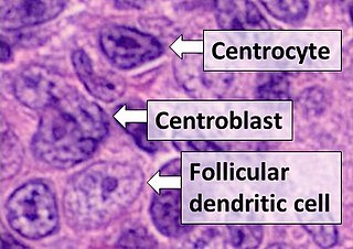
Follicular dendritic cells (FDC) are cells of the immune system found in primary and secondary lymph follicles of the B cell areas of the lymphoid tissue. Unlike dendritic cells (DC), FDCs are not derived from the bone-marrow hematopoietic stem cell, but are of mesenchymal origin. Possible functions of FDC include: organizing lymphoid tissue's cells and microarchitecture, capturing antigen to support B cell, promoting debris removal from germinal centers, and protecting against autoimmunity. Disease processes that FDC may contribute include primary FDC-tumor, chronic inflammatory conditions, HIV-1 infection development, and neuroinvasive scrapie.

<span class="mw-page-title-main">CD80</span> Mammalian protein found in Homo sapiens

The Cluster of differentiation 80 is a B7, type I membrane protein in the immunoglobulin superfamily, with an extracellular immunoglobulin constant-like domain and a variable-like domain required for receptor binding. It is closely related to CD86, another B7 protein (B7-2), and often works in tandem. Both CD80 and CD86 interact with costimulatory receptors CD28, CTLA-4 (CD152) and the p75 neurotrophin receptor.

An immunoreceptor tyrosine-based inhibitory motif (ITIM), is a conserved sequence of amino acids that is found intracellularly in the cytoplasmic domains of many inhibitory receptors of the non-catalytic tyrosine-phosphorylated receptor family found on immune cells. These immune cells include T cells, B cells, NK cells, dendritic cells, macrophages and mast cells. ITIMs have similar structures of S/I/V/LxYxxI/V/L, where x is any amino acid, Y is a tyrosine residue that can be phosphorylated, S is the amino acide Serine, I is the amino acid Isoleucine, and V is the amino acid Valine. ITIMs recruit SH2 domain-containing phosphatases, which inhibit cellular activation. ITIM-containing receptors often serve to target Immunoreceptor tyrosine-based activation motif(ITAM)-containing receptors, resulting in an innate inhibition mechanism within cells. ITIM bearing receptors have important role in regulation of immune system allowing negative regulation at different levels of the immune response.

<span class="mw-page-title-main">CD5 (protein)</span> Protein-coding gene in the species Homo sapiens

CD5 is a cluster of differentiation expressed on the surface of T cells and in a subset of murine B cells known as B-1a. The expression of this receptor in human B cells has been a controversial topic and to date there is no consensus regarding the role of this receptor as a marker of human B cells. B-1 cells have limited diversity of their B-cell receptor due to their lack of the enzyme terminal deoxynucleotidyl transferase (TdT) and are potentially self-reactive. CD5 serves to mitigate activating signals from the BCR so that the B-1 cells can only be activated by very strong stimuli and not by normal tissue proteins. CD5 was used as a T-cell marker until monoclonal antibodies against CD3 were developed.

<span class="mw-page-title-main">PD-L1</span> Mammalian protein found in Homo sapiens

Programmed death-ligand 1 (PD-L1) also known as cluster of differentiation 274 (CD274) or B7 homolog 1 (B7-H1) is a protein that in humans is encoded by the CD274 gene.

<span class="mw-page-title-main">LILRB1</span> Protein-coding gene in the species Homo sapiens

Leukocyte immunoglobulin-like receptor subfamily B member 1 is a protein that in humans is encoded by the LILRB1 gene.

<span class="mw-page-title-main">CD83</span>

CD83 is a human protein encoded by the CD83 gene.

<span class="mw-page-title-main">CD69</span>

CD69 is a human transmembrane C-Type lectin protein encoded by the CD69 gene. It is an early activation marker that is expressed in hematopoietic stem cells, T cells, and many other cell types in the immune system. It is also implicated in T cell differentiation as well as lymphocyte retention in lymphoid organs.

<span class="mw-page-title-main">CD244</span> Protein found in humans

CD244 also known as 2B4 or SLAMF4 is a protein that in humans is encoded by the CD244 gene.

<span class="mw-page-title-main">CD226</span> Protein-coding gene in the species Homo sapiens

CD226, PTA1 or DNAM-1 is a ~65 kDa immunoglobulin-like transmembrane glycoprotein expressed on the surface of natural killer cells, NK T cell, B cells, dendritic cells, hematopoietic precursor cells, platelets, monocytes and T cells.

<span class="mw-page-title-main">CD200</span> Protein-coding gene in the species Homo sapiens

OX-2 membrane glycoprotein, also named CD200 is a human protein encoded by the CD200 gene. CD200 gene is in human located on chromosome 3 in proximity to genes encoding other B7 proteins CD80/CD86. In mice CD200 gene is on chromosome 16.

<span class="mw-page-title-main">HAVCR2</span> Protein-coding gene in the species Homo sapiens

Hepatitis A virus cellular receptor 2 (HAVCR2), also known as T-cell immunoglobulin and mucin-domain containing-3 (TIM-3), is a protein that in humans is encoded by the HAVCR2 (TIM-3)gene. HAVCR2 was first described in 2002 as a cell surface molecule expressed on IFNγ producing CD4+ Th1 and CD8+ Tc1 cells. Later, the expression was detected in Th17 cells, regulatory T-cells, and innate immune cells. HAVCR2 receptor is a regulator of the immune response.

<span class="mw-page-title-main">KLRG1</span> Protein-coding gene in humans

Killer cell lectin-like receptor subfamily G member 1 is a protein that in humans is encoded by the KLRG1 gene.

<span class="mw-page-title-main">FCGR2B</span>

Fc fragment of IgG receptor IIb is a low affinity inhibitory receptor for the Fc region of immunoglobulin gamma (IgG). FCGR2B participates in the phagocytosis of immune complexes and in the regulation of antibody production by B lymphocytes.

<span class="mw-page-title-main">FCAR</span> Mammalian protein found in Homo sapiens

Fc fragment of IgA receptor (FCAR) is a human gene that codes for the transmembrane receptor FcαRI, also known as CD89. FcαRI binds the heavy-chain constant region of Immunoglobulin A (IgA) antibodies. FcαRI is present on the cell surface of myeloid lineage cells, including neutrophils, monocytes, macrophages, and eosinophils, though it is notably absent from intestinal macrophages and does not appear on mast cells. FcαRI plays a role in both pro- and anti-inflammatory responses depending on the state of IgA bound. Inside-out signaling primes FcαRI in order for it to bind its ligand, while outside-in signaling caused by ligand binding depends on FcαRI association with the Fc receptor gamma chain.

<span class="mw-page-title-main">NKG2D</span> Protein-coding gene in the species Homo sapiens

NKG2D is an activating receptor (transmembrane protein) belonging to the NKG2 family of C-type lectin-like receptors. NKG2D is encoded by KLRK1 (killer cell lectin like receptor K1) gene which is located in the NK-gene complex (NKC) situated on chromosome 6 in mice and chromosome 12 in humans. In mice, it is expressed by NK cells, NK1.1+ T cells, γδ T cells, activated CD8+ αβ T cells and activated macrophages. In humans, it is expressed by NK cells, γδ T cells and CD8+ αβ T cells. NKG2D recognizes induced-self proteins from MIC and RAET1/ULBP families which appear on the surface of stressed, malignant transformed, and infected cells.

<span class="mw-page-title-main">Immune checkpoint</span> Regulators of the immune system

Immune checkpoints are regulators of the immune system. These pathways are crucial for self-tolerance, which prevents the immune system from attacking cells indiscriminately. However, some cancers can protect themselves from attack by stimulating immune checkpoint targets.

<span class="mw-page-title-main">CD28 family receptor</span> Group of regulatory cell surface receptors

CD28 family receptors are a group of regulatory cell surface receptors expressed on immune cells. The CD28 family in turn is a subgroup of the immunoglobulin superfamily.

References

  1. 1 2 3 GRCh38: Ensembl release 89: ENSG00000186265 - Ensembl, May 2017
  2. 1 2 3 GRCm38: Ensembl release 89: ENSMUSG00000052013 - Ensembl, May 2017
  3. "Human PubMed Reference:". National Center for Biotechnology Information, U.S. National Library of Medicine.
  4. "Mouse PubMed Reference:". National Center for Biotechnology Information, U.S. National Library of Medicine.
  5. "Entrez Gene: BTLA B and T lymphocyte associated".
  6. Watanabe N, Gavrieli M, Sedy JR, Yang J, Fallarino F, Loftin SK, et al. (July 2003). "BTLA is a lymphocyte inhibitory receptor with similarities to CTLA-4 and PD-1". Nature Immunology. 4 (7): 670–9. doi:10.1038/ni944. PMID   12796776. S2CID   11943145.
  7. 1 2 3 4 5 6 7 8 9 10 Yu X, Zheng Y, Mao R, Su Z, Zhang J (2019). "BTLA/HVEM Signaling: Milestones in Research and Role in Chronic Hepatitis B Virus Infection". Frontiers in Immunology. 10: 617. doi: 10.3389/fimmu.2019.00617 . PMC   6449624 . PMID   30984188.
  8. 1 2 Pasero C, Olive D (March 2013). "Interfering with coinhibitory molecules: BTLA/HVEM as new targets to enhance anti-tumor immunity". Immunology Letters. 151 (1–2): 71–5. doi:10.1016/j.imlet.2013.01.008. PMID   23439006.
  9. 1 2 3 4 5 6 7 8 9 10 11 12 Ning Z, Liu K, Xiong H (2021-03-29). "Roles of BTLA in Immunity and Immune Disorders". Frontiers in Immunology. 12: 654960. doi: 10.3389/fimmu.2021.654960 . PMC   8043046 . PMID   33859648.
  10. Derré L, Rivals JP, Jandus C, Pastor S, Rimoldi D, Romero P, et al. (January 2010). "BTLA mediates inhibition of human tumor-specific CD8+ T cells that can be partially reversed by vaccination". The Journal of Clinical Investigation. 120 (1): 157–67. doi:10.1172/JCI40070. PMC   2799219 . PMID   20038811.| *Lay summary in: "Researchers Identify Protein that Inhibits Tumor-Targeting Immune Cells When Activated". Genetic Engineering & Biotechnology News. December 29, 2009.
  11. Song J, Wu L (2020). "Friend or Foe: Prognostic and Immunotherapy Roles of BTLA in Colorectal Cancer". Frontiers in Molecular Biosciences. 7: 148. doi: 10.3389/fmolb.2020.00148 . PMC   7385242 . PMID   32793631.
  12. Breloer M, Hartmann W, Blankenhaus B, Eschbach ML, Pfeffer K, Jacobs T (February 2015). "Cutting Edge: the BTLA-HVEM regulatory pathway interferes with protective immunity to intestinal Helminth infection". Journal of Immunology. 194 (4): 1413–6. doi: 10.4049/jimmunol.1402510 . PMID   25595777.

Further reading

This article incorporates text from the United States National Library of Medicine, which is in the public domain.