Central serous chorioretinopathy

Last updated
Central serous chorioretinopathy
Central serous retinopathy.jpg
An occurrence of central serous retinopathy of the fovea centralis imaged using optical coherence tomography.
Specialty Ophthalmology   OOjs UI icon edit-ltr-progressive.svg

Central serous chorioretinopathy (CSC or CSCR), also known as central serous retinopathy (CSR), is an eye disease that causes visual impairment, often temporary, usually in one eye. [1] [2] When the disorder is active it is characterized by leakage of fluid under the retina that has a propensity to accumulate under the central macula. This results in blurred or distorted vision (metamorphopsia). A blurred or gray spot in the central visual field is common when the retina is detached. Reduced visual acuity may persist after the fluid has disappeared. [1]

Contents

The disease is considered of unknown cause. It mostly affects white males in the age group 20 to 50 (male:female ratio 6:1) [3] and occasionally other groups. The condition is believed to be exacerbated by stress or corticosteroid use. [4]

Pathophysiology

Recently, central serous chorioretinopathy has been understood to be part of the pachychoroid spectrum. [5] [6] In pachychoroid spectrum disorders, of which CSR represents stage II, the choroid, the highly vascularized layer below the retina, is thickened and congested with increased blood vessel diameter, especially in the deep choroid (the so-called Haller's layer). This results in increased pressure from the deep choroid against the superficial choroid close to the retina, damaging the fine blood vessels (capillaries) needed to supply oxygen and nutrients to the retinal pigment epithelium and retina. Additionally, fluid can leak from these damaged vessels and accumulate under the retina.

Different stages of the pachychoroid are defined depending on the amount of cumulative damage. [5] [6] If there are defects in the retinal pigment epithelium without accumulation of fluid below the retina, a pachychoroid pigmentepitheliopathy (PPE) is present. Accumulation of fluid results in central serous chorioretinopathy (CSR). The development of secondary blood vessels, so-called choroidal neovascularization (CNV) leads to pachychoroid neovasculopathy (PNV). If parts of these new vessels bulge outward, so-called aneurysms develop within this CNV, defining pachychoroid aneurysmal type 1 CNV (or, still widely used, polypoidal choroidal vasculopathy (PCV)).

Since the individual stages develop one after the other from the respective preliminary stage, pachychoroidal diseases of the macula are divided into 4 stages according to Siedlecki, Schworm and Priglinger: [7]

Pachychoroid spectrum disorders of the macula (after Siedlecki et al. [7] )
0Uncomplicated pachychoroid (UCP)
IPachychoroid pigmentepitheliopathy (PPE)
IICentral serous chorioretinopathy (CSC)
IIIPachychoroid neovasculopathy (PNV)
a) with neurosensory detachment (subretinal fluid)
b) without neurosensory detachment (no subretinal fluid)
IVPachychoroid aneurysmal type 1 choroidal neovascularization (PAT1)

(also polypoidal choroidal vasculopathy, PCV)

Risk factors

CSR is sometimes called idiopathic CSR which means that its cause is unknown. Nevertheless, stress appears to play an important role. An oft-cited but potentially inaccurate conclusion is that persons in stressful occupations, such as airplane pilots, have a higher incidence of CSR.

CSR has also been associated with cortisol and corticosteroids. Persons with CSR have higher levels of cortisol. [8] Cortisol is a hormone secreted by the adrenal cortex which allows the body to deal with stress, which may explain the CSR-stress association. There is extensive evidence to the effect that corticosteroids (e.g. cortisone), commonly used to treat inflammations, allergies, skin conditions and even certain eye conditions, can trigger CSR, aggravate it and cause relapses. [9] [10] [11] In a case report, a young male was using Prednisolone and began to display subretinal fluid indicative of CSR. With the discontinuation of the steroid drop the subretinal fluid resolved and did not show any sign of recurrence. Thus indicating the steroid was the probable cause of the CSR. [12] A study of 60 persons with Cushing's syndrome found CSR in 3 (5%). [13] Cushing's syndrome is characterized by very high cortisol levels. Certain sympathomimetic drugs have also been associated with causing the disease. [14]

Evidence has also implicated helicobacter pylori (see gastritis) as playing a role. [15] [16] It would appear that the presence of the bacteria is well correlated with visual acuity and other retinal findings following an attack.

Evidence also shows that people with MPGN type II kidney disease can develop retinal abnormalities including CSR caused by deposits of the same material that originally damaged the glomerular basement membrane in the kidneys. [17]

Diagnosis

Optical coherence tomography imaging of central serous retinopathy Central serous chorioretinopathy.jpg
Optical coherence tomography imaging of central serous retinopathy
Indocyanine green angiography (left) and laser Doppler imaging (right) of the macula in central serous retinopathy, revealing choroidal vessels. Blue and red correspond to low and high blood flow respectively. ICG vs LDH in CSCR.jpg
Indocyanine green angiography (left) and laser Doppler imaging (right) of the macula in central serous retinopathy, revealing choroidal vessels. Blue and red correspond to low and high blood flow respectively.

The diagnosis usually starts with a dilated examination of the retina, followed with confirmation by optical coherence tomography and fluorescein angiography. The angiography test will usually show one or more fluorescent spots with fluid leakage. In 10%-15% of the cases these will appear in a "classic" smokestack shape.[ citation needed ] Differential diagnosis should be immediately performed to rule out retinal detachment, which is a medical emergency. A clinical record should be taken to keep a timeline of the detachment. The affected eye will sometimes exhibit a refractive spectacle prescription that is more far-sighted than the fellow eye due to the decreased focal length caused by the raising of the retina.

Indocyanine green angiography or laser Doppler imaging can be used to reveal the underlying swollen choroidal vessels under the retinal pigment epithelium and assess the health of the retina in the affected area which can be useful in making a treatment decision.

Treatment

Any ongoing corticosteroid treatment should be tapered and stopped, where possible. It is important to check current medication, including nasal sprays and creams, for ingredients of corticosteroids, if found seek advice from a medical practitioner for an alternative.

Most eyes with CSR undergo spontaneous resorption of subretinal fluid within 3–4 months. Recovery of visual acuity usually follows. Treatment should be considered if resorption does not occur within 3–4 months, [19] spontaneously or as the result of counselling. [1] The available evidence suggests that half-dose (or half-fluence) photodynamic therapy is the treatment of choice for CSR with subretinalfluid for longer than 3–4 months. [20]

Due to the natural disease course of CSR - in which spontaneous resolution of subretinal fluid may occur - retrospective studies may erroneously report positive treatment outcomes and should, therefore, be evaluated with caution.

Laser treatments

Full-dose photodynamic therapy (PDT) with verteporfin was first described in CSR in 2003. [21] Later, reduced-settings PDT (half-dose, half-fluence, and half-time) was found to have the same efficacy and a lower chance of complications. Follow-up studies have confirmed the treatment's long-term effectiveness [22] including its effectiveness for the chronic variant of the disease. [23] In the PLACE trial, half-dose photodynamic therapy was found to be superior compared to high-density subthreshold micropulse laser, both with regard to anatomical and functional outcomes. [24] Indocyanine green angiography can be used to predict how the patient will respond to PDT. [19] [25]

Laser photocoagulation, which effectively burns the leak area shut, may be considered in cases where there is little improvement in a 3- to 4-month duration, and the leakage is confined to a single or a few sources of leakage at a safe distance from the fovea. Laser photocoagulation is not indicated for cases where the leak is very near the central macula or for cases where the leakage is widespread and its source is difficult to identify. Laser photocoagulation can permanently damage vision where applied. Carefully tuned lasers can limit this damage. [26] Even so, laser photocoagulation is not a preferred treatment for leaks in the central vision and is considered an outdated treatment by some doctors. [19] Foveal attenuation has been associated with more than 4 months' duration of symptoms, however a better long-term outcome has not been demonstrated with laser photocoagulation than without photocoagulation. [1]

In chronic cases, transpupillary thermotherapy has been suggested as an alternative to laser photocoagulation where the leak is in the central macula. [27]

Yellow micropulse laser has shown promise in very limited retrospective trials. [4]

A Cochrane review seeking to compare the effectiveness of various treatment for CSR found low quality evidence that half-dose PDT treatment resulted in improved visual acuity and less recurrence of CSR in patients with acute CSR, compared to patients in the control group. [28] The review also found benefits in micropulse laser treatments, where patients with acute and chronic CSR had improved visual acuity compared to control patients. [28]

Oral medications

Spironolactone is a mineralocorticoid receptor antagonist that may help reduce the fluid associated with CSR. In a retrospective study noted by Acta Ophthalmologica, spironolactone improved visual acuity in CSR patients over the course of 8 weeks. [29]

Eplerenone is another mineralocorticoid receptor antagonist that has been thought to reduce the subretinal fluid that is present with CSR. In a study noted in International Journal of Ophthalmology, results showed Epleronone decreased the subretinal fluid both horizontally and vertically over time. [30] However, the most recent and largest randomized controlled trial showed that eplerenone has no significant effect on chronic CSR. [31] [32]

Low dosage ibuprofen has been shown to quicken recovery in some cases. [33]

Topical treatment

Though no topical treatment has been proven to be effective in the treatment of CSR. Some doctors have attempted to use nonsteroidal topical medications to reduce the subretinal fluid associated with CSR. The nonsteroidal topical medications that are sometimes used to treat CSR are, Ketorolac, Diclofenac, or Bromfenac. [34]

Lifestyle changes

People who have irregular sleep patterns, type A personalities, sleep apnea, or systemic hypertension are more susceptible to CSR, as stated in Medscape. "The pathogenesis here is thought to be elevated circulating cortisol and epinephrine, which affect the autoregulation of the choroidal circulation." [35] With management of these lifestyle patterns and associated cortisol and epinephrine levels, it has been shown that the fluid associated with CSR can spontaneously resolve. Melatonin has been shown to help regulate sleep in people who have irregular sleep patterns (such as 3rd shift workers, or overnight employees), in turn, better regulating cortisol and epinephrine levels to manage CSR.

Prognosis

The prognosis for CSR is generally excellent. While immediate vision loss may be as poor as 20/200 in the affected eye, clinically, over 90% of patients regain 20/25 vision or better within 45 days. [1] Once the fluid has resolved, either spontaneously or through treatment, distortion is reduced and visual acuity improves as the eye heals. However, some visual abnormalities can remain even where visual acuity is measured at 20/20. This includes localized reductions in light sensitivity as assessed by visual field testing (microperimetry). [36] Lasting problems include decreased night vision, reduced color discrimination, and localized distortion caused by scarring of the sub-retinal layers. [37]

Complications include subretinal neovascularization and pigment epithelial detachment. [38]

The disease can re-occur causing progressive vision loss. There is also a chronic form, titled as type II central serous retinopathy, which occurs in approximately 5% of cases. This exhibits diffuse rather than localized abnormality of the pigment epithelium, producing a persistent subretinal fluid. The serous fluid in these cases tends to be shallow rather than dome shaped. The prognosis for this condition is less favorable and continued clinical consultation is advised.[ citation needed ]

See also

Related Research Articles

<span class="mw-page-title-main">Retinopathy</span> Medical condition

Retinopathy is any damage to the retina of the eyes, which may cause vision impairment. Retinopathy often refers to retinal vascular disease, or damage to the retina caused by abnormal blood flow. Age-related macular degeneration is technically included under the umbrella term retinopathy but is often discussed as a separate entity. Retinopathy, or retinal vascular disease, can be broadly categorized into proliferative and non-proliferative types. Frequently, retinopathy is an ocular manifestation of systemic disease as seen in diabetes or hypertension. Diabetes is the most common cause of retinopathy in the U.S. as of 2008. Diabetic retinopathy is the leading cause of blindness in working-aged people. It accounts for about 5% of blindness worldwide and is designated a priority eye disease by the World Health Organization.

<span class="mw-page-title-main">Diabetic retinopathy</span> Medical condition

Diabetic retinopathy, is a medical condition in which damage occurs to the retina due to diabetes. It is a leading cause of blindness in developed countries.

<span class="mw-page-title-main">Vitrectomy</span> Surgery to remove vitreous humor from the eye

Vitrectomy is a surgery to remove some or all of the vitreous humor from the eye.

<span class="mw-page-title-main">Retinoschisis</span> Eye disease involving splitting of the retina

Retinoschisis is an eye disease characterized by the abnormal splitting of the retina's neurosensory layers, usually in the outer plexiform layer. Retinoschisis can be divided into degenerative forms which are very common and almost exclusively involve the peripheral retina and hereditary forms which are rare and involve the central retina and sometimes the peripheral retina. The degenerative forms are asymptomatic and involve the peripheral retina only and do not affect the visual acuity. Some rarer forms result in a loss of vision in the corresponding visual field.

<span class="mw-page-title-main">Macular edema</span> Medical condition

Macular edema occurs when fluid and protein deposits collect on or under the macula of the eye and causes it to thicken and swell (edema). The swelling may distort a person's central vision, because the macula holds tightly packed cones that provide sharp, clear, central vision to enable a person to see detail, form, and color that is directly in the centre of the field of view.

<span class="mw-page-title-main">Retinal detachment</span> Medical condition of the eye

Retinal detachment is a disorder of the eye in which the retina peels away from its underlying layer of support tissue. Initial detachment may be localized, but without rapid treatment the entire retina may detach, leading to vision loss and blindness. It is a surgical emergency.

<span class="mw-page-title-main">Macular degeneration</span> Medical condition associated with vision loss

Macular degeneration, also known as age-related macular degeneration, is a medical condition which may result in blurred or no vision in the center of the visual field. Early on there are often no symptoms. Over time, however, some people experience a gradual worsening of vision that may affect one or both eyes. While it does not result in complete blindness, loss of central vision can make it hard to recognize faces, drive, read, or perform other activities of daily life. Visual hallucinations may also occur.

<span class="mw-page-title-main">Optic disc drusen</span> Medical condition

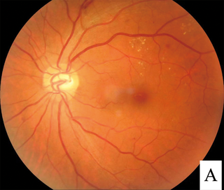
Optic disc drusen (ODD) are globules of mucoproteins and mucopolysaccharides that progressively calcify in the optic disc. They are thought to be the remnants of the axonal transport system of degenerated retinal ganglion cells. ODD have also been referred to as congenitally elevated or anomalous discs, pseudopapilledema, pseudoneuritis, buried disc drusen, and disc hyaline bodies.

<span class="mw-page-title-main">Optic pit</span> Medical condition

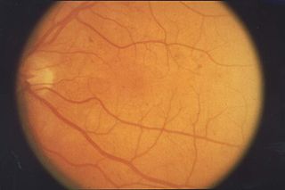
Optic pit, optic nerve pit, or optic disc pit (ODP) is rare a congenital excavation (or regional depression) of the optic disc (also optic nerve head), resulting from a malformation during development of the eye. The incidence of ODP is 1 in 10,000 people with no predilection for either gender. There is currently no known risk factors for their development. Optic pits are important because they are associated with posterior vitreous detachments (PVD) and even serous retinal detachments.

<span class="mw-page-title-main">Angioid streaks</span> Medical condition

Angioid streaks, also called Knapp streaks or Knapp striae, are small breaks in Bruch's membrane, an elastic tissue containing membrane of the retina that may become calcified and crack. Up to 50% of angioid streak cases are idiopathic. It may occur secondary to blunt trauma, or it may be associated with many systemic diseases. The condition is usually asymptomatic, but decrease in vision may occur due to choroidal neovascularization.

<span class="mw-page-title-main">Intraocular hemorrhage</span> Medical condition

Intraocular hemorrhage is bleeding inside the eye. Bleeding can occur from any structure of the eye where there is vasculature or blood flow, including the anterior chamber, vitreous cavity, retina, choroid, suprachoroidal space, or optic disc.

<span class="mw-page-title-main">Macular telangiectasia</span> Disease of the retina affecting central vision

Macular telangiectasia is a condition of the retina, the light-sensing tissue at the back of the eye that causes gradual deterioration of central vision, interfering with tasks such as reading and driving.

<span class="mw-page-title-main">Laser coagulation</span> Procedure widely used in eye surgery

Laser coagulation or laser photocoagulation surgery is used to treat a number of eye diseases and has become widely used in recent decades. During the procedure, a laser is used to finely cauterize ocular blood vessels to attempt to bring about various therapeutic benefits.

<span class="mw-page-title-main">Familial exudative vitreoretinopathy</span> Retinal vascular disease

Familial exudative vitreoretinopathy is a genetic disorder affecting the growth and development of blood vessels in the retina of the eye. This disease can lead to visual impairment and sometimes complete blindness in one or both eyes. FEVR is characterized by incomplete vascularization of the peripheral retina. This can lead to the growth of new blood vessels which are prone to leakage and hemorrhage and can cause retinal folds, tears, and detachments. Treatment involves laser photocoagulation of the avascular portions of the retina to reduce new blood vessel growth and risk of complications including leakage of retinal blood vessels and retinal detachments.

<span class="mw-page-title-main">Radiation retinopathy</span> Medical condition

Radiation retinopathy is damage to retina due to exposure to ionizing radiation. Radiation retinopathy has a delayed onset, typically after months or years of radiation, and is slowly progressive. In general, radiation retinopathy is seen around 18 months after treatment with external-beam radiation and with brachytherapy. The time of onset of radiation retinopathy is between 6 months to 3 years.

Joan Whitten Miller is a Canadian-American ophthalmologist and scientist who has made notable contributions to the treatment and understanding of eye disorders. She is credited for developing photodynamic therapy (PDT) with verteporfin (Visudyne), the first pharmacologic therapy for retinal disease. She also co-discovered the role of vascular endothelial growth factor (VEGF) in eye disease and demonstrated the therapeutic potential of VEGF inhibitors, forming the scientific basis of anti-VEGF therapy for age-related macular degeneration (AMD), diabetic retinopathy, and related conditions.

<span class="mw-page-title-main">Pachychoroid disorders of the macula</span>

Pachychoroid disorders of the macula represent a group of diseases affecting the central part of the retina of the eye, the macula. Due to thickening and congestion of the highly vascularized layer underneath the macula, the choroid, damage to the retinal pigment epithelium and the retinal photoreceptor cells ensues. This leads to impaired vision. The best known representative of the pachychoroid disease spectrum, central serous chorioretinopathy, is the fourth most common cause of irreversible damage to the macula:.

<span class="mw-page-title-main">Indocyanine green angiography</span> Diagnostic procedure

Indocyanine green angiography (ICGA) is a diagnostic procedure used to examine choroidal blood flow and associated pathology. Indocyanine green (ICG) is a water soluble cyanine dye which shows fluorescence in near-infrared (790–805 nm) range, with peak spectral absorption of 800-810 nm in blood. The near infrared light used in ICGA penetrates ocular pigments such as melanin and xanthophyll, as well as exudates and thin layers of sub-retinal vessels. Age-related macular degeneration is the third main cause of blindness worldwide, and it is the leading cause of blindness in industrialized countries. Indocyanine green angiography is widely used to study choroidal neovascularization in patients with exudative age-related macular degeneration. In nonexudative AMD, ICGA is used in classification of drusen and associated subretinal deposits.

Sickle cell retinopathy can be defined as retinal changes due to blood vessel damage in the eye of a person with a background of sickle cell disease. It can likely progress to loss of vision in late stages due to vitreous hemorrhage or retinal detachment. Sickle cell disease is a structural red blood cell disorder leading to consequences in multiple systems. It is characterized by chronic red blood cell destruction, vascular injury, and tissue ischemia causing damage to the brain, eyes, heart, lungs, kidneys, spleen, and musculoskeletal system.

Polypoidal choroidal vasculopathy (PCV) is an eye disease primarily affecting the choroid. It may cause sudden blurring of vision or a scotoma in the central field of vision. Since Indocyanine green angiography gives better imaging of choroidal structures, it is more preferred in diagnosing PCV. Treatment options of PCV include careful observation, photodynamic therapy, thermal laser, intravitreal injection of anti-VEGF therapy, or combination therapy.

References

  1. 1 2 3 4 5 Wang M, Munch IC, Hasler PW, Prünte C, Larsen M (March 2008). "Central serous chorioretinopathy". Acta Ophthalmologica. 86 (2): 126–45. doi: 10.1111/j.1600-0420.2007.00889.x . PMID   17662099. S2CID   42537355.
  2. Quillen DA, Gass DM, Brod RD, Gardner TW, Blankenship GW, Gottlieb JL (January 1996). "Central serous chorioretinopathy in women". Ophthalmology. 103 (1): 72–9. doi:10.1016/s0161-6420(96)30730-6. PMID   8628563.
  3. Sartini F, Figus M, Nardi M, Casini G, Posarelli C (July 2019). "Non-resolving, recurrent and chronic central serous chorioretinopathy: available treatment options". Eye. 33 (7): 1035–1043. doi:10.1038/s41433-019-0381-7. PMC   6707196 . PMID   30824822.
  4. 1 2 André Maia (February 2010). "A New Treatment for Chronic Central Serous Retinopathy". Retina Today. Archived from the original on 2012-08-09. Retrieved 2013-08-11.{{cite journal}}: Cite journal requires |journal= (help)
  5. 1 2 Cheung CM, Lee WK, Koizumi H, Dansingani K, Lai TY, Freund KB (January 2019). "Pachychoroid disease". Eye. 33 (1): 14–33. doi:10.1038/s41433-018-0158-4. PMC   6328576 . PMID   29995841.
  6. 1 2 Akkaya S (October 2018). "Spectrum of pachychoroid diseases". International Ophthalmology. 38 (5): 2239–2246. doi:10.1007/s10792-017-0666-4. PMID   28766279. S2CID   4022900.
  7. 1 2 Siedlecki J, Schworm B, Priglinger SG (December 2019). "The Pachychoroid Disease Spectrum-and the Need for a Uniform Classification System". Ophthalmology. Retina. 3 (12): 1013–1015. doi:10.1016/j.oret.2019.08.002. PMID   31810570. S2CID   208813687.
  8. Garg SP, Dada T, Talwar D, Biswas NR (November 1997). "Endogenous cortisol profile in patients with central serous chorioretinopathy". The British Journal of Ophthalmology. 81 (11): 962–4. doi:10.1136/bjo.81.11.962. PMC   1722041 . PMID   9505819.
  9. Pizzimenti JJ, Daniel KP (August 2005). "Central serous chorioretinopathy after epidural steroid injection". Pharmacotherapy. 25 (8): 1141–6. doi:10.1592/phco.2005.25.8.1141. PMID   16207106. S2CID   23535867.
  10. Bevis T, Ratnakaram R, Smith MF, Bhatti MT (August 2005). "Visual loss due to central serous chorioretinopathy during corticosteroid treatment for giant cell arteritis". Clinical & Experimental Ophthalmology. 33 (4): 437–9. doi:10.1111/j.1442-9071.2005.01017.x. PMID   16033370. S2CID   1224694.
  11. Fernández Hortelano A, Sádaba LM, Heras Mulero H, García Layana A (April 2005). "Coroidopatía serosa central como complicación de epitelitis en tratamiento con corticoides" [Central serous chorioretinopathy as a complication of epitheliopathy under treatment with glucocorticoids]. Archivos de la Sociedad Española de Oftalmología. 80 (4): 255–8. doi: 10.4321/S0365-66912005000400010 . PMID   15852168.
  12. Shah SP, Desai CK, Desai MK, Dikshit RK (September 2011). "Steroid-induced central serous retinopathy". Indian Journal of Pharmacology. 43 (5): 607–8. doi: 10.4103/0253-7613.84985 . PMC   3195140 . PMID   22022013.
  13. Bouzas EA, Scott MH, Mastorakos G, Chrousos GP, Kaiser-Kupfer MI (September 1993). "Central serous chorioretinopathy in endogenous hypercortisolism". Archives of Ophthalmology. 111 (9): 1229–33. doi:10.1001/archopht.1993.01090090081024. PMID   8363466.
  14. Michael JC, Pak J, Pulido J, de Venecia G (July 2003). "Central serous chorioretinopathy associated with administration of sympathomimetic agents". American Journal of Ophthalmology. 136 (1): 182–5. doi:10.1016/S0002-9394(03)00076-X. PMID   12834690.
  15. Ahnoux-Zabsonre A, Quaranta M, Mauget-Faÿsse M (December 2004). "[Prevalence of Helicobacter pylori in central serous chorioretinopathy and diffuse retinal epitheliopathy: a complementary study]" [Prevalence of Helicobacter pylori in central serous chorioretinopathy and diffuse retinal epitheliopathy: a complementary study]. Journal Français d'Ophtalmologie (in French). 27 (10): 1129–33. doi:10.1016/S0181-5512(04)96281-X. PMID   15687922.
  16. Cotticelli L, Borrelli M, D'Alessio AC, Menzione M, Villani A, Piccolo G, et al. (2006). "Central serous chorioretinopathy and Helicobacter pylori". European Journal of Ophthalmology. 16 (2): 274–8. doi:10.1177/112067210601600213. PMID   16703546. S2CID   37258065.
  17. Colville D, Guymer R, Sinclair RA, Savige J (August 2003). "Visual impairment caused by retinal abnormalities in mesangiocapillary (membranoproliferative) glomerulonephritis type II ("dense deposit disease")". American Journal of Kidney Diseases. 42 (2): E2-5. doi:10.1016/S0272-6386(03)00665-6. PMID   12900843.
  18. Puyo, Léo, Michel Paques, Mathias Fink, José-Alain Sahel, and Michael Atlan. "Choroidal vasculature imaging with laser Doppler holography." Biomedical optics express 10, no. 2 (2019): 995-1012.
  19. 1 2 3 Boscia F (April 2010). "When to Treat and Not to Treat Patients With Central Serous Retinopathy". Retina Today. Archived from the original on 2012-10-17.
  20. van Rijssen, TJ (2019). "Central serous chorioretinopathy: Towards an evidence-based treatment guideline". Prog Retin Eye Res. 73: 100770. doi: 10.1016/j.preteyeres.2019.07.003 . hdl: 2066/215584 . PMID   31319157.
  21. Yannuzzi, L. A. (2003). "Indocyanine green angiography-guided photodynamic therapy for treatment of chronic central serous chorioretinopathy: a pilot study". Retina. 23 (3): 288–98. doi:10.1097/00006982-200306000-00002. PMID   12824827.
  22. Chan WM, Lai TY, Lai RY, Liu DT, Lam DS (October 2008). "Half-dose verteporfin photodynamic therapy for acute central serous chorioretinopathy: one-year results of a randomized controlled trial". Ophthalmology. 115 (10): 1756–65. doi:10.1016/j.ophtha.2008.04.014. PMID   18538401.
  23. Karakus SH, Basarir B, Pinarci EY, Kirandi EU, Demirok A (May 2013). "Long-term results of half-dose photodynamic therapy for chronic central serous chorioretinopathy with contrast sensitivity changes". Eye. 27 (5): 612–20. doi:10.1038/eye.2013.24. PMC   3650272 . PMID   23519277.
  24. van Dijk, Elon (2018-05-20). "Half-Dose Photodynamic Therapy versus High-Density Subthreshold Micropulse Laser Treatment in Patients with Chronic Central Serous Chorioretinopathy: The PLACE Trial". Ophthalmology. 125 (10): 1547–1555. doi: 10.1016/j.ophtha.2018.04.021 . hdl: 2066/196622 . PMID   29776672.
  25. Inoue R, Sawa M, Tsujikawa M, Gomi F (March 2010). "Association between the efficacy of photodynamic therapy and indocyanine green angiography findings for central serous chorioretinopathy". American Journal of Ophthalmology. 149 (3): 441–6.e1–2. doi:10.1016/j.ajo.2009.10.011. PMID   20172070.
  26. Roider J, Brinkmann R, Wirbelauer C, Laqua H, Birngruber R (August 1999). "Retinal sparing by selective retinal pigment epithelial photocoagulation". Archives of Ophthalmology. 117 (8): 1028–34. doi: 10.1001/archopht.117.8.1028 . PMID   10448745.
  27. Wei SY, Yang CM (2005). "Transpupillary thermotherapy in the treatment of central serous chorioretinopathy". Ophthalmic Surgery, Lasers & Imaging. 36 (5): 412–5. doi:10.3928/1542-8877-20050901-11. PMID   16238041.
  28. 1 2 Salehi M, Wenick AS, Law HA, Evans JR, Gehlbach P (December 2015). "Interventions for central serous chorioretinopathy: a network meta-analysis". The Cochrane Database of Systematic Reviews. 2015 (12): CD011841. doi:10.1002/14651858.CD011841.pub2. PMC   5030073 . PMID   26691378.
  29. Lee J.Y. (2016). "Spironolactone in the treatment of non-resolving central serous chorioretinopathy: a comparative analysis". Acta Ophthalmologica. 94. doi: 10.1111/j.1755-3768.2016.0285 .
  30. Singh RP, Sears JE, Bedi R, Schachat AP, Ehlers JP, Kaiser PK (2015). "Oral eplerenone for the management of chronic central serous chorioretinopathy". International Journal of Ophthalmology. 8 (2): 310–4. doi:10.3980/j.issn.2222-3959.2015.02.17. PMC   4413566 . PMID   25938046.
  31. Lotery A, Sivaprasad S, O'Connell A, Harris RA, Culliford L, Ellis L, et al. (January 2020). "Eplerenone for chronic central serous chorioretinopathy in patients with active, previously untreated disease for more than 4 months (VICI): a randomised, double-blind, placebo-controlled trial". Lancet. 395 (10220): 294–303. doi: 10.1016/S0140-6736(19)32981-2 . hdl: 1983/c40782ba-12d7-428b-8724-bbffb45d7bcf . PMID   31982075.
  32. Trials Centre, Bristol. "VICI Trial YouTube video". Twitter.
  33. Pecora JL (November 1978). "Ibuprofen in the treatment of central serous chorioretinopathy". Annals of Ophthalmology. 10 (11): 1481–3. PMID   727624.
  34. "Retinal Physician - Central Serous Chorioretinopathy and Topical NSAIDs". Retinal Physician. Archived from the original on 30 August 2017. Retrieved 15 October 2017.
  35. "Central Serous Chorioretinopathy: Background, Pathophysiology, Epidemiology". 16 March 2017. Archived from the original on 16 October 2017. Retrieved 15 October 2017 via eMedicine.{{cite journal}}: Cite journal requires |journal= (help)
  36. Pfau M, van Dijk EHC, van Rijssen TJ, Schmitz-Valckenberg S, Holz FG, Fleckenstein M; et al. (2021). "Estimation of current and post-treatment retinal function in chronic central serous chorioretinopathy using artificial intelligence". Sci Rep. 11 (1): 20446. Bibcode:2021NatSR..1120446P. doi:10.1038/s41598-021-99977-4. PMC   8516921 . PMID   34650220.{{cite journal}}: CS1 maint: multiple names: authors list (link)
  37. Baran NV, Gürlü VP, Esgin H (August 2005). "Long-term macular function in eyes with central serous chorioretinopathy". Clinical & Experimental Ophthalmology. 33 (4): 369–72. doi:10.1111/j.1442-9071.2005.01027.x. PMID   16033348. S2CID   21817531.
  38. Kanyange ML, De Laey JJ (2002). "Long-term follow-up of central serous chorioretinopathy (CSCR)". Bulletin de la Société Belge d'Ophtalmologie (284): 39–44. PMID   12161989.