Auer rod

Last updated
Myeloblast with an Auer rod (to the left of the nucleus). Myeloblast with Auer rod.jpg
Myeloblast with an Auer rod (to the left of the nucleus).

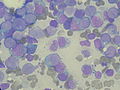
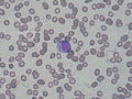
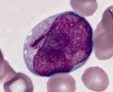
Auer rods (or Auer bodies) are large, crystalline cytoplasmic inclusion bodies sometimes observed in myeloid blast cells during acute myeloid leukemia, acute promyelocytic leukemia, high-grade myelodysplastic syndromes and myeloproliferative disorders. Composed of fused lysosomes and rich in lysosomal enzymes, Auer rods are azurophilic and can resemble needles, commas, diamonds, rectangles, corkscrews, or (rarely) granules. [1]

Contents

Eponym

Although Auer rods are named for American physiologist John Auer, [2] they were first described in 1905 by Canadian physician Thomas McCrae, then at Johns Hopkins Hospital, [3] as Auer himself acknowledged in his 1906 paper. Both McCrae and Auer mistakenly thought that the cells containing the rods were lymphoblasts. [4]

Additional images

Related Research Articles

<span class="mw-page-title-main">Leukemia</span> Blood cancers forming in the bone marrow

Leukemia is a group of blood cancers that usually begin in the bone marrow and result in high numbers of abnormal blood cells. These blood cells are not fully developed and are called blasts or leukemia cells. Symptoms may include bleeding and bruising, bone pain, fatigue, fever, and an increased risk of infections. These symptoms occur due to a lack of normal blood cells. Diagnosis is typically made by blood tests or bone marrow biopsy.

<span class="mw-page-title-main">Myelodysplastic syndrome</span> Diverse collection of blood-related cancers

A myelodysplastic syndrome (MDS) is one of a group of cancers in which immature blood cells in the bone marrow do not mature, and as a result, do not develop into healthy blood cells. Early on, no symptoms typically are seen. Later, symptoms may include fatigue, shortness of breath, bleeding disorders, anemia, or frequent infections. Some types may develop into acute myeloid leukemia.

<span class="mw-page-title-main">Chronic myelogenous leukemia</span> Medical condition

Chronic myelogenous leukemia (CML), also known as chronic myeloid leukemia, is a cancer of the white blood cells. It is a form of leukemia characterized by the increased and unregulated growth of myeloid cells in the bone marrow and the accumulation of these cells in the blood. CML is a clonal bone marrow stem cell disorder in which a proliferation of mature granulocytes and their precursors is found. It is a type of myeloproliferative neoplasm associated with a characteristic chromosomal translocation called the Philadelphia chromosome.

<span class="mw-page-title-main">Tumors of the hematopoietic and lymphoid tissues</span> Medical condition

Tumors of the hematopoietic and lymphoid tissues or tumours of the haematopoietic and lymphoid tissues are tumors that affect the blood, bone marrow, lymph, and lymphatic system. Because these tissues are all intimately connected through both the circulatory system and the immune system, a disease affecting one will often affect the others as well, making aplasia, myeloproliferation and lymphoproliferation closely related and often overlapping problems. While uncommon in solid tumors, chromosomal translocations are a common cause of these diseases. This commonly leads to a different approach in diagnosis and treatment of hematological malignancies. Hematological malignancies are malignant neoplasms ("cancer"), and they are generally treated by specialists in hematology and/or oncology. In some centers "hematology/oncology" is a single subspecialty of internal medicine while in others they are considered separate divisions. Not all hematological disorders are malignant ("cancerous"); these other blood conditions may also be managed by a hematologist.

<span class="mw-page-title-main">Acute lymphoblastic leukemia</span> Blood cancer characterised by overproduction of lymphoblasts

Acute lymphoblastic leukemia (ALL) is a cancer of the lymphoid line of blood cells characterized by the development of large numbers of immature lymphocytes. Symptoms may include feeling tired, pale skin color, fever, easy bleeding or bruising, enlarged lymph nodes, or bone pain. As an acute leukemia, ALL progresses rapidly and is typically fatal within weeks or months if left untreated.

<span class="mw-page-title-main">Acute promyelocytic leukemia</span> Subtype of acute myeloid leukaemia characterised by accumulation of promyelocytes

Acute promyelocytic leukemia is a subtype of acute myeloid leukemia (AML), a cancer of the white blood cells. In APL, there is an abnormal accumulation of immature granulocytes called promyelocytes. The disease is characterized by a chromosomal translocation involving the retinoic acid receptor alpha (RARA) gene and is distinguished from other forms of AML by its responsiveness to all-trans retinoic acid therapy. Acute promyelocytic leukemia was first characterized in 1957 by French and Norwegian physicians as a hyperacute fatal illness, with a median survival time of less than a week. Today, prognoses have drastically improved; 10-year survival rates are estimated to be approximately 80-90% according to one study.

<span class="mw-page-title-main">Myeloid sarcoma</span> Medical condition

A myeloid sarcoma is a solid tumor composed of immature white blood cells called myeloblasts. A chloroma is an extramedullary manifestation of acute myeloid leukemia; in other words, it is a solid collection of leukemic cells occurring outside of the bone marrow.

Acute leukemia or acute leukaemia is a family of serious medical conditions relating to an original diagnosis of leukemia. In most cases, these can be classified according to the lineage, myeloid or lymphoid, of the malignant cells that grow uncontrolled, but some are mixed and for those such an assignment is not possible.

<span class="mw-page-title-main">Acute myeloid leukemia</span> Cancer of the myeloid line of blood cells

Acute myeloid leukemia (AML) is a cancer of the myeloid line of blood cells, characterized by the rapid growth of abnormal cells that build up in the bone marrow and blood and interfere with normal blood cell production. Symptoms may include feeling tired, shortness of breath, easy bruising and bleeding, and increased risk of infection. Occasionally, spread may occur to the brain, skin, or gums. As an acute leukemia, AML progresses rapidly, and is typically fatal within weeks or months if left untreated.

<span class="mw-page-title-main">Acute monocytic leukemia</span> Medical condition

Acute monocytic leukemia is a type of acute myeloid leukemia. In AML-M5 >80% of the leukemic cells are of monocytic lineage. This cancer is characterized by a dominance of monocytes in the bone marrow. There is an overproduction of monocytes that the body does not need in the periphery. These overproduced monocytes interfere with normal immune cell production which causes many health complications for the affected individual.

<span class="mw-page-title-main">Chronic myelomonocytic leukemia</span> Medical condition

Chronic myelomonocytic leukemia (CMML) is a type of leukemia, which are cancers of the blood-forming cells of the bone marrow. In adults, blood cells are formed in the bone marrow, by a process that is known as haematopoiesis. In CMML, there are increased numbers of monocytes and immature blood cells (blasts) in the peripheral blood and bone marrow, as well as abnormal looking cells (dysplasia) in at least one type of blood cell.

<span class="mw-page-title-main">John Auer</span> American physiologist and pharmacologist

John Auer was an American physiologist and pharmacologist. He published nearly 150 papers during his career and is credited with the first description of Auer rods. Auer also contributed to the study of anaphylaxis and helped develop modern thoracic surgery. During World War I, he conducted wartime research with the Rockefeller Institute for Medical Research.

<span class="mw-page-title-main">Acute myeloblastic leukemia with maturation</span> Medical condition

Acute myeloblastic leukemia with maturation (M2) is a subtype of acute myeloid leukemia (AML).

<span class="mw-page-title-main">CD135</span> Protein-coding gene in the species Homo sapiens

Cluster of differentiation antigen 135 (CD135) also known as fms like tyrosine kinase 3, receptor-type tyrosine-protein kinase FLT3, or fetal liver kinase-2 (Flk2) is a protein that in humans is encoded by the FLT3 gene. FLT3 is a cytokine receptor which belongs to the receptor tyrosine kinase class III. CD135 is the receptor for the cytokine Flt3 ligand (FLT3L).

Biphenotypic acute leukaemia (BAL) is an uncommon type of leukemia which arises in multipotent progenitor cells which have the ability to differentiate into both myeloid and lymphoid lineages. It is a subtype of "leukemia of ambiguous lineage".

<span class="mw-page-title-main">Minimally differentiated acute myeloblastic leukemia</span> Medical condition

Minimally differentiated acute myeloblastic leukemia is a subtype of AML. It is classified as M0 by FAB. It represents 2–3% of all cases of AML. Although minimally differentiated AML was recognized earlier, criteria for FAB M0 were developed in 1987. The blasts in these cases cannot be recognized as myeloid based on morphology and cytochemistry, but immunophenotyping demonstrates myeloid antigens.

Leukostasis is a medical emergency most commonly seen in patients with acute myeloid leukemia. It is characterized by an extremely elevated blast cell count and symptoms of decreased tissue perfusion. The pathophysiology of leukostasis is not well understood, but inadequate delivery of oxygen to the body's cells is the result. Leukostasis is diagnosed when white cell plugs are seen in the microvasculature. The most common symptoms are dyspnea and hypoxia, usually accompanied by visual changes, headaches, dizziness, confusion, somnolence, and coma. Prompt treatment is required since, if left untreated, it has a very high mortality rate. Treatments aim to rapidly reduce white blood cell counts while also treating the underlying disorder.

Mixed-phenotype acute leukemia (MPAL) is a group of blood cancers (leukemia) which have combined features of myeloid and lymphoid cancers. It is a rare disease, constituting about 2–5% of all leukemia cases. It mostly involve myeloid with either of T lymphocyte or B lymphocyte progenitors, but in rare cases all the three cell lineages. Knowledge on the cause, clinical features and cellular mechanism is poor, making the treatment and management (prognosis) difficult.

Christopher Hourigan is a physician-scientist known for work on measurable residual disease in acute myeloid leukemia.

The LL-100 panel is a group of 100 human leukemia and lymphoma cell line, can be used in model of biomedical research.

References

  1. Ackerman, G. Adolph (1950). "Microscopic and Histochemical Studies on the Auer Bodies in Leukemic Cells". Blood. 5 (9): 847–863. doi: 10.1182/blood.V5.9.847.847 . PMID   15434012.
  2. Auer, John (1906). "Some hitherto undescribed structures found in the large lymphocytes of a case of acute leukaemia". American Journal of the Medical Sciences . 131 (6): 1002–1015. doi:10.1097/00000441-190606000-00008. ISSN   0002-9629. S2CID   71853154.
  3. McCrae, Thomas (February 1905). "Acute lymphatic leukaemia with a report of five cases". British Medical Journal . 1 (2304): 404–408. doi:10.1136/bmj.1.2304.404. PMC   2319598 . PMID   20761949.
  4. Bain, Barbara (August 2011). "Auer rods or McCrae rods?". American Journal of Hematology . 86 (8): 689. doi: 10.1002/ajh.21978 . PMID   21761434.