Hereditary stomatocytosis

Last updated
Hereditary stomatocytosis
Stomatocyten1x63.jpg
Stomatocytes
Specialty Hematology   OOjs UI icon edit-ltr-progressive.svg
Stomatocyte compared to other forms of poikilocytosis. Poikilocytes - Red blood cell types.jpg
Stomatocyte compared to other forms of poikilocytosis.

Hereditary stomatocytosis describes a number of inherited, mostly autosomal dominant human conditions which affect the red blood cell and create the appearance of a slit-like area of central pallor (stomatocyte) among erythrocytes on peripheral blood smear. The erythrocytes' cell membranes may abnormally 'leak' sodium and/or potassium ions, causing abnormalities in cell volume. [1] Hereditary stomatocytosis should be distinguished from acquired causes of stomatocytosis, including dilantin toxicity and alcoholism, as well as artifact from the process of preparing peripheral blood smears. [2] :237

Contents

Signs and symptoms

Stomatocytosis may present with signs and symptoms consistent with hemolytic anemia as a result of extravascular hemolysis and often intravascular hemolysis. These include fatigue and pallor, as well as signs of jaundice, splenomegaly and gallstone formation from prolonged hemolysis. [1] [3] Certain cases of hereditary stomatocytosis associated with genetic syndromes have additional symptoms that are unrelated to the hemolytic anemia. [1]

Pathophysiology

The two varieties of stomatocytosis classified with respect to cellular hydration status are overhydrated (hydrocytosis) and dehydrated (xerocytosis). [2] :225–226 Hereditary xerocytosis is characterized by autosomal dominant mutations in PIEZO1 , which encodes a cation channel whose mechanosensitive properties enable erythrocytes to deform as they pass through narrow capillaries by decreasing their intracellular volume. [4] More rarely, hereditary xerocytosis may be caused by mutations in KCNN4 , which encodes a calcium ion-sensitive potassium channel that mediates the potassium efflux triggered by a rise in intracellular Ca2+ via activated PIEZO1 channels. [4] Hereditary xerocytosis occurs more commonly in African populations, [2] [4] and it exhibits complex interactions with other hereditary alterations of red blood cells, including sickle cell disease [4] [5] and malaria resistance. [4] [6]

Osmosis leads to the red blood cell having a constant tendency to swell and burst. This tendency is countered by manipulating the flow of sodium and potassium ions. A 'pump' forces sodium out of the cell and potassium in, and this action is balanced by a process called 'the passive leak'. In overhydrated hereditary stomatocytoses, the passive leak is increased and the erythrocyte becomes swamped with salt and water. The affected erythrocytes have increased osmotic fragility. [4] Haemolytic anaemia results. For as yet unknown reasons, the cells take on the shape of a cup, with a 'mouth-shaped' (stoma) area of central pallor.

Overhydrated hereditary stomatocytosis is frequently linked to mutations in genes that encode components of the band 3 complex, such as RHAG . [4] It is the altered band 3 protein complex which mediates the cation leaks that are characteristic of hydrocytotic hereditary stomatocytosis. [7]

Rare cases of hereditary spherocytosis can occur without cation leaks. These include cases of phytosterolemia nonleaky stomatocytosis, a disorder of lipid metabolism associated with mutations in ABCG5 and/or ABCG8 , which encode sterol transporters. [1] The resulting abnormal sterol composition of erythrocyte cell membranes causes them to appear as deformed stomatocytes on peripheral blood smear. [1]

Diagnosis

Ektacytometry may be helpful in distinguishing different subtypes of hereditary stomatocytosis. [4]

Variants

Haematologists have identified a number of variants. These can be classified as below.

There are other families that do not fall neatly into any of these classifications. [9]

Stomatocytosis is also found as a hereditary disease in Alaskan malamute and miniature schnauzer dogs. [10]

Treatment

At present there is no specific treatment. Many patients with hemolytic anemia take folic acid (vitamin B9) since the greater turnover of cells consumes this vitamin. During crises transfusion may be required. Clotting problems can occur for which anticoagulation may be needed. Unlike hereditary spherocytosis, splenectomy is contraindicated. [11]

Related Research Articles

<span class="mw-page-title-main">Hemolysis</span> Rupturing of red blood cells and release of their contents

Hemolysis or haemolysis, also known by several other names, is the rupturing (lysis) of red blood cells (erythrocytes) and the release of their contents (cytoplasm) into surrounding fluid. Hemolysis may occur in vivo or in vitro.

<span class="mw-page-title-main">Spherocytosis</span> Medical condition

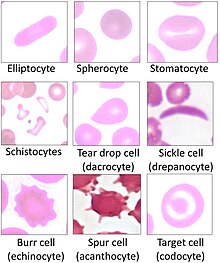
Spherocytosis is the presence of spherocytes in the blood, i.e. erythrocytes that are sphere-shaped rather than bi-concave disk shaped as normal. Spherocytes are found in all hemolytic anemias to some degree. Hereditary spherocytosis and autoimmune hemolytic anemia are characterized by having only spherocytes.

<span class="mw-page-title-main">Hereditary spherocytosis</span> Medical condition

Hereditary spherocytosis (HS) is a congenital hemolytic disorder wherein a genetic mutation coding for a structural membrane protein phenotype causes the red blood cells to be sphere-shaped (spherocytosis), rather than the normal biconcave disk shape. This abnormal shape interferes with the cells' ability to flex during blood circulation, and also makes them more prone to rupture under osmotic stress, mechanical stress, or both. Cells with the dysfunctional proteins are degraded in the spleen, which leads to a shortage of erythrocytes and results in hemolytic anemia.

<span class="mw-page-title-main">Paroxysmal nocturnal hemoglobinuria</span> Medical condition

Paroxysmal nocturnal hemoglobinuria (PNH) is a rare, acquired, life-threatening disease of the blood characterized by destruction of red blood cells by the complement system, a part of the body's innate immune system. This destructive process occurs due to deficiency of the red blood cell surface protein DAF, which normally inhibits such immune reactions. Since the complement cascade attacks the red blood cells within the blood vessels of the circulatory system, the red blood cell destruction (hemolysis) is considered an intravascular hemolytic anemia. There is ongoing research into other key features of the disease, such as the high incidence of venous blood clot formation. Research suggests that PNH thrombosis is caused by both the absence of GPI-anchored complement regulatory proteins on PNH platelets and the excessive consumption of nitric oxide (NO).

<span class="mw-page-title-main">Hemolytic anemia</span> Medical condition

Hemolytic anemia or haemolytic anaemia is a form of anemia due to hemolysis, the abnormal breakdown of red blood cells (RBCs), either in the blood vessels or elsewhere in the human body (extravascular). This most commonly occurs within the spleen, but also can occur in the reticuloendothelial system or mechanically. Hemolytic anemia accounts for 5% of all existing anemias. It has numerous possible consequences, ranging from general symptoms to life-threatening systemic effects. The general classification of hemolytic anemia is either intrinsic or extrinsic. Treatment depends on the type and cause of the hemolytic anemia.

<span class="mw-page-title-main">Pyruvate kinase deficiency</span> Medical condition

Pyruvate kinase deficiency is an inherited metabolic disorder of the enzyme pyruvate kinase which affects the survival of red blood cells. Both autosomal dominant and recessive inheritance have been observed with the disorder; classically, and more commonly, the inheritance is autosomal recessive. Pyruvate kinase deficiency is the second most common cause of enzyme-deficient hemolytic anemia, following G6PD deficiency.

<span class="mw-page-title-main">Band 3 anion transport protein</span> Mammalian protein found in Homo sapiens

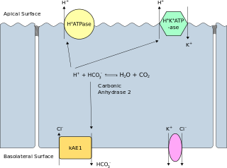
Band 3 anion transport protein, also known as anion exchanger 1 (AE1) or band 3 or solute carrier family 4 member 1 (SLC4A1), is a protein that is encoded by the SLC4A1 gene in humans.

Southeast Asian ovalocytosis is a blood disorder that is similar to, but distinct from hereditary elliptocytosis. It is common in some communities in Malaysia and Papua New Guinea, as it confers some resistance to cerebral Falciparum Malaria.

<span class="mw-page-title-main">Hereditary pyropoikilocytosis</span> Medical condition

Hereditary pyropoikilocytosis (HPP) is an autosomal recessive form of hemolytic anemia characterized by an abnormal sensitivity of red blood cells to heat and erythrocyte morphology similar to that seen in thermal burns or from prolonged exposure of a healthy patient's blood sample to high ambient temperatures. Patients with HPP tend to experience severe hemolysis and anemia in infancy that gradually improves, evolving toward typical elliptocytosis later in life. However, the hemolysis can lead to rapid sequestration and destruction of red cells. Splenectomy is curative when this occurs.

The Kell antigen system is a human blood group system, that is, a group of antigens on the human red blood cell surface which are important determinants of blood type and are targets for autoimmune or alloimmune diseases which destroy red blood cells. The Kell antigens are K, k, Kpa, Kpb, Jsa and Jsb. The Kell antigens are peptides found within the Kell protein, a 93-kilodalton transmembrane zinc-dependent endopeptidase which is responsible for cleaving endothelin-3.

<span class="mw-page-title-main">Ankyrin</span> Protein family

Ankyrins are a family of proteins that mediate the attachment of integral membrane proteins to the spectrin-actin based membrane cytoskeleton. Ankyrins have binding sites for the beta subunit of spectrin and at least 12 families of integral membrane proteins. This linkage is required to maintain the integrity of the plasma membranes and to anchor specific ion channels, ion exchangers and ion transporters in the plasma membrane. The name is derived from the Greek word ἄγκυρα (ankyra) for "anchor".

<span class="mw-page-title-main">Protein 4.2</span> Protein-coding gene in the species Homo sapiens

Erythrocyte membrane protein band 4.2 is a protein that in humans is encoded by the EPB42 gene. It is part of the red blood cell cytoskeleton.

<span class="mw-page-title-main">Aldolase A deficiency</span> Medical condition

Aldolase A deficiency is an autosomal recessive metabolic disorder resulting in a deficiency of the enzyme aldolase A; the enzyme is found predominantly in red blood cells and muscle tissue. The deficiency may lead to hemolytic anaemia as well as myopathy associated with exercise intolerance and rhabdomyolysis in some cases.

<span class="mw-page-title-main">Spectrin, alpha 1</span> Protein-coding gene in the species Homo sapiens

Spectrin alpha chain, erythrocyte is a protein that in humans is encoded by the SPTA1 gene.

<span class="mw-page-title-main">Hexokinase deficiency</span> Medical condition

Hexokinase deficiency is an extremely rare autosomal recessive condition that falls under the category of erythroenzymopathies, or defects in red cell enzymes. Hexokinase deficiency manifests is associated with chronic nonspherocytic hemolytic anemia. Hemolytic anemia seems to be the only clinical sign of hexokinase deficiency. In 1967 the first case of hexokinase deficiency was described by Valentine et al, since then, less than 50 cases have been reported.

Congenital hemolytic anemia (CHA) is a diverse group of rare hereditary conditions marked by decreased life expectancy and premature removal of erythrocytes from blood flow. Defects in erythrocyte membrane proteins and red cell enzyme metabolism, as well as changes at the level of erythrocyte precursors, lead to impaired bone marrow erythropoiesis. CAH is distinguished by variable anemia, chronic extravascular hemolysis, decreased erythrocyte life span, splenomegaly, jaundice, biliary lithiasis, and iron overload. Immune-mediated mechanisms may play a role in the pathogenesis of these uncommon diseases, despite the paucity of data regarding the immune system's involvement in CHAs.

Mechanical hemolytic anemia is a form of hemolytic anemia due to mechanically induced damage to red blood cells. Red blood cells, while flexible, may in some circumstances succumb to physical shear and compression. This may result in hemoglobinuria. The damage is induced through repetitive mechanical motions such as prolonged marching and marathon running. Mechanical damage can also be induced through the chronic condition microangiopathic hemolytic anemia or due to prosthetic heart valves.

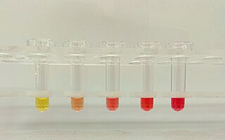
Erythrocyte fragility refers to the propensity of erythrocytes to hemolyse (rupture) under stress. It can be thought of as the degree or proportion of hemolysis that occurs when a sample of red blood cells are subjected to stress. Depending on the application as well as the kind of fragility involved, the amount of stress applied and/or the significance of the resultant hemolysis may vary.

<span class="mw-page-title-main">Stomatin</span> Mammalian protein found in Homo sapiens

Stomatin also known as human erythrocyte integral membrane protein band 7 is a protein that in humans is encoded by the STOM gene.

<span class="mw-page-title-main">Yoda1</span> Chemical compound

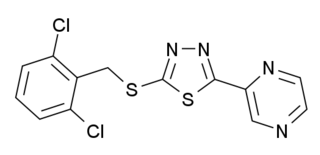
Yoda1 is a chemical compound which is the first agonist developed for the mechanosensitive ion channel PIEZO1. This protein is involved in regulation of blood pressure and red blood cell volume, and Yoda1 is used in scientific research in these areas.

References

  1. 1 2 3 4 5 6 7 8 9 Andolfo I, Russo R, Gambale A, Iolascon A (January 2018). "Hereditary stomatocytosis: An underdiagnosed condition". American Journal of Hematology. 93 (1): 107–121. doi: 10.1002/ajh.24929 . PMID   28971506.
  2. 1 2 3 Tyrrell, L; Rose, G; Shukri, A; Kawash, SB (2021). "Morphologic changes in red blood cells: An illustrated review of clinically important light microscopic findings" (PDF). The Malaysian Journal of Pathology. 43 (2): 219–239. PMID   34448787 . Retrieved 12 September 2022.
  3. Phillips J, Henderson AC (September 2018). "Hemolytic Anemia: Evaluation and Differential Diagnosis". American Family Physician. 98 (6): 354–361. PMID   30215915 . Retrieved 5 September 2022.
  4. 1 2 3 4 5 6 7 8 Risinger, Mary; Kalfa, Theodosia A. (2020). "Red cell membrane disorders: Structure meets function". Blood. 136 (11): 1250–1261. doi: 10.1182/blood.2019000946 . PMC   7483429 . PMID   32702754.
  5. Ilboudo, Yann; Bartolucci, Pablo; Garrett, Melanie E.; Ashley-Koch, Allison; Telen, Marilyn; Brugnara, Carlo; Galactéros, Frédéric; Lettre, Guillaume (2018). "A common functional PIEZO1 deletion allele associates with red blood cell density in sickle cell disease patients". American Journal of Hematology. 93 (11): E362–E365. doi: 10.1002/ajh.25245 . PMID   30105803.
  6. Nguetse, Christian N.; Purington, Natasha; Ebel, Emily R.; Shakya, Bikash; Tetard, Marilou; Kremsner, Peter G.; Velavan, Thirumalaisamy P.; Egan, Elizabeth S. (2020). "A common polymorphism in the mechanosensitive ion channel PIEZO1 is associated with protection from severe malaria in humans". Proceedings of the National Academy of Sciences. 117 (16): 9074–9081. Bibcode:2020PNAS..117.9074N. doi: 10.1073/pnas.1919843117 . PMC   7183233 . PMID   32265284.
  7. Bruce LJ, Robinson HC, Guizouarn H, Borgese F, Harrison P, King MJ, et al. (November 2005). "Monovalent cation leaks in human red cells caused by single amino-acid substitutions in the transport domain of the band 3 chloride-bicarbonate exchanger, AE1". Nature Genetics. 37 (11): 1258–1263. doi:10.1038/ng1656. PMID   16227998. S2CID   23554234.
  8. 1 2 Coles SE, Stewart GW (October 1999). "Temperature effects on cation transport in hereditary stomatocytosis and allied disorders". International Journal of Experimental Pathology. 80 (5): 251–258. doi:10.1046/j.1365-2613.1999.00120.x. PMC   2517829 . PMID   10607015.
  9. Oski FA, Naiman JL, Blum SF, Zarkowsky HS, Whaun J, Shohet SB, et al. (April 1969). "Congenital hemolytic anemia with high-sodium, low-potassium red cells. Studies of three generations of a family with a new variant". The New England Journal of Medicine. 280 (17): 909–916. doi:10.1056/NEJM196904242801701. PMID   4237839.
  10. Thrall MA, Baker DC (2006). Veterinary Hematology and Clinical Chemistry. Blackwell Publishing. pp. 71–72. ISBN   978-0-7817-5799-7. OCLC   954019258.
  11. Picard V, Guitton C, Thuret I, Rose C, Bendelac L, Ghazal K, et al. (August 2019). "Clinical and biological features in PIEZO1-hereditary xerocytosis and Gardos channelopathy: a retrospective series of 126 patients". Haematologica. 104 (8): 1554–1564. doi:10.3324/haematol.2018.205328. PMC   6669138 . PMID   30655378.

Further reading