Sideroblastic anemia

Last updated
Sideroblastic anemia
Sideroblast.png
A ring sideroblast visualized by Prussian blue stain
Specialty Hematology   OOjs UI icon edit-ltr-progressive.svg

Sideroblastic anemia, or sideroachrestic anemia, is a form of anemia in which the bone marrow produces ringed sideroblasts rather than healthy red blood cells (erythrocytes). [1] In sideroblastic anemia, the body has iron available but cannot incorporate it into hemoglobin, which red blood cells need in order to transport oxygen efficiently. The disorder may be caused either by a genetic disorder or indirectly as part of myelodysplastic syndrome, [2] which can develop into hematological malignancies (especially acute myeloid leukemia).

Contents

Sideroblasts ( sidero- + -blast ) are nucleated erythroblasts (precursors to mature red blood cells) with granules of iron accumulated in the mitochondria surrounding the nucleus. [3] Normally, sideroblasts are present in the bone marrow, and enter the circulation after maturing into a normal erythrocyte. The presence of sideroblasts per se does not define sideroblastic anemia. Only the finding of ring (or ringed) sideroblasts characterizes sideroblastic anemia.

Ring sideroblasts are named so because iron-laden mitochondria form a ring around the nucleus. It is a subtype of basophilic granules of the erythrocyte, but which can only be seen in bone marrow. To count a cell as a ring sideroblast, the ring must encircle a third or more of the nucleus and contain five or more iron granules, according to the 2008 WHO classification of the tumors of the hematopoietic and lymphoid tissues. [4]

Types

The WHO International Working Group on Morphology of MDS (IWGM-MDS) defined three types of sideroblasts:[ citation needed ]

  1. Type 1 sideroblasts: fewer than 5 siderotic granules in the cytoplasm
  2. Type 2 sideroblasts: 5 or more siderotic granules, but not in a perinuclear distribution
  3. Type 3 or ring sideroblasts: 5 or more granules in a perinuclear position, surrounding the nucleus or encompassing at least one third of the nuclear circumference.

Type 1 and type 2 are found in non-sideroblastic anemias. Type 3 is found only in sideroblastic anemia.[ citation needed ]

Symptoms and signs

Symptoms of sideroblastic anemia include skin paleness, fatigue, dizziness, and enlarged spleen and liver. Heart disease, liver damage, and kidney failure can result from iron buildup in these organs. [5]

Symptoms of sideroblastic anemia usually resemble the common symptoms of anemia. In addition to the symptoms listed above, patients with sideroblastic anemia may experience shortness of breath, heart palpitations, and headache. Some patients may have bronze-colored skin due to an overload of iron. Patients with syndromic hereditary sideroblastic anemia may experience diabetes mellitus and deafness. [6]

Causes

Causes of sideroblastic anemia can be categorized into three groups: congenital sideroblastic anemia, acquired clonal sideroblastic anemia, and acquired reversible sideroblastic anemia. All cases involve dysfunctional heme synthesis or processing. This leads to granular deposition of iron in the mitochondria that form a ring around the nucleus of the developing red blood cell. Congenital forms often present with normocytic or microcytic anemia while acquired forms of sideroblastic anemia are often normocytic or macrocytic.[ citation needed ]

Diagnosis

Bone marrow aspirate: ring sideroblasts Ring Sideroblast smear 2010-01-13.JPG
Bone marrow aspirate: ring sideroblasts

Ringed sideroblasts are seen in the bone marrow.

On the peripheral blood smear can be found erythrocytes with basophilic stippling (cytoplasmic granules of RNA precipitates) and Pappenheimer bodies (cytoplasmic granules of iron). [13]

The anemia is moderate to severe and dimorphic. Microscopic viewing of the red blood cells will reveal marked unequal cell size and abnormal cell shape. Basophilic stippling is marked and target cells are common. The mean cell volume is commonly decreased (i.e., a microcytic anemia), but it may also be normal or even high. The RDW is increased with the red blood cell histogram shifted to the left. Leukocytes and platelets are normal. Bone marrow shows erythroid hyperplasia with a maturation arrest.In excess of 40% of the developing erythrocytes are ringed sideroblasts. Serum iron, percentage saturation and ferritin are increased. The total iron-binding capacity of the cells is normal to decreased. Stainable marrow hemosiderin is increased.[ citation needed ]

Classification

Sideroblastic anemia is typically divided into subtypes based on its cause.

OMIM NameGene
300751 X-linked sideroblastic anemia (XLSA) ALAS2
301310 sideroblastic anemia with spinocerebellar ataxia (ASAT) ABCB7
205950 pyridoxine-refractory autosomal recessive sideroblastic anemia SLC25A38
206000 pyridoxine-responsive sideroblastic anemia(vitamin B6 deficiency; pyridoxal phosphate required for heme synthesis)

GLRX5 has also been implicated. [15]

Laboratory findings

Treatment

Occasionally, the anemia is so severe that support with transfusion is required. These patients usually do not respond to erythropoietin therapy. [16] Some cases have been reported that the anemia is reversed or heme level is improved through use of moderate to high doses of pyridoxine (vitamin B6). In severe cases of SBA, bone marrow transplant is also an option with limited information about the success rate. Some cases are listed on MedLine and various other medical sites. In the case of isoniazid-induced sideroblastic anemia, the addition of B6 is sufficient to correct the anemia. Deferoxamine, a chelating agent, is used to treat iron overload from transfusions. Therapeutic phlebotomy can be used to manage iron overload. [17]

Prognosis

Sideroblastic anemias are often described as responsive or non-responsive in terms of increased hemoglobin levels to pharmacological doses of vitamin B6.[ citation needed ]

1- Congenital: 80% are responsive, though the anemia does not completely resolve.

2- Acquired clonal: 40% are responsive, but the response may be minimal.

3- Acquired reversible: 60% are responsive, but course depends on treatment of the underlying cause.

Severe refractory sideroblastic anemias requiring regular transfusions and/or that undergo leukemic transformation (5–10%) significantly reduce life expectancy.

See also

Related Research Articles

<span class="mw-page-title-main">Anemia</span> Medical condition

Anemia or anaemia is a blood disorder in which the blood has a reduced ability to carry oxygen due to a lower than normal number of red blood cells, a reduction in the amount of hemoglobin or hemoglobin abnormalities. The name is derived from Ancient Greek: ἀναιμία anaimia, meaning 'lack of blood', from ἀν- an-, 'not' and αἷμα haima, 'blood'. When anemia comes on slowly, the symptoms are often vague, such as tiredness, weakness, shortness of breath, headaches, and a reduced ability to exercise. When anemia is acute, symptoms may include confusion, feeling like one is going to pass out, loss of consciousness, and increased thirst. Anemia must be significant before a person becomes noticeably pale. Symptoms of anemia depend on how quickly hemoglobin decreases. Additional symptoms may occur depending on the underlying cause. Preoperative anemia can increase the risk of needing a blood transfusion following surgery. Anemia can be temporary or long term and can range from mild to severe.

<span class="mw-page-title-main">Myelodysplastic syndrome</span> Diverse collection of blood-related cancers

A myelodysplastic syndrome (MDS) is one of a group of cancers in which immature blood cells in the bone marrow do not mature, and as a result, do not develop into healthy blood cells. Early on, no symptoms typically are seen. Later, symptoms may include fatigue, shortness of breath, bleeding disorders, anemia, or frequent infections. Some types may develop into acute myeloid leukemia.

<span class="mw-page-title-main">Fanconi anemia</span> Medical condition

Fanconi anemia (FA) is a rare, AR, genetic disease resulting in impaired response to DNA damage in the FA/BRCA pathway. Although it is a very rare disorder, study of this and other bone marrow failure syndromes has improved scientific understanding of the mechanisms of normal bone marrow function and development of cancer. Among those affected, the majority develop cancer, most often acute myelogenous leukemia (AML), MDS, and liver tumors. 90% develop aplastic anemia by age 40. About 60–75% have congenital defects, commonly short stature, abnormalities of the skin, arms, head, eyes, kidneys, and ears, and developmental disabilities. Around 75% have some form of endocrine problem, with varying degrees of severity. 60% of FA is FANC-A, 16q24.3, which has later onset bone marrow failure.

<span class="mw-page-title-main">Paroxysmal nocturnal hemoglobinuria</span> Medical condition

Paroxysmal nocturnal hemoglobinuria (PNH) is a rare, acquired, life-threatening disease of the blood characterized by destruction of red blood cells by the complement system, a part of the body's innate immune system. This destructive process occurs due to deficiency of the red blood cell surface protein DAF, which normally inhibits such immune reactions. Since the complement cascade attacks the red blood cells within the blood vessels of the circulatory system, the red blood cell destruction (hemolysis) is considered an intravascular hemolytic anemia. Other key features of the disease, such as the high incidence of venous blood clot formation, are incompletely understood.

<span class="mw-page-title-main">Erythropoiesis</span> Process which produces red blood cells

Erythropoiesis is the process which produces red blood cells (erythrocytes), which is the development from erythropoietic stem cell to mature red blood cell.

<span class="mw-page-title-main">Aminolevulinic acid synthase</span> Class of enzymes

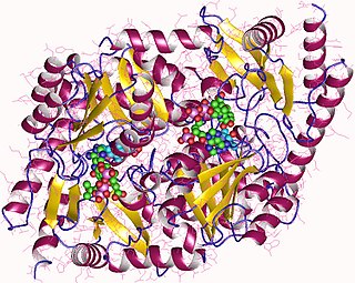
Aminolevulinic acid synthase (ALA synthase, ALAS, or delta-aminolevulinic acid synthase) is an enzyme (EC 2.3.1.37) that catalyzes the synthesis of δ-aminolevulinic acid (ALA) the first common precursor in the biosynthesis of all tetrapyrroles such as hemes, cobalamins and chlorophylls. The reaction is as follows:

<span class="mw-page-title-main">Chromosome 5q deletion syndrome</span> Human disease

Chromosome 5q deletion syndrome is an acquired, hematological disorder characterized by loss of part of the long arm of human chromosome 5 in bone marrow myelocyte cells. This chromosome abnormality is most commonly associated with the myelodysplastic syndrome.

Pancytopenia is a medical condition in which there is significant reduction in the number of almost all blood cells.

<span class="mw-page-title-main">Microcytic anemia</span> Medical condition

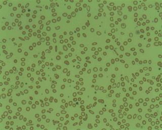
Microcytic anaemia is any of several types of anemia characterized by smaller than normal red blood cells. The normal mean corpuscular volume is approximately 80–100 fL. When the MCV is <80 fL, the red cells are described as microcytic and when >100 fL, macrocytic. The MCV is the average red blood cell size.

<span class="mw-page-title-main">Howell–Jolly body</span> Cluster of DNA in red blood cells

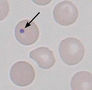
A Howell–Jolly body is a cytopathological finding of basophilic nuclear remnants in circulating erythrocytes. During maturation in the bone marrow, late erythroblasts normally expel their nuclei; but, in some cases, a small portion of DNA remains. The presence of Howell–Jolly bodies usually signifies a damaged or absent spleen, because a healthy spleen would normally filter such erythrocytes.

<span class="mw-page-title-main">Myeloblast</span>

The myeloblast is a unipotent stem cell which differentiates into the effectors of the granulocyte series. It is found in the bone marrow. Stimulation of myeloblasts by G-CSF and other cytokines triggers maturation, differentiation, proliferation and cell survival.

<span class="mw-page-title-main">GATA1</span> Protein-coding gene in humans

GATA-binding factor 1 or GATA-1 is the founding member of the GATA family of transcription factors. This protein is widely expressed throughout vertebrate species. In humans and mice, it is encoded by the GATA1 and Gata1 genes, respectively. These genes are located on the X chromosome in both species.

<span class="mw-page-title-main">Chronic myelomonocytic leukemia</span> Medical condition

Chronic myelomonocytic leukemia (CMML) is a type of leukemia, which are cancers of the blood-forming cells of the bone marrow. In adults, blood cells are formed in the bone marrow, by a process that is known as haematopoiesis. In CMML, there are increased numbers of monocytes and immature blood cells (blasts) in the peripheral blood and bone marrow, as well as abnormal looking cells (dysplasia) in at least one type of blood cell.

<span class="mw-page-title-main">Copper deficiency</span> Insufficient level of copper in the body, leading to anaemia and nervous symptoms

Copper deficiency, or hypocupremia, is defined either as insufficient copper to meet the needs of the body, or as a serum copper level below the normal range. Symptoms may include fatigue, decreased red blood cells, early greying of the hair, and neurological problems presenting as numbness, tingling, muscle weakness, and ataxia. The neurodegenerative syndrome of copper deficiency has been recognized for some time in ruminant animals, in which it is commonly known as "swayback". Copper deficiency can manifest in parallel with vitamin B12 and other nutritional deficiencies.

<span class="mw-page-title-main">Pappenheimer bodies</span> Abnormal iron deposits in red blood cells

Pappenheimer bodies are abnormal basophilic granules of iron found inside red blood cells on routine blood stain. They are a type of inclusion body composed of ferritin aggregates, or mitochondria or phagosomes containing aggregated ferritin. They appear as dense, blue-purple granules within the red blood cell and there are usually only one or two, located in the cell periphery. They stain on a Romanowsky stain because clumps of ribosomes are co‐precipitated with the iron‐containing organelles.

Normocytic anemia is a type of anemia and is a common issue that occurs for men and women typically over 85 years old. Its prevalence increases with age, reaching 44 percent in men older than 85 years. The most common type of normocytic anemia is anemia of chronic disease.

<span class="mw-page-title-main">ALAS2</span> Protein-coding gene in humans

Delta-aminolevulinate synthase 2 also known as ALAS2 is a protein that in humans is encoded by the ALAS2 gene. ALAS2 is an aminolevulinic acid synthase.

<span class="mw-page-title-main">Basophilic stippling</span> Cytoplasmic granules in basophils

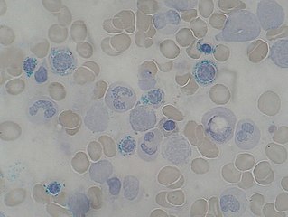
Basophilic stippling, also known as punctate basophilia, is the presence of numerous basophilic granules that are dispersed through the cytoplasm of erythrocytes in a peripheral blood smear. They can be demonstrated to be RNA. They are composed of aggregates of ribosomes; degenerating mitochondria and siderosomes may be included in the aggregates. In contrast to Pappenheimer bodies, they are negative with Perls' acid ferrocyanide stain for iron. Basophilic stippling is indicative of disturbed erythropoiesis. It can also be found in some normal individuals.

<span class="mw-page-title-main">GLRX5</span> Protein-coding gene in the species Homo sapiens

Glutaredoxin 5, also known as GLRX5, is a protein which in humans is encoded by the GLRX5 gene located on chromosome 14. This gene encodes a mitochondrial protein, which is evolutionarily conserved. It is involved in the biogenesis of iron- sulfur clusters, which are required for normal iron homeostasis. Mutations in this gene are associated with autosomal recessive pyridoxine-refractory sideroblastic anemia.

Bone marrow failure occurs in individuals who produce an insufficient amount of red blood cells, white blood cells or platelets. Red blood cells transport oxygen to be distributed throughout the body's tissue. White blood cells fight off infections that enter the body. Bone marrow also contains platelets, which trigger clotting, and thus help stop the blood flow when a wound occurs.

References

  1. Caudill JS, Imran H, Porcher JC, Steensma DP (October 2008). "Congenital sideroblastic anemia associated with germline polymorphisms reducing expression of FECH". Haematologica. 93 (10): 1582–4. doi: 10.3324/haematol.12597 . PMID   18698088.
  2. Sideroblastic Anemias: Anemias Caused by Deficient Erythropoiesis at Merck Manual of Diagnosis and Therapy Professional Edition
  3. "Ring sideroblasts". Blood. 107 (5): 1746. 2006-03-01. doi: 10.1182/blood.V107.5.1746.1746 . ISSN   0006-4971.
  4. Mufti, GJ; Bennett, JM; Goasguen, J; Bain, BJ; Baumann, I; Brunning, R; Cazzola, M; Fenaux, P; Germing, U; Hellström-Lindberg, E; Jinnai, I; Manabe, A; Matsuda, A; Niemeyer, CM; Sanz, G; Tomonaga, M; Vallespi, T; Yoshimi, A; International Working Group on Morphology of Myelodysplastic, Syndrome (Nov 2008). "Diagnosis and classification of myelodysplastic syndrome: International Working Group on Morphology of myelodysplastic syndrome (IWGM-MDS) consensus proposals for the definition and enumeration of myeloblasts and ring sideroblasts". Haematologica. 93 (11): 1712–7. doi: 10.3324/haematol.13405 . PMID   18838480.
  5. Genetics Home Reference: Genetic Conditions > X-linked sideroblastic anemia Reviewed October 2006. Retrieved on 5 Mars, 2009
  6. Ashorobi, Damilola; Chhabra, Anil (18 July 2022). Sideroblastic Anemia. StatPearls Publishing. PMID   30855871 . Retrieved 20 February 2023.
  7. Aivado M, Gattermann N, Rong A, et al. (2006). "X-linked sideroblastic anemia associated with a novel ALAS2 mutation and unfortunate skewed X-chromosome inactivation patterns". Blood Cells Mol. Dis. 37 (1): 40–5. doi:10.1016/j.bcmd.2006.04.003. PMID   16735131.
  8. Shander, Petra Seeber, Aryeh (2013). Basics of blood management (2nd ed.). Chichester, West Sussex: Wiley-Blackwell. p. 46. ISBN   978-0-470-67070-5.{{cite book}}: CS1 maint: multiple names: authors list (link)
  9. Lubran, MM (1980). "Lead toxicity and heme biosynthesis". Annals of Clinical and Laboratory Science. 10 (5): 402–13. PMID   6999974.
  10. 1 2 Canada, editors, John P. Greer; Daniel A. Arber; Bertil Glader; Alan F. List; Robert T. Means; Frixos Paraskevas; George M. Rodgers; John Foerster (2014). Wintrobe's clinical hematology (Thirteenth ed.). p. 656. ISBN   978-1451172683.{{cite book}}: |first1= has generic name (help)CS1 maint: multiple names: authors list (link)
  11. Forman, W.B. (1990). "Zinc abuse: an unsuspected cause of sideroblastic anemia". West J Med. 152 (2): 190–2. PMC   1002314 . PMID   2400417.
  12. Saini, N; Jacobson, JO; Jha, S; Saini, V; Weinger, R (April 2012). "The perils of not digging deep enough--uncovering a rare cause of acquired anemia". American Journal of Hematology. 87 (4): 413–6. doi: 10.1002/ajh.22235 . PMID   22120958.
  13. Rodak, Bernadette F. (2007). Hematology : clinical principles and applications (3rd ed.). Philadelphia: Saunders. p. 535. ISBN   978-1416030065.
  14. X-linked sideroblastic anemia at NLM Genetics Home Reference
  15. Camaschella C (September 2008). "Recent advances in the understanding of inherited sideroblastic anaemia". Br. J. Haematol. 143 (1): 27–38. doi: 10.1111/j.1365-2141.2008.07290.x . PMID   18637800.
  16. Papadakis, Maxine A.; Tierney, Lawrence M.; McPhee, Stephen J. (2005). "Sideroblastic Anemia" . Current Medical Diagnosis & Treatment, 2006. McGraw-Hill Medical. ISBN   978-0-07-145410-0.
  17. Peto, T. E. A., Pippard, M. J., Weatherall, D. J. Iron overload in mild sideroblastic anaemias" Lancet 321: 375-378, 1983. Note: Originally Volume I.