Sickle cell trait

Last updated
Sickle cell trait
Sickle cells.jpg
Sickle cells in human blood: both normal red blood cells and sickle-shaped cells are present
Specialty Hematology   OOjs UI icon edit-ltr-progressive.svg

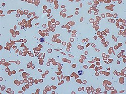
Sickle cell trait describes a condition in which a person has one abnormal allele of the hemoglobin beta gene (is heterozygous), but does not display the severe symptoms of sickle cell disease that occur in a person who has two copies of that allele (is homozygous). Those who are heterozygous for the sickle cell allele produce both normal and abnormal hemoglobin (the two alleles are codominant with respect to the actual concentration of hemoglobin in the circulating cells).

Contents

Sickle cell disease is a blood disorder wherein there is a single amino acid substitution in the hemoglobin protein of the red blood cells, which causes these cells to assume a sickle shape, especially when under low oxygen tension. Sickling and sickle cell disease also confer some resistance to malaria parasitization of red blood cells, so that individuals with sickle-cell trait (heterozygotes) have a selective advantage in environments where malaria is present.

Symptoms and signs

Sickle cell trait is a hemoglobin genotype AS and is generally regarded as a benign condition. [1] However, individuals with sickle cell trait may have rare complications. For example, in November 2010, Dr. Jeffery K. Taubenberger of the National Institutes of Health discovered the earliest proof of sickle-cell disease while looking for the virus of the 1918 flu during the autopsy of an African-American soldier. Taubenberger's autopsy results showed that the soldier had had a sickle-cell crisis that contributed to his death even though he had only one copy of the gene. [2] There have been calls to reclassify sickle cell trait as a disease state, based on its malignant clinical presentations. [3] Significance may be greater during exercise. [4]

Association with other medical conditions

Malaria

The sickle cell trait provides a survival advantage against malaria fatality over people with normal hemoglobin in regions where malaria is endemic. The trait is known to cause significantly fewer deaths due to malaria, especially when Plasmodium falciparum is the causative organism. This is a prime example of natural selection, evidenced by the fact that the geographical distribution of the gene for hemoglobin S and the distribution of malaria in Africa virtually overlap. Because of the unique survival advantage, people with the trait become increasingly numerous as the number of malaria-infected people increases. Conversely, people who have normal hemoglobin are more likely to succumb to the complications of malaria.[ citation needed ]

The way in which sickle cell protects against malaria is attributed to several different things. One of the more common explanations is that the sickle hemoglobin inhibits the plasmodium parasite from infecting the red blood cells which reduces the number of malaria parasites to infect the host. Another factor is the production of heme oxygenase-1 (HO-1) enzyme, which is highly present in the sickle hemoglobin. This enzyme produces carbon monoxide which has been proven to protect against cerebral malaria. [5]

Established associations

Suggested

There have been reports of pulmonary venous thromboembolism in pregnant women with sickle cell trait, [18] or men during prolonged airflight, and mild strokes and abnormalities on PET scans in children with the trait.[ citation needed ]

Sickle cell trait appears to worsen the complications seen in diabetes mellitus type 2 (retinopathy, nephropathy and proteinuria) [19] and provoke hyperosmolar diabetic coma nephropathy, especially in male patients.

Genetics

Normally, a person inherits two copies of the gene that produces beta-globin, a protein needed to produce normal hemoglobin (hemoglobin A, genotype AA). A person with sickle cell trait inherits one normal allele and one abnormal allele encoding hemoglobin S (hemoglobin genotype AS).[ citation needed ]

The sickle cell trait can be used to demonstrate the concepts of co-dominance and incomplete dominance. An individual with the sickle cell trait shows incomplete dominance when the shape of the red blood cell is considered. This is because the sickling happens only at low oxygen concentrations. With regards to the actual concentration of hemoglobin in the circulating cells, the alleles demonstrate co-dominance as both 'normal' and mutant forms co-exist in the bloodstream. Thus it is an ambiguous condition showing both incomplete dominance and co-dominance.[ citation needed ]

Unlike the sickle-cell trait, sickle-cell disease is passed on in a recessive manner. Sickle cell anemia affects about 72,000 people in the United States. Most Americans who have sickle cell anemia are of African descent. The disease also affects Americans from the Caribbean, Central America, and parts of South America, Turkey, Greece, Italy, the Middle East and East India.

Sickle-cell disease and the associated trait are most prevalent in Africa and Central America, which is attributed to natural selection: the sickle-cell trait confers a survival advantage in areas with a high occurrence of malaria, which has a high death rate among individuals without the trait.[ citation needed ]

There also have been studies that show changes in the globin genes. There have been noted changes in the beta-globin sequence, to what is known as the sickle hemoglobin.

The significance of the sickle-cell trait is that it does not show any symptoms, nor does it cause any major difference in blood cell count. The trait confers about 30% protection against malaria [ clarification needed ] and its occurrence appears to have risen tremendously in Africa, India and the Middle East. Some findings also show the reduction of the sickle-cell trait in those who retain much more fetal hemoglobin than usual in adulthood. Fetal hemoglobin likely plays a role in the prevention of sickling. Elevated fetal hemoglobin levels have been observed in populations where sickle-cell disease is prevalent. [20] [5] [21]

Whole genome sequence analysis has identified a single origin of the sickle trait, with one haplotype ancestral to all sickle-cell variants. This haplotype is thought to have originated in the Sahara during the Holocene Wet Phase around 7,300 years ago. Sickle cell variants descended from this ancestral haplotype comprise five haplotypes named after toponyms or ethnolinguistic groups (the Arabian/Indian, Benin, Cameroon, Central African Republic/Bantu, and Senegal variants), and another designation earmarked for atypical sickle-cell haplotypes. [22] [23] Their clinical importance is because some are associated with higher HbF levels (e.g., Senegal and Saudi-Asian variants tend to have milder disease). [24]

In athletes

In some cases, athletes with sickle cell trait do not achieve the same level of performance as elite athletes with normal hemoglobin (AA). Athletes with sickle cell trait and their instructors must be aware of the dangers of the condition during anaerobic exertion especially in hot and dehydrated conditions. [25] In rare cases, exercise-induced dehydration or exhaustion may cause healthy red blood cells to turn sickle-shaped, which can cause death during sporting activities. [26]

While more research is necessary on the topic, the correlation found between individuals with sickle cell trait and an increased risk of sudden death appears to be related to microcirculatory disorders, during exercise. [27] In recent years the NCAA has partnered with the ACSM and issued a joint statement, warning athletes about both the prevalence and the potential risk factors of sickle cell trait. [28] The NCAA has also recently encouraged athletes to become aware of their sickle cell trait status, as the trait itself does not typically result in symptoms under normal conditions but can become dangerous during extreme physical activity similar to the daily training that athletes undergo.[ citation needed ]

Normal hemoglobin (and hemoglobin S in the presence of oxygen) contains a deformability characteristic that allows erythrocytes to essentially squeeze their way into smaller vessels, including those involved in microcirculation to the capillaries within muscle tissue as well as blood supply embedded within organ tissues. When hemoglobin S is deprived of oxygen, it can polymerize, which is what is proposed to cause the "sickled" cells. [27] The sickled erythrocytes present a decreased deformability when compared to normal erythrocytes, leading to distress in circulation into the smaller vessels involved in microcirculation, particularly, in this case, the capillaries embedded in muscle tissue.[ citation needed ]

The resulting microvasculatory distress in capillaries specific to muscle tissue can cause acute rhabdomyolysis and necrosis within the muscle cells. [28] [29] The inflammation and leakage of intracellular material resulting from muscle cell necrosis releases a particular protein, myoglobin, into the blood stream. While necessary in muscle tissue to bind iron and oxygen, myoglobin circulating through the bloodstream can break down into smaller compounds that damage kidney cells, leading to various complications, such as those seen in sickle cell trait athletes during high levels of physical exertion. [30]

Because of the link between deformability and sickled cells, deformability can be used to evaluate the amount of sickled cells in the blood. Deformability of the erythrocytes that cause the microcirculatory distress can be demonstrated through various other hemorheological characteristics. [27] In order to determine the deformability of erythrocytes multiple factors including blood and plasma viscosity and hematocrit (a calculation of the percent of red blood cells present in the blood) are measured. [25] [27]

Alpha-thalassemia

Alpha-thalassemia, like sickle cell trait, is typically inherited in areas with increased exposure to malaria. It manifests itself as a decreased expression of alpha-globin chains, causing an imbalance and excess of beta-globin chains, and can occasionally result in anemic symptoms. The abnormal hemoglobin can cause the body to destroy red blood cells, essentially causing anemia. [31]

In endurance-trained individuals with sickle cell trait the presence of alpha-thalassemia has been shown to act protectively against microvasculatory distress before, during, and after exercise. [27]

Signs, symptoms, and prevention

Because of the microcirculatory distress, a telltale sign or symptom of a potential sickling collapse is cramping. Specifically to sickle cell trait, cramping occurs in the lower extremities and back in athletes undergoing intense physical activity or exertion. [29] In comparison to heat cramps, sickling cramps are less intense in terms of pain and have a weakness and fatigue associated with them, as opposed to tightly contracted muscles that lock up during heat cramps.[ citation needed ]

A sickling collapse comes on slowly, following cramps, weakness, general body aches and fatigue. [29] [30] Individuals with known positive sickle cell trait status experiencing significant muscle weakness or fatigue during exercise should take extra time to recover and hydrate before returning to activity in order to prevent further symptoms. [32] Another common side effect or symptom is depression especially if the disease is left untreated. [33] [ failed verification ]

A collapse can be prevented by taking steps to ensure sufficient oxygen levels in the blood. Among these preventative measures are proper hydration [25] and gradual acclimation to conditions such as heat, humidity, and decreased air pressure due to higher altitude. [28] [29] [32] Gradual progression of exertion levels also helps athletes' bodies adjust and compensate, gaining fitness slowly over the course of several weeks. [28] [29] [34]

See also

Related Research Articles

<span class="mw-page-title-main">Hemoglobinopathy</span> Any of various genetic disorders of blood

Hemoglobinopathy is the medical term for a group of inherited blood disorders involving the hemoglobin, the protein of red blood cells. They are single-gene disorders and, in most cases, they are inherited as autosomal co-dominant traits.

<span class="mw-page-title-main">Thalassemia</span> Family of inherited blood disorders

Thalassemias are inherited blood disorders that result in abnormal hemoglobin. Symptoms depend on the type of thalassemia and can vary from none to severe. Often there is mild to severe anemia as thalassemia can affect the production of red blood cells and also affect how long the red blood cells live. Symptoms of anemia include feeling tired and having pale skin. Other symptoms of thalassemia include bone problems, an enlarged spleen, yellowish skin, pulmonary hypertension, and dark urine. Slow growth may occur in children. Symptoms and presentations of thalassemia can change over time. Thalassemia is also known as cooley's anemia or Mediterranean anemia.

<span class="mw-page-title-main">Fetal hemoglobin</span> Oxygen carrier protein in the human fetus

Fetal hemoglobin, or foetal haemoglobin is the main oxygen carrier protein in the human fetus. Hemoglobin F is found in fetal red blood cells, and is involved in transporting oxygen from the mother's bloodstream to organs and tissues in the fetus. It is produced at around 6 weeks of pregnancy and the levels remain high after birth until the baby is roughly 2–4 months old. Hemoglobin F has a different composition than adult forms of hemoglobin, allowing it to bind oxygen more strongly; this in turn enables the developing fetus to retrieve oxygen from the mother's bloodstream, which occurs through the placenta found in the mother's uterus.

<span class="mw-page-title-main">Hemoglobin A</span> Normal human hemoglobin in adults

Hemoglobin A (HbA), also known as adult hemoglobin, hemoglobin A1 or α2β2, is the most common human hemoglobin tetramer, accounting for over 97% of the total red blood cell hemoglobin. Hemoglobin is an oxygen-binding protein, found in erythrocytes, which transports oxygen from the lungs to the tissues. Hemoglobin A is the most common adult form of hemoglobin and exists as a tetramer containing two alpha subunits and two beta subunits (α2β2). Hemoglobin A2 (HbA2) is a less common adult form of hemoglobin and is composed of two alpha and two delta-globin subunits. This hemoglobin makes up 1-3% of hemoglobin in adults.

Hemoglobin C is an abnormal hemoglobin in which glutamic acid residue at the 6th position of the β-globin chain is replaced with a lysine residue due to a point mutation in the HBB gene. People with one copy of the gene for hemoglobin C do not experience symptoms, but can pass the abnormal gene on to their children. Those with two copies of the gene are said to have hemoglobin C disease and can experience mild anemia. It is possible for a person to have both the gene for hemoglobin S and the gene for hemoglobin C; this state is called hemoglobin SC disease, and is generally more severe than hemoglobin C disease, but milder than sickle cell anemia.

<span class="mw-page-title-main">Alpha-thalassemia</span> Thalassemia involving the genes HBA1and HBA2 hemoglobin genes

Alpha-thalassemia is a form of thalassemia involving the genes HBA1 and HBA2. Thalassemias are a group of inherited blood conditions which result in the impaired production of hemoglobin, the molecule that carries oxygen in the blood. Normal hemoglobin consists of two alpha chains and two beta chains; in alpha-thalassemia, there is a quantitative decrease in the amount of alpha chains, resulting in fewer normal hemoglobin molecules. Furthermore, alpha-thalassemia leads to the production of unstable beta globin molecules which cause increased red blood cell destruction. The degree of impairment is based on which clinical phenotype is present.

<span class="mw-page-title-main">Beta thalassemia</span> Thalassemia characterized by the reduced or absent synthesis of the beta globin chains of hemoglobin

Beta thalassemias are a group of inherited blood disorders. They are forms of thalassemia caused by reduced or absent synthesis of the beta chains of hemoglobin that result in variable outcomes ranging from severe anemia to clinically asymptomatic individuals. Global annual incidence is estimated at one in 100,000. Beta thalassemias occur due to malfunctions in the hemoglobin subunit beta or HBB. The severity of the disease depends on the nature of the mutation.

<span class="mw-page-title-main">Hemoglobin subunit beta</span> Mammalian protein found in Homo sapiens

Hemoglobin subunit beta is a globin protein, coded for by the HBB gene, which along with alpha globin (HBA), makes up the most common form of haemoglobin in adult humans, hemoglobin A (HbA). It is 147 amino acids long and has a molecular weight of 15,867 Da. Normal adult human HbA is a heterotetramer consisting of two alpha chains and two beta chains.

<span class="mw-page-title-main">Hemoglobin variants</span> Forms of hemoglobin caused by variations in genetics

Hemoglobin variants are different types of hemoglobin molecules, by different combinations of its subunits and/or mutations thereof. Hemoglobin variants are a part of the normal embryonic and fetal development. They may also be pathologic mutant forms of hemoglobin in a population, caused by variations in genetics. Some well-known hemoglobin variants, such as sickle-cell anemia, are responsible for diseases and are considered hemoglobinopathies. Other variants cause no detectable pathology, and are thus considered non-pathological variants.

In medical genetics, compound heterozygosity is the condition of having two or more heterogeneous recessive alleles at a particular locus that can cause genetic disease in a heterozygous state; that is, an organism is a compound heterozygote when it has two recessive alleles for the same gene, but with those two alleles being different from each other. Compound heterozygosity reflects the diversity of the mutation base for many autosomal recessive genetic disorders; mutations in most disease-causing genes have arisen many times. This means that many cases of disease arise in individuals who have two unrelated alleles, who technically are heterozygotes, but both the alleles are defective.

<span class="mw-page-title-main">Hemoglobin E</span> Medical condition

Hemoglobin E (HbE) is an abnormal hemoglobin with a single point mutation in the β chain. At position 26 there is a change in the amino acid, from glutamic acid to lysine (E26K). Hemoglobin E is very common among people of Southeast Asian, Northeast Indian, Sri Lankan and Bangladeshi descent.

<span class="mw-page-title-main">Sickle cell disease</span> Group of genetic blood disorders

Sickle cell disease (SCD), also simply called sickle cell, is a group of hemoglobin-related blood disorders typically inherited. The most common type is known as sickle cell anaemia. It results in an abnormality in the oxygen-carrying protein haemoglobin found in red blood cells. This leads to a rigid, sickle-like shape under certain circumstances. Problems in sickle cell disease typically begin around 5 to 6 months of age. A number of health problems may develop, such as attacks of pain, anemia, swelling in the hands and feet, bacterial infections, and stroke. Long-term pain may develop as people get older. The average life expectancy in the developed world is 40 to 60 years. All the major organs are affected by sickle cell disease. The liver, heart, kidneys gallbladder, eyes, bones, and joints also can suffer damage from the abnormal functions of the sickle cells, and their inability to flow through the small blood vessels correctly.

Congenital hemolytic anemia (CHA) is a diverse group of rare hereditary conditions marked by decreased life expectancy and premature removal of erythrocytes from blood flow. Defects in erythrocyte membrane proteins and red cell enzyme metabolism, as well as changes at the level of erythrocyte precursors, lead to impaired bone marrow erythropoiesis. CAH is distinguished by variable anemia, chronic extravascular hemolysis, decreased erythrocyte life span, splenomegaly, jaundice, biliary lithiasis, and iron overload. Immune-mediated mechanisms may play a role in the pathogenesis of these uncommon diseases, despite the paucity of data regarding the immune system's involvement in CHAs.

Sickle cell nephropathy is a type of nephropathy associated with sickle cell disease which causes kidney complications as a result of sickling of red blood cells in the small blood vessels. The hypertonic and relatively hypoxic environment of the renal medulla, coupled with the slow blood flow in the vasa recta, favors sickling of red blood cells, with resultant local infarction. Functional tubule defects in patients with sickle cell disease are likely the result of partial ischemic injury to the renal tubules.

Human genetic resistance to malaria refers to inherited changes in the DNA of humans which increase resistance to malaria and result in increased survival of individuals with those genetic changes. The existence of these genotypes is likely due to evolutionary pressure exerted by parasites of the genus Plasmodium which cause malaria. Since malaria infects red blood cells, these genetic changes are most common alterations to molecules essential for red blood cell function, such as hemoglobin or other cellular proteins or enzymes of red blood cells. These alterations generally protect red blood cells from invasion by Plasmodium parasites or replication of parasites within the red blood cell.

<span class="mw-page-title-main">Hemoglobin Lepore syndrome</span> Medical condition

Hemoglobin Lepore syndrome is typically an asymptomatic hemoglobinopathy, which is caused by an autosomal recessive genetic mutation. The Hb Lepore variant, consisting of two normal alpha globin chains (HBA) and two delta-beta globin fusion chains which occurs due to a "crossover" between the delta (HBD) and beta globin (HBB) gene loci during meiosis and was first identified in the Lepore family, an Italian-American family, in 1958. There are three varieties of Hb Lepore, Washington, Baltimore and Hollandia. All three varieties show similar electrophoretic and chromatographic properties and hematological findings bear close resemblance to those of the beta-thalassemia trait; a blood disorder that reduces the production of the iron-containing protein hemoglobin which carries oxygen to cells and which may cause anemia.

Within the medical specialty of hematology, Hemoglobin D-Punjab, also known as hemoglobin D-Los Angeles, D-North Carolina, D-Portugal, D-Oak Ridge, and D-Chicago, is a hemoglobin variant. It originates from a point mutation in the human β-globin locus and is one of the most common hemoglobin variants worldwide. It is so named because of its higher prevalence in the Punjab region of India and Pakistan, along with northern China, and North America. It is also the most frequent hemoglobin variant in Xinjiang Uyghur Autonomous Region of China, with a 1997 study indicating that Hemoglobin D-Punjab accounts for 55.6% of the total hemoglobin variants.

Sickle cell-beta thalassemia is an inherited blood disorder. The disease may range in severity from being relatively benign and like sickle cell trait to being similar to sickle cell disease.

Hemoglobin H disease, also called alpha-thalassemia intermedia, is a disease affecting hemoglobin, the oxygen carrying molecule within red blood cells. It is a form of Alpha-thalassemia which most commonly occurs due to deletion of 3 out of 4 of the α-globin genes.

<span class="mw-page-title-main">Genetic history of the African diaspora</span>

The genetic history of the African diaspora is composed of the overall genetic history of the African diaspora, within regions outside of Africa, such as North America, Central America, the Caribbean, South America, Europe, Asia, and Australia; this includes the genetic histories of African Americans, Afro-Canadians, Afro-Caribbeans, Afro-Latinos, Afro-Europeans, Afro-Asians, and African Australians.

References

  1. Roach, E. S. (2005). "Sickle Cell Trait". Archives of Neurology. 62 (11): 1781–2. doi:10.1001/archneur.62.11.1781. PMID   16286558.
  2. "From 1918 Autopsy, A First Glimpse of Sickle Cell — and a Warning". www.wired.com.
  3. Ajayi, A.A. Leslie (2005). "Should the sickle cell trait be reclassified as a disease state?". European Journal of Internal Medicine. 16 (6): 463. doi:10.1016/j.ejim.2005.02.010. PMID   16198915.
  4. Connes, Philippe; Reid, Harvey; Hardy-Dessources, Marie-Dominique; Morrison, Errol; Hue, Olivier (2008). "Physiological Responses of Sickle Cell Trait Carriers during Exercise". Sports Medicine. 38 (11): 931–46. doi:10.2165/00007256-200838110-00004. PMID   18937523. S2CID   29469035.
  5. 1 2 Ferreira, Ana; Marguti, Ivo; Bechmann, Ingo; Jeney, Viktória; Chora, Ângelo; Palha, Nuno R.; Rebelo, Sofia; Henri, Annie; Beuzard, Yves; Soares, Miguel P. (2011). "Sickle Hemoglobin Confers Tolerance to Plasmodium Infection". Cell. 145 (3): 398–409. doi: 10.1016/j.cell.2011.03.049 . PMID   21529713. S2CID   8567718.
  6. 1 2 Zadeii, Gino; Lohr James W. (1997). "Renal Papillary Necrosis in a Patient with Sickle Cell Trait". Journal of the American Society of Nephrology. 8 (6): 1034–9. doi: 10.1681/ASN.V861034 . PMID   9189873.
  7. Gupta AK, Kirchner KA, Nicholson R, et al. (December 1991). "Effects of alpha-thalassemia and sickle polymerization tendency on the urine-concentrating defect of individuals with sickle cell trait". J. Clin. Invest. 88 (6): 1963–8. doi:10.1172/JCI115521. PMC   295777 . PMID   1752955.
  8. Davis, Charles J.; Mostofi, F. K.; Sesterhenn, Isabell A. (1995). "Renal Medullary Carcinoma The Seventh Sickle Cell Nephropathy". The American Journal of Surgical Pathology. 19 (1): 1–11. doi:10.1097/00000478-199501000-00001. PMID   7528470. S2CID   23101326.
  9. Mary Louise Turgeon (2005). Clinical Hematology: Theory and Procedures. Lippincott Williams & Wilkins. pp. 179–. ISBN   978-0-7817-5007-3 . Retrieved 6 May 2010.
  10. Amit K. Ghosh (13 June 2008). Mayo Clinic Internal Medicine Review: Eighth Edition. Informa Health Care. pp. 425–. ISBN   978-1-4200-8478-8 . Retrieved 6 May 2010.
  11. Sheikha Anwar (2005). "Splenic syndrome in patients at high altitude with unrecognized sickle cell trait: splenectomy is often unnecessary". Canadian Journal of Surgery. 48 (5): 377–81. PMC   3211898 . PMID   16248136.
  12. Kark, John A.; Posey, David M.; Schumacher, Harold R.; Ruehle, Charles J. (1987). "Sickle-Cell Trait as a Risk Factor for Sudden Death in Physical Training". New England Journal of Medicine. 317 (13): 781–7. doi:10.1056/NEJM198709243171301. PMID   3627196.
  13. Mitchell, Bruce L. (2007). "Sickle Cell Trait and Sudden Death—Bringing it Home". Journal of the National Medical Association. 99 (3): 300–5. PMC   2569637 . PMID   17393956.
  14. Betty Pace (2007). Renaissance of Sickle Cell Disease Research in the Genome Era. Imperial College Press. pp. 62–. ISBN   978-1-86094-645-5 . Retrieved 6 May 2010.
  15. 1 2 3 Tsaras G, Owusu-Ansah A, Boateng FO, Amoateng-Adjepong Y (June 2009). "Complications associated with sickle cell trait: a brief narrative review". Am. J. Med. 122 (6): 507–12. doi:10.1016/j.amjmed.2008.12.020. PMID   19393983.
  16. Birnbaum BF, Pinzone JJ (2008). "Sickle cell trait and priapism: a case report and review of the literature". Cases J. 1: 429. doi: 10.1186/1757-1626-1-429 . PMC   2628646 . PMID   19116025.
  17. Osuntokun, B. O.; Osuntokun, O. (1972). "Complicated Migraine and Haemoglobin AS in Nigerians". BMJ. 2 (5814): 621–2. doi:10.1136/bmj.2.5814.621. PMC   1788370 . PMID   5031686.
  18. Austin H, Key NS, Benson JM, et al. (August 2007). "Sickle cell trait and the risk of venous thromboembolism among blacks". Blood. 110 (3): 908–12. doi: 10.1182/blood-2006-11-057604 . ISSN   0006-4971. PMID   17409269. S2CID   6604541.
  19. Ajayi AA, Kolawole BA (August 2004). "Sickle cell trait and gender influence type 2 diabetic complications in African patients". Eur. J. Intern. Med. 15 (5): 312–315. doi:10.1016/j.ejim.2004.06.003. ISSN   0953-6205. PMID   15450989.
  20. Pasricha,S.,Hall, W., Hall, E. (2018).How our red blood cells keep evolving to fight malaria. Quartzafrica, Retrieved from https://qz.com/africa/1450731/to-beat-malaria-red-blood-cells-keep-evolving-like-sickle-cell/
  21. Griffiths, Anthony JF; Gelbart, William M.; Miller, Jeffrey H.; Lewontin, Richard C. (1999). "Interactions Between the Alleles of One Gene". Modern Genetic Analysis.
  22. Daniel Shriner; Charles N. Rotimi. "Whole genome sequence-based haplotypes reveal single origin of the sickle allele during the 2 Holocene Wet Phase". bioRxiv   10.1101/187419 .
  23. Okpala, Iheanyi (2008). Practical Management of Haemoglobinopathies. John Wiley & Sons. p. 21. ISBN   978-1-4051-4020-1 . Retrieved 2 June 2016.
  24. Green NS, Fabry ME, Kaptue-Noche L, Nagel RL (Oct 1993). "Senegal haplotype is associated with higher HbF than Benin and Cameroon haplotypes in African children with sickle cell anemia". Am. J. Hematol. 44 (2): 145–6. doi:10.1002/ajh.2830440214. ISSN   0361-8609. PMID   7505527. S2CID   27341091.
  25. 1 2 3 Tripette, J.; Loko, G.; Samb, A.; Gogh, B. D.; Sewade, E.; Seck, D.; Hue, O.; Romana, M.; Diop, S.; Diaw, M.; Brudey, K.; Bogui, P.; Cisse, F.; Hardy-Dessources, M.-D.; Connes, P. (2010). "Effects of hydration and dehydration on blood rheology in sickle cell trait carriers during exercise". AJP: Heart and Circulatory Physiology. 299 (3): H908–14. doi:10.1152/ajpheart.00298.2010. PMID   20581085. S2CID   20311789.
  26. Eichner, ER (2007). "Sickle cell trait". Journal of Sport Rehabilitation. 16 (3): 197–203. doi:10.1123/jsr.16.3.197. PMID   17923725.
  27. 1 2 3 4 5 Monchanin, Geraldine; Connes, Philippe; Wouassi, Dieudonne; Francina, Alain; Djoda, Bernard; Banga, Pierre Edmond; Owona, Francois Xavier; Thiriet, Patrice; Massarelli, Raphael; Martin, Cyril (2005). "Hemorheology, Sickle Cell Trait, and α-Thalassemia in Athletes: Effects of Exercise". Medicine and Science in Sports and Exercise. 37 (7): 1086–92. doi: 10.1249/01.mss.0000170128.78432.96 . PMID   16015123.
  28. 1 2 3 4 "ACSM and NCAA Joint Statement on Sickle Cell Trait and Exercise", NCAA.
  29. 1 2 3 4 5 "Consensus Statement: Sickle Cell Trait and the Athlete", National Athletic Trainers' Association.
  30. 1 2 MedlinePlus Encyclopedia : Rhabdomyolysis
  31. ""Sickle Cell Disease and Thalassemia", American Society of Hematology.
  32. 1 2 "Sickle Cell Disease Association of America, Inc". Archived from the original on 2016-07-29. Retrieved 2015-05-26.[ full citation needed ]
  33. Jenerette, Coretta; Funk, Marjorie; Murdaugh, Carolyn (January 2005). "Sickle Cell Disease: A Stigmatizing Condition That May Lead to Depression". Issues in Mental Health Nursing. 26 (10): 1081–1101. doi:10.1080/01612840500280745. ISSN   0161-2840. PMID   16284000.
  34. "Sickle Cell Trait". MetroHealth. Archived from the original on October 23, 2017. Retrieved January 31, 2016.