Acute erythroid leukemia

Last updated
Acute erythrocyte leukemia
Other namesDi Guglielmo syndrome
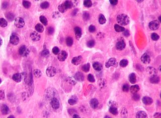
AML-M6, multinucleated erythroblast.jpg
Bone marrow smear from a case of erythroleukemia
Specialty Hematology, oncology

Acute erythrocyte leukemia is a rare form of acute myeloid leukemia (less than 5% of AML cases [1] ) where the myeloproliferation is of erythrocytic precursors. It is defined as type "M6" under the FAB classification.

Contents

Signs and symptoms

The most common symptoms of AEL are related to pancytopenia (a shortage of all types of blood cells), including fatigue, infections, and mucocutaneous bleeding. [2] Almost half of people with AEL exhibit weight loss, fever and night sweats at the time of diagnosis. [2] Almost all people with AEL are anemic, and 77% have a hemoglobin level under 10.0 g/dl. [2] Signs of thrombocytopenia are found in about half of people with AEL. [2]

Causes

The causes of AEL are unknown. [3] Prior to a 2008 reclassification by the World Health Organization, cases that evolved from myelodysplastic syndromes, myeloproliferative neoplasms, chemotherapy for other cancers or exposure to toxins were defined as secondary AEL. [1] These cases are now likely to instead be classified as acute myeloid leukemia with myelodysplasia-related changes or therapy-related AML. [1] +

Diagnosis

Acute erythroid leukemias can be classified as follows:

M6a (Erythroleukemia)

50% or more of all nucleated bone marrow cells are erythroblasts, dyserythropoiesis is prominent and 20% or more of the remaining cells (non- erythroid) are myeloblasts. [4] [5]

M6b (Pure erythroid leukemia)

In rare cases the erythroid lineage is the only obvious component of an acute leukemia; a myeloblast component is not apparent. The erythroid component consists predominantly or exclusively of proerythroblasts and early basophilic erythroblasts. These cells may constitute 90% or more of the marrow elements. Despite this lack of myeloblasts, these cases should be considered acute leukemias. In a WHO proposal the blastic leukemias that are limited to the erythroid series are designated pure erythroid malignancies. [6]

M6c (Erythroleukemia and Pure erythroid leukemia)

Myeloblast- and proerythroblast-rich mixed variant. [7]

Treatment

Treatment for erythroleukemia generally follows that for other types of AML, not otherwise specified. [1] It consists of chemotherapy, frequently consisting of cytarabine, daunorubicin, and idarubicin. [8] It can also involve bone marrow transplantation. [1]

Prognosis

Information on prognosis is limited by the rarity of the condition. Prognosis appears to be no different from AML in general, taking into account other risk factors. [9] [10] Acute erythroid leukemia (M6) has a relatively poor prognosis. A 2010 study of 124 patients found a median overall survival of 8 months. [10] A 2009 study on 91 patients found a median overall survival for erythroleukemia patients of 36 weeks, with no statistically significant difference to other AML patients. AEL patients did have a significantly shorter disease-free survival period, a median of 32 weeks, but this effect was explained by other prognostic factors. That is, AEL is often associated with other risk factors, like monosomal karyotypes and a history of myelodysplastic syndrome. [9] Prognosis is worse in elderly patients, those with a history of myelodysplastic syndrome, and in patients who had previously received chemotherapy for the treatment of a different neoplasm. [1] [11]

Epidemiology

Acute erythroid leukemia is rare, accounting for only 3–5% of all acute myeloid leukemia cases. [2] One study estimated an occurrence rate of 0.077 cases per 100,000 people each year. [12] 64–70% of people with this condition are male, and most are elderly, with a median age of 65. [2]

History

The first known case of acute erythroid leukemia was described in 1912 by M. Copelli under the name erythromatosis. [2] [13] In 1917, Italian hematologist Giovanni Di Guglielmo (1886–1962), expanded on the description, coining the name "eritroleucemia" (Italian for erythroleukemia). [2] [14] Di Guglielmo was the first to recognize the leukemic nature of the condition, and it is sometimes referred to as Di Guglielmo's syndrome in recognition of his work. [2] Ernst Neumark was widely credited for introducing Di Guglielmo's syndrome to English pathology. [15]

Chris Squire, bassist from the progressive rock group Yes, died from complications related to acute erythroid leukemia on June 27, 2015. [16]

Related Research Articles

<span class="mw-page-title-main">Myelodysplastic syndrome</span> Diverse collection of blood-related cancers

A myelodysplastic syndrome (MDS) is one of a group of cancers in which immature blood cells in the bone marrow do not mature, and as a result, do not develop into healthy blood cells. Early on, no symptoms typically are seen. Later, symptoms may include fatigue, shortness of breath, bleeding disorders, anemia, or frequent infections. Some types may develop into acute myeloid leukemia.
<span class="mw-page-title-main">Tumors of the hematopoietic and lymphoid tissues</span> Medical condition

Tumors of the hematopoietic and lymphoid tissues or tumours of the haematopoietic and lymphoid tissues are tumors that affect the blood, bone marrow, lymph, and lymphatic system. Because these tissues are all intimately connected through both the circulatory system and the immune system, a disease affecting one will often affect the others as well, making aplasia, myeloproliferation and lymphoproliferation closely related and often overlapping problems. While uncommon in solid tumors, chromosomal translocations are a common cause of these diseases. This commonly leads to a different approach in diagnosis and treatment of hematological malignancies. Hematological malignancies are malignant neoplasms ("cancer"), and they are generally treated by specialists in hematology and/or oncology. In some centers "hematology/oncology" is a single subspecialty of internal medicine while in others they are considered separate divisions. Not all hematological disorders are malignant ("cancerous"); these other blood conditions may also be managed by a hematologist.

<span class="mw-page-title-main">Myeloid sarcoma</span> Medical condition

A myeloid sarcoma is a solid tumor composed of immature white blood cells called myeloblasts. A chloroma is an extramedullary manifestation of acute myeloid leukemia; in other words, it is a solid collection of leukemic cells occurring outside of the bone marrow.

Acute leukemia or acute leukaemia is a family of serious medical conditions relating to an original diagnosis of leukemia. In most cases, these can be classified according to the lineage, myeloid or lymphoid, of the malignant cells that grow uncontrolled, but some are mixed and for those such an assignment is not possible.

<span class="mw-page-title-main">Acute myeloid leukemia</span> Cancer of the myeloid line of blood cells

Acute myeloid leukemia (AML) is a cancer of the myeloid line of blood cells, characterized by the rapid growth of abnormal cells that build up in the bone marrow and blood and interfere with normal blood cell production. Symptoms may include feeling tired, shortness of breath, easy bruising and bleeding, and increased risk of infection. Occasionally, spread may occur to the brain, skin, or gums. As an acute leukemia, AML progresses rapidly, and is typically fatal within weeks or months if left untreated.

<span class="mw-page-title-main">Chronic myelomonocytic leukemia</span> Medical condition

Chronic myelomonocytic leukemia (CMML) is a type of leukemia, which are cancers of the blood-forming cells of the bone marrow. In adults, blood cells are formed in the bone marrow, by a process that is known as haematopoiesis. In CMML, there are increased numbers of monocytes and immature blood cells (blasts) in the peripheral blood and bone marrow, as well as abnormal looking cells (dysplasia) in at least one type of blood cell.

<span class="mw-page-title-main">Acute myeloblastic leukemia with maturation</span> Medical condition

Acute myeloblastic leukemia with maturation (M2) is a subtype of acute myeloid leukemia (AML).

The French–American–British (FAB) classification systems refers to a series of classifications of hematologic diseases. It is based on the presence of dysmyelopoiesis and the quantification of myeloblasts and erythroblasts.

<span class="mw-page-title-main">HOXA9</span> Protein-coding gene in humans

Homeobox protein Hox-A9 is a protein that in humans is encoded by the HOXA9 gene.

Refractory anemia with excess of blasts (RAEB) is a type of myelodysplastic syndrome with a marrow blast percentage of 5% to 19%.

<span class="mw-page-title-main">Acute megakaryoblastic leukemia</span> Medical condition

Acute megakaryoblastic leukemia (AMKL) is life-threatening leukemia in which malignant megakaryoblasts proliferate abnormally and injure various tissues. Megakaryoblasts are the most immature precursor cells in a platelet-forming lineage; they mature to promegakaryocytes and, ultimately, megakaryocytes which cells shed membrane-enclosed particles, i.e. platelets, into the circulation. Platelets are critical for the normal clotting of blood. While malignant megakaryoblasts usually are the predominant proliferating and tissue-damaging cells, their similarly malignant descendants, promegakaryocytes and megakaryocytes, are variable contributors to the malignancy.

Acute myelomonocytic leukemia (AMML) is a form of acute myeloid leukemia that involves a proliferation of CFU-GM myeloblasts and monoblasts. AMML occurs with a rapid increase amount in white blood cell count and is defined by more than 20% of myeloblast in the bone marrow. It is classified under "M4" in the French-American-British classification (FAB). It is classified under "AML, not otherwise classified" in the WHO classification.

Biphenotypic acute leukaemia (BAL) is an uncommon type of leukemia which arises in multipotent progenitor cells which have the ability to differentiate into both myeloid and lymphoid lineages. It is a subtype of "leukemia of ambiguous lineage".

Acute eosinophilic leukemia (AEL) is a rare subtype of acute myeloid leukemia with 50 to 80 percent of eosinophilic cells in the blood and marrow. It can arise de novo or may develop in patients having the chronic form of a hypereosinophilic syndrome. Patients with acute eosinophilic leukemia have a propensity for developing bronchospasm as well as symptoms of the acute coronary syndrome and/or heart failure due to eosinophilic myocarditis and eosinophil-based endomyocardial fibrosis. Hepatomegaly and splenomegaly are more common than in other variants of AML.

Acute panmyelosis with myelofibrosis (APMF) is a poorly defined disorder that arises as either a clonal disorder, or following toxic exposure to the bone marrow.

A hypomethylating agent is a drug that inhibits DNA methylation: the modification of DNA nucleotides by addition of a methyl group. Because DNA methylation affects cellular function through successive generations of cells without changing the underlying DNA sequence, treatment with a hypomethylating agent is considered a type of epigenetic therapy.

<span class="mw-page-title-main">Childhood leukemia</span> Medical condition

Childhood leukemia is leukemia that occurs in a child and is a type of childhood cancer. Childhood leukemia is the most common childhood cancer, accounting for 29% of cancers in children aged 0–14 in 2018. There are multiple forms of leukemia that occur in children, the most common being acute lymphoblastic leukemia (ALL) followed by acute myeloid leukemia (AML). Survival rates vary depending on the type of leukemia, but may be as high as 90% in ALL.

In haematology atypical localization of immature precursors (ALIP) refers to finding of atypically localized precursors on bone marrow biopsy. In healthy humans, precursors are rare and are found localized near the endosteum, and consist of 1-2 cells. In some cases of myelodysplastic syndromes, immature precursors might be located in the intertrabecular region and occasionally aggregate as clusters of 3 ~ 5 cells. The presence of ALIPs is associated with worse prognosis of MDS. Recently, in bone marrow sections of patients with acute myeloid leukemia cells similar to ALIPs were defined as ALIP-like clusters. The presence of ALIP-like clusters in AML patients within remission was reported to be associated with early relapse of the disease.

FLAG is a chemotherapy regimen used for relapsed and refractory acute myeloid leukemia (AML). The acronym incorporates the three primary ingredients of the regimen:

  1. Fludarabine: an antimetabolite that, while not active toward AML, increases formation of an active cytarabine metabolite, ara-CTP, in AML cells;
  2. Arabinofuranosyl cytidine : an antimetabolite that has been proven to be the most active toward AML among various cytotoxic drugs in single-drug trials; and
  3. Granulocyte colony-stimulating factor (G-CSF): a glycoprotein that shortens the duration and severity of neutropenia.

Microtransplantation (MST) is an advanced technology to treat malignant hematological diseases and tumors by infusing patients with granulocyte colony-stimulating factor (G-CSF) mobilized human leukocyte antigen (HLA)-mismatched allogeneic peripheral blood stem cells following a reduced-intensity chemotherapy or targeted therapy. The term "microtransplantation" comes from its mechanism of reaching donor cell microchimerism.

References

  1. 1 2 3 4 5 6 Zuo Z, Polski JM, Kasyan A, Medeiros LJ (2010). "Acute erythroid leukemia". Arch. Pathol. Lab. Med. 134 (9): 1261–70. doi: 10.5858/2009-0350-RA.1 . PMID   20807044.
  2. 1 2 3 4 5 6 7 8 9 Santos FP, Bueso-Ramos CE, Ravandi F (2010). "Acute erythroleukemia: diagnosis and management". Expert Rev Hematol. 3 (6): 705–18. doi:10.1586/ehm.10.62. PMID   21091147. S2CID   8987729.
  3. Naiem F, Rao PN (2009). "Acute Myeloid Leukemia". Hematopathology: Morphology, Immunophenotype, Cytogenetics, and Molecular Approaches. Academic Press. p. 236. ISBN   978-0-08-091948-5.
  4. Schwab, Manfred (2011-09-23). Encyclopedia of Cancer. Springer Science & Business Media. p. 1313. ISBN   978-3-642-16482-8.
  5. Cheng, Liang; Bostwick, David G. (2011-03-18). Essentials of Anatomic Pathology. Springer Science & Business Media. p. 764. ISBN   978-1-4419-6043-6.
  6. Armitage, James O. (2004). Atlas of Clinical Hematology. Lippincott Williams & Wilkins. p. 148. ISBN   978-0-7817-5128-5.
  7. Raghavan, Derek; Brecher, Martin L.; Johnson, David H.; Meropol, Neal J.; Moots, Paul L.; Rose, Peter G. (2006-07-11). Textbook of Uncommon Cancer. John Wiley & Sons. p. 546. ISBN   978-0-470-03055-4.
  8. Erythroleukemia ~treatment at eMedicine
  9. 1 2 Santos FP, Faderl S, Garcia-Manero G, et al. (December 2009). "Adult acute erythroleukemia: an analysis of 91 patients treated at a single institution". Leukemia. 23 (12): 2275–80. doi:10.1038/leu.2009.181. PMC   4217206 . PMID   19741728.
  10. 1 2 Hasserjian RP, Zuo Z, Garcia C, Tang G, Kasyan A, Luthra R, Abruzzo LV, Kantarjian HM, Medeiros LJ, Wang SA (2010). "Acute erythroid leukemia: a reassessment using criteria refined in the 2008 WHO classification". Blood. 115 (10): 1985–92. doi:10.1182/blood-2009-09-243964. PMC   2942006 . PMID   20040759.
  11. Orazi, Attilio; O'Malley, Dennis P.; Arber, Daniel A. (2006-07-20). Illustrated Pathology of the Bone Marrow. Cambridge University Press. p. 59. ISBN   978-1-139-45552-7.
  12. Wells AW, Bown N, Reid MM, Hamilton PJ, Jackson GH, Taylor PR (2001). "Erythroleukaemia in the north of England: a population based study". J. Clin. Pathol. 54 (8): 608–12. doi:10.1136/jcp.54.8.608. PMC   1731487 . PMID   11477115.
  13. Kasyan A, Medeiros LJ, Zuo Z, Santos FP, Ravandi-Kashani F, Miranda R, Vadhan-Raj S, Koeppen H, Bueso-Ramos CE (2010). "Acute erythroid leukemia as defined in the World Health Organization classification is a rare and pathogenetically heterogeneous disease". Mod. Pathol. 23 (8): 1113–26. doi:10.1038/modpathol.2010.96. PMC   5846338 . PMID   20473273.
  14. Di Guglielmo G. (1917). "Richerche di ematologia. I. Un caso di eritroleucemia. Megacariociti in circolo e loro funzione piastrinopoietico". Folia Medica (Pavia). 13: 386.
  15. "Obituary Notices" (PDF). British Medical Journal. 1967-07-29. Retrieved 2022-03-03.
  16. "Yes bass guitarist Chris Squire dies aged 67". BBC News. 28 June 2015. Retrieved 22 February 2020.