Properdin deficiency

Last updated
Properdin deficiency
X-linked recessive.svg
This condition is inherited in an x-linked recessive manner

Properdin deficiency is a rare X-linked disease in which properdin, an important complement factor responsible for the stabilization of the alternative C3 convertase, is deficient. [1] There are three forms of properdin deficiencies: Type I, which is identified by the total absence of the properdin protein in the plasma, Type II, which is a low but detectable amount of the properdin protein in the plasma, and Type III, which is a rare case of normal levels of properdin protein, but a dysfunctional variant. [2] One of the first studied cases of properdin deficiency was in 1980 by Davis and Forrestal. [3] These families had members with only partial deficiencies which resulted in a lowered consumption of the C3 protein. [3] Properdin deficiency was studied again shortly after in 1982 by Sjoholm in which all of the subjects were deceased shortly after the study because of their disease. [3] The largest study of properdin deficiency was in 1989 by Fijen which included nine males across three generations. [3] Out of the 46 family members in Fijen's study, the 9 who were affected were found to be more susceptible to diseases from the Neisseria genus. [3]

Contents

Signs and symptoms

As a protein involved in the function of the immune system, no external changes in physiology or aberrant physical characteristics are expressed by individuals possessing a properdin deficiency. However, individuals that have a properdin deficiency do have a heightened susceptibility to bacterial infections, most notably caused by bacteria within the Neisseria genus, though there have also been studied cases of individuals with recurrent pneumococcus bacteremia as a result of Streptococcus pneumoniae , another species of bacteria from an entirely different phylum. [3] [4] Due to a heightened susceptibility to Neisseria bacterium, individuals with properdin deficiency are far more likely to succumb to bacterial infection such as meningitis, resulting in inflammation of the brain and spinal cord, which causes severe headaches, fevers, and neck stiffness, and may result in further development of other meningococcal diseases and extreme complications such as sepsis. [5] Individuals with properdin deficiency are also more likely to catch the sexually transmitted disease, gonorrhea, as it is also caused by Neisseria bacterium, resulting in swelling, itching, pain, and formation of pus on the mucous membranes, including, but not limited to, the genitals, mouth, and rectum. [6]

Diagnosis

As mentioned before, there are no external indications of properdin deficiency, and as such, properdin deficiency can only be reliably detected by lab tests. [3] The typical tests for complement deficiencies, such as the measurement of C3 and C4, do not detect low levels of the absence of properdin. [2] These pathways are typically unaltered by any of the three types of properdin deficiencies, but even when they are affected, it is typically within normal levels and is not cause for concern. [2] Instead, histories of infection with anything from the Neisseria genus as well as family history can be indicators, but only specialist centers can screen for properdin deficiencies using immunochemical assays. [2] In particular, the use of ELISA proves to be one of the most effective methods of detecting properdin deficiency, as the average healthy male is expected to show properdin antigen levels of around 128.0 ELISA units/ml, and obligate carrier females (recall that properdin deficiency is an X-linked disease) tend to show an average of 45.6 units/ml. [3] An individual with properdin deficiency should, by definition, show very little to no properdin antigen levels at all, as they do not possess the requisite gene to produce the protein. While properdin deficiencies are rare, they have only been diagnosed in Caucasians, but no other race/ethnicity. [2]

Management

Pertaining to complement deficiencies, there is no cure and the treatments for complement deficiencies vary widely. The best course of action for management is usually for a patient to treat the complement deficiency as an immune deficiency and get immunized against the microbe associated with their deficiency (or best candidate). [7] As mentioned earlier, individuals with properdin deficiency are increasingly susceptible to Neisseria bacterium. Recent studies have indicated that individuals with properdin deficiency respond well when they are immunized with tetravalent polysaccharide meningococcal vaccine, which generates anti capsular antibodies and bactericidal anti-meningococcal activity against serotypes covered by the given vaccine. [2] The vaccine has been reported to lower the chances of reinfection by meningococci in individuals who have undergone the treatment, however the vaccine does not protect against group B meningococci and chemotherapy is recommended if full protection from all meningococci variants is desired. [2]

Genetics

Properdin deficiency is caused by an X-linked recessive allele, meaning that the gene responsible is only present on the X chromosome. Given that it is recessive, this means that the condition can only be inherited if the alleles for both X-chromosomes have the deficiency if the patient is female (xx) or if patient only has one X chromosome, as with male patients (xY). This means that a female could be heterozygous for the allele (Xx) and not express the weakened immune system, as individuals with partial properdin deficiency have been shown to function effectively the same as healthy individuals. [3] This also means that any male patient that receives the recessive allele from their mother will inherit the deficiency, regardless of the genotype of their father, as the father must have passed the Y chromosome to them, which plays no part in genes with x-linked inheritance.

The gene responsible for the production of properdin, Complement Factor Properdin (CFP), lies on the X-chromosome at the coordinates: GRCh38: X:47,623,281-47,630,304. [8] There are three types of properdin deficiency, which are caused by different mutations in the exons of the CFP gene. [8]

Type I properdin deficiency can be the result of a nonsense mutation on exon 5 of the CDP gene, caused by a C-to-T transition at position 2767, which results in an early termination of the sequence and the production of an immature properdin protein. [8] Type I can also be the result of a G-T transversion at position 3511 in exon 7, causing an amino acid substitution from gly271 to valine, which renders the protein non-functional. [8] Finally, Type I can alternatively be caused by a C-to-G transversion on exon 6 at position 3041, converting a serine codon to a stop codon. [8]

Type II properdin deficiency is thought to be caused by one or both of two mutations on the CFP gene. [8] One of the mutations is a C-to-T transition at position 2124, located on exon 4, which converted an arginine to a tryptophan, resulting in an improper protein fold. [8] The second mutation is a G-to-A transition at position 827 in intron 3, which is hypothesized to cause a cryptic splice site, resulting in improper splicing of the mRNA. [8]

Type III properdin deficiency is caused by a T-to-G mutation on exon 9, resulting in a conversion of tyr387 to aspartic acid, resulting in an ineffective protein. [8]

Type I and type II properdin deficiency result in an absence or extremely low presence of properdin, whereas type III results in the presence of a dysfunctional properdin protein structure. [3] Ultimately, all three types have the same basic effect, reducing defensive capabilities against bacteria, especially those previously mentioned.

Epidemiology

Complement deficiencies are rare and currently not well characterized, so there has been difficulty detecting them. Currently, complement deficiencies only comprise approximately 2% of all primary immunodeficiency disorders. While the frequency of properdin deficiency has not been assessed worldwide, the risk of meningococcal infection in an individual with properdin deficiency has been calculated to be around 50%. [9]

Related Research Articles

<span class="mw-page-title-main">Dominance (genetics)</span> One gene variant masking the effect of another in the other copy of the gene

In genetics, dominance is the phenomenon of one variant (allele) of a gene on a chromosome masking or overriding the effect of a different variant of the same gene on the other copy of the chromosome. The first variant is termed dominant and the second is called recessive. This state of having two different variants of the same gene on each chromosome is originally caused by a mutation in one of the genes, either new or inherited. The terms autosomal dominant or autosomal recessive are used to describe gene variants on non-sex chromosomes (autosomes) and their associated traits, while those on sex chromosomes (allosomes) are termed X-linked dominant, X-linked recessive or Y-linked; these have an inheritance and presentation pattern that depends on the sex of both the parent and the child. Since there is only one copy of the Y chromosome, Y-linked traits cannot be dominant or recessive. Additionally, there are other forms of dominance, such as incomplete dominance, in which a gene variant has a partial effect compared to when it is present on both chromosomes, and co-dominance, in which different variants on each chromosome both show their associated traits.

<span class="mw-page-title-main">Leigh syndrome</span> Mitochondrial metabolism disease characterized by progressive loss of mental and movement abilities

Leigh syndrome is an inherited neurometabolic disorder that affects the central nervous system. It is named after Archibald Denis Leigh, a British neuropsychiatrist who first described the condition in 1951. Normal levels of thiamine, thiamine monophosphate, and thiamine diphosphate are commonly found, but there is a reduced or absent level of thiamine triphosphate. This is thought to be caused by a blockage in the enzyme thiamine-diphosphate kinase, and therefore treatment in some patients would be to take thiamine triphosphate daily.

<span class="mw-page-title-main">X-linked recessive inheritance</span> Mode of inheritance

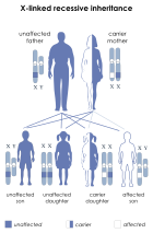
X-linked recessive inheritance is a mode of inheritance in which a mutation in a gene on the X chromosome causes the phenotype to be always expressed in males and in females who are homozygous for the gene mutation, see zygosity. Females with one copy of the mutated gene are carriers.

<i>Neisseria meningitidis</i> Species of bacterium that can cause meningitis

Neisseria meningitidis, often referred to as the meningococcus, is a Gram-negative bacterium that can cause meningitis and other forms of meningococcal disease such as meningococcemia, a life-threatening sepsis. The bacterium is referred to as a coccus because it is round, and more specifically a diplococcus because of its tendency to form pairs.

<span class="mw-page-title-main">X-linked agammaglobulinemia</span> Medical condition

X-linked agammaglobulinemia (XLA) is a rare genetic disorder discovered in 1952 that affects the body's ability to fight infection. As the form of agammaglobulinemia that is X-linked, it is much more common in males. In people with XLA, the white blood cell formation process does not generate mature B cells, which manifests as a complete or near-complete lack of proteins called gamma globulins, including antibodies, in their bloodstream. B cells are part of the immune system and normally manufacture antibodies, which defend the body from infections by sustaining a humoral immunity response. Patients with untreated XLA are prone to develop serious and even fatal infections. A mutation occurs at the Bruton's tyrosine kinase (Btk) gene that leads to a severe block in B cell development and a reduced immunoglobulin production in the serum. Btk is particularly responsible for mediating B cell development and maturation through a signaling effect on the B cell receptor BCR. Patients typically present in early childhood with recurrent infections, in particular with extracellular, encapsulated bacteria. XLA is deemed to have a relatively low incidence of disease, with an occurrence rate of approximately 1 in 200,000 live births and a frequency of about 1 in 100,000 male newborns. It has no ethnic predisposition. XLA is treated by infusion of human antibody. Treatment with pooled gamma globulin cannot restore a functional population of B cells, but it is sufficient to reduce the severity and number of infections due to the passive immunity granted by the exogenous antibodies.

Hypogammaglobulinemia is an immune system disorder in which not enough gamma globulins are produced in the blood. This results in a lower antibody count, which impairs the immune system, increasing risk of infection. Hypogammaglobulinemia may result from a variety of primary genetic immune system defects, such as common variable immunodeficiency, or it may be caused by secondary effects such as medication, blood cancer, or poor nutrition, or loss of gamma globulins in urine, as in nonselective glomerular proteinuria. Patients with hypogammaglobulinemia have reduced immune function; important considerations include avoiding use of live vaccines, and take precautionary measures when traveling to regions with endemic disease or poor sanitation such as receiving immunizations, taking antibiotics abroad, drinking only safe or boiled water, arranging appropriate medical cover in advance of travel, and ensuring continuation of any immunoglobulin infusions needed.

<span class="mw-page-title-main">Biotinidase deficiency</span> Medical condition

Biotinidase deficiency is an autosomal recessive metabolic disorder in which biotin is not released from proteins in the diet during digestion or from normal protein turnover in the cell. This situation results in biotin deficiency.

<span class="mw-page-title-main">Meningococcal disease</span> Medical condition

Meningococcal disease describes infections caused by the bacterium Neisseria meningitidis. It has a high mortality rate if untreated but is vaccine-preventable. While best known as a cause of meningitis, it can also result in sepsis, which is an even more damaging and dangerous condition. Meningitis and meningococcemia are major causes of illness, death, and disability in both developed and under-developed countries.

<span class="mw-page-title-main">Properdin</span> Protein-coding gene in the species Homo sapiens

Properdin is a protein that in humans is encoded by the CFP gene.

<span class="mw-page-title-main">Alström syndrome</span> Rare genetic disorder involving childhood obesity and multiple organ dysfunction

Alström syndrome (AS), also called Alström–Hallgren syndrome, is a very rare autosomal recessive genetic disorder characterised by childhood obesity and multiple organ dysfunction. Symptoms include early-onset type 2 diabetes, cone-rod dystrophy resulting in blindness, sensorineural hearing loss and dilated cardiomyopathy. Endocrine disorders typically also occur, such as hypergonadotrophic hypogonadism and hypothyroidism, as well as acanthosis nigricans resulting from hyperinsulinemia. Developmental delay is seen in almost half of people with Alström syndrome.

The Kidd antigen system are proteins found in the Kidd's blood group, which act as antigens, i.e., they have the ability to produce antibodies under certain circumstances. The Jk antigen is found on a protein responsible for urea transport in the red blood cells and the kidney. They are important in transfusion medicine. People with two Jk(a) antigens, for instance, may form antibodies against donated blood containing two Jk(b) antigens. This can lead to hemolytic anemia, in which the body destroys the transfused blood, leading to low red blood cell counts. Another disease associated with the Jk antigen is hemolytic disease of the newborn, in which a pregnant woman's body creates antibodies against the blood of her fetus, leading to destruction of the fetal blood cells. Hemolytic disease of the newborn associated with Jk antibodies is typically mild, though fatal cases have been reported.

<span class="mw-page-title-main">Complement component 2</span> Protein-coding gene in the species Homo sapiens

Complement C2 is a protein that in humans is encoded by the C2 gene. The protein encoded by this gene is part of the classical pathway of the complement system, acting as a multi-domain serine protease. Deficiency of C2 has been associated with certain autoimmune diseases.

<span class="mw-page-title-main">Mannan-binding lectin</span> Mammalian protein found in Homo sapiens

Mannose-binding lectin (MBL), also called mannan-binding lectin or mannan-binding protein (MBP), is a lectin that is instrumental in innate immunity as an opsonin and via the lectin pathway.

George D. Heist (1886–1920) was an immunologist specializing in the study of infections of meningococcal bacteria that often result in meningococcal disease, which is well known as highly lethal and debilitating, and extremely difficult to treat.

<span class="mw-page-title-main">Complement deficiency</span> Medical condition

Complement deficiency is an immunodeficiency of absent or suboptimal functioning of one of the complement system proteins. Because of redundancies in the immune system, many complement disorders are never diagnosed. Some studies estimate that less than 10% are identified. Hypocomplementemia may be used more generally to refer to decreased complement levels, while secondary complement disorder means decreased complement levels that are not directly due to a genetic cause but secondary to another medical condition.

<span class="mw-page-title-main">Autosomal recessive polycystic kidney disease</span> Medical condition

Autosomal recessive polycystic kidney disease (ARPKD) is the recessive form of polycystic kidney disease. It is associated with a group of congenital fibrocystic syndromes. Mutations in the PKHD1 cause ARPKD.

NmVac4-A/C/Y/W-135 is the commercial name of the polysaccharide vaccine against the bacterium that causes meningococcal meningitis. The product, by JN-International Medical Corporation, is designed and formulated to be used in developing countries for protecting populations during meningitis disease epidemics.

Surfactant metabolism dysfunction is a condition where pulmonary surfactant is insufficient for adequate respiration. Surface tension at the liquid-air interphase in the alveoli makes the air sacs prone to collapsing post expiration. This is due to the fact that water molecules in the liquid-air surface of alveoli are more attracted to one another than they are to molecules in the air. For sphere-like structures like alveoli, water molecules line the inner walls of the air sacs and stick tightly together through hydrogen bonds. These intermolecular forces put great restraint on the inner walls of the air sac, tighten the surface all together, and unyielding to stretch for inhalation. Thus, without something to alleviate this surface tension, alveoli can collapse and cannot be filled up again. Surfactant is essential mixture that is released into the air-facing surface of inner walls of air sacs to lessen the strength of surface tension. This mixture inserts itself among water molecules and breaks up hydrogen bonds that hold the tension. Multiple lung diseases, like ISD or RDS, in newborns and late-onsets cases have been linked to dysfunction of surfactant metabolism.

<span class="mw-page-title-main">BENTA disease</span> Medical condition

BENTA disease is a rare genetic disorder of the immune system. BENTA stands for "B cell expansion with NF-κB and T cell anergy" and is caused by germline heterozygous gain-of-function mutations in the gene CARD11. This disorder is characterized by polyclonal B cell lymphocytosis with onset in infancy, splenomegaly, lymphadenopathy, mild immunodeficiency, and increased risk of lymphoma. Investigators Andrew L. Snow and Michael J. Lenardo at the National Institute of Allergy and Infectious Diseases at the U.S. National Institutes of Health first characterized BENTA disease in 2012. Dr. Snow's current laboratory at the Uniformed Services University of the Health Sciences is now actively studying this disorder.

PGM3 deficiency is a rare genetic disorder of the immune system associated with diminished phosphoglucomutase 3 function. PGM3 is an enzyme which in humans is encoded by gene PGM3. This disorder manifests as severe atopy, immune deficiency, autoimmunity, intellectual disability, and hypomyelination. In 2014, Investigators Atfa Sassi at the Pasteur Institute of Tunis, Sandra Lazaroski at the University Medical Center Freiburg, and Gang Wu at the Imperial College London, identified PGM3 mutations in nine patients from four consanguineous families. In the same year, a researchers from the laboratories of Joshua Milner and Helen Su at the National Institute of Allergy and Infectious Disease at the U.S. National Institutes of Health described PGM3 deficiency in eight additional patients from two families.

References

  1. van den Bogaard R, Fijen CA, Schipper MG, de Galan L, Kuijper EJ, Mannens MM (July 2000). "Molecular characterisation of 10 Dutch properdin type I deficient families: mutation analysis and X-inactivation studies". Eur. J. Hum. Genet. 8 (7): 513–8. doi: 10.1038/sj.ejhg.5200496 . PMID   10909851.
  2. 1 2 3 4 5 6 7 Linton, S. M.; Morgan, B. P. (November 1999). "Properdin deficiency and meningococcal disease--identifying those most at risk". Clinical and Experimental Immunology. 118 (2): 189–191. doi:10.1046/j.1365-2249.1999.01057.x. ISSN   0009-9104. PMC   1905414 . PMID   10540177.
  3. 1 2 3 4 5 6 7 8 9 10 "OMIM Entry - # 312060 - PROPERDIN DEFICIENCY, X-LINKED; CFPD". www.omim.org. Retrieved 2022-03-23.
  4. Sexton, Daniel J. "Invasive pneumococcal (Streptococcus pneumoniae) infections and bacteremia". www.uptodate.com. Retrieved 2022-03-24.
  5. "Meningitis", Wikipedia, 2022-03-20, retrieved 2022-03-24
  6. "Gonorrhea", Wikipedia, 2022-03-22, retrieved 2022-03-24
  7. "Complement Deficiencies | Immune Deficiency Foundation". primaryimmune.org. Retrieved 2022-03-24.
  8. 1 2 3 4 5 6 7 8 9 "OMIM Entry - * 300383 - COMPLEMENT FACTOR PROPERDIN; CFP". www.omim.org. Retrieved 2022-03-24.
  9. editor., Leung, Donald Y. M., 1949- (19 October 2020). Pediatric allergy : principles and practice. Elsevier. ISBN   978-0-323-67462-1. OCLC   1202992443.{{cite book}}: |last= has generic name (help)CS1 maint: multiple names: authors list (link)