ABCA4 | |||||||||||||||||||||||||||||||||||||||||||||||||||
---|---|---|---|---|---|---|---|---|---|---|---|---|---|---|---|---|---|---|---|---|---|---|---|---|---|---|---|---|---|---|---|---|---|---|---|---|---|---|---|---|---|---|---|---|---|---|---|---|---|---|---|
Identifiers | |||||||||||||||||||||||||||||||||||||||||||||||||||
Aliases | ABCA4 , AW050280, Abc10, Abcr, D430003I15Rik, RmP, ARMD2, CORD3, FFM, RP19, STGD, STGD1, ATP binding cassette subfamily A member 4 | ||||||||||||||||||||||||||||||||||||||||||||||||||
External IDs | OMIM: 601691; MGI: 109424; HomoloGene: 298; GeneCards: ABCA4; OMA:ABCA4 - orthologs | ||||||||||||||||||||||||||||||||||||||||||||||||||
| |||||||||||||||||||||||||||||||||||||||||||||||||||
| |||||||||||||||||||||||||||||||||||||||||||||||||||
| |||||||||||||||||||||||||||||||||||||||||||||||||||
| |||||||||||||||||||||||||||||||||||||||||||||||||||
| |||||||||||||||||||||||||||||||||||||||||||||||||||
Wikidata | |||||||||||||||||||||||||||||||||||||||||||||||||||
|
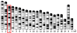
ATP-binding cassette, sub-family A (ABC1), member 4, also known as ABCA4 or ABCR, is a protein which in humans is encoded by the ABCA4 gene. [5] [6] [7]
ABCA4 is a member of the ATP-binding cassette transporter gene sub-family A (ABC1) found exclusively in multicellular eukaryotes. [5] The gene was first cloned and characterized in 1997 as a gene that causes Stargardt disease, an autosomal recessive disease that causes macular degeneration. [8] The ABCA4 gene transcribes a large retina-specific protein with two transmembrane domains (TMD), two glycosylated extracellular domains (ECD), and two nucleotide-binding domains (NBD). The ABCA4 protein is almost exclusively expressed in retina localizing in outer segment disk edges of rod photoreceptors. [9]
Previously known as the photoreceptor rim protein RmP or ABCR, the recently proposed ABCA4 structure consists of two transmembrane domains (TMDs), two large glycosylated extracytosolic domains (ECD), and two internal nucleotide binding domains (NBDs). One TMD spans across membranes with six units of protein linked together to form a domain. The TMDs are usually not conserved across genomes due to its specificity and diversity in function as channels or ligand-binding controllers. However, NBDs are highly conserved across different genomes—an observation consistent with which it binds and hydrolyzes ATP. NBD binds adenosine triphosphate molecules (ATP) to utilize the high-energy inorganic phosphate to carry out change in conformation of the ABC transporter. Transcribed ABCA4 forms into a heterodimer: the two dimerized compartments of the channel are different from each other. When TMDs are situated in a membrane, they form a barrel-like structure permeable to retinoid ligands and control channel access to its binding sites. [10] Once an ATP is hydrolized at the NBDs of the channel, NBDs are brought together to tilt and modify TMDs to modulate ligand binding to the channel. [11] A recently proposed model of retinoid transfer occurring as a result of alternating exposure of external and internal TMD ligand binding sites, all controlled by binding of ATP, is based on recent structural analyses of bacterial ABC transporters.
ABCR is localized to outer segment disk edges of rods and cones. ABCR is expressed much less than rhodopsin, approximately at 1:120. Comparisons between mammalian ABCA4 to other ABCs, cellular localization of ABCA4, and analyses of ABCA4 knockout mice suggest that ABCA4 may function as an inward-directed retinoid flippase. [12] Flippase is a transmembrane protein that "flips" its conformation to transport materials across a membrane. In the case of ABCA4, the flippase facilitates transfer of N-retinyl-phosphatidylethanolamine (NR-PE), a covalent adduct of all-trans retinaldehyde (ATR) with phosphatidylethanolamine (PE), trapped inside the disk as charged species out to the cytoplasmic surface. [13] Once transported, ATR is reduced to vitamin A and then transferred to retinal pigment epithelium to be recycled into 11-cis-retinal. This alternating access-release model for ABCA4 has four steps: (1) binding of ATP to an NBD to bring two NBDs together and expose outer vestibule high affinity binding site located in TMD, (2) binding of NR-PE/ATR on extracellular side of the channel, (3) ATP hydrolysis promoting gate opening and movement of NR-PE/ATR across the membrane to the low-affinity binding site on the intracellular portion of TMD, and (4) release of adenosine diphosphate (ADP) and inorganic phosphate (Pi) to release the bound ligand. The channel is then ready to transfer another molecule of NR-PE/ATR again.
The ABCR -/- knockout mouse has delayed dark adaptation but normal final rod threshold relative to controls. [12] This suggests bulk transmembrane diffusion pathways that remove ATR/NR-PE from extracellular membranes. After bleaching the retina with strong light, ATR/NR-PE accumulates significantly in outer segments. This accumulation leads to formation of toxic cationic bis-pyridinium salt, N-retinylidene-N-retinyl-ethanolamine (A2E), which causes human dry and wet age-related macular degeneration. [14] From this experiment, it was concluded that ABCR has a significant role in clearing accumulation of ATR/NR-PE to prevent formation of A2E in extracellular photoreceptor surfaces during bleach recovery.
Mutations in ABCA4 gene are known to cause the autosomal-recessive disease Stargardt macular dystrophy (STGD), which is a hereditary juvenile macular degeneration disease causing progressive loss of photoreceptor cells. STGD is characterized by reduced visual acuity and color vision, loss of central (macular) vision, delayed dark adaptation, and accumulation of autofluorescent RPE lipofuscin. [14] Removal of NR-PE/ATR appears to be significant in normal bleach recovery and to mitigate persistent opsin signaling that causes photoreceptors to degenerate. ABCA4 also mitigates long-term effects of accumulation of ATR that results in irreversible ATR binding to a second molecule of ATR and NR-PE to form dihydro-N-retinylidene-N-retinyl-phosphatidyl-ethanolamine (A2PE-H2). A2PE-H2 traps ATR and accumulates in outer segments to further oxidize into N-retinylidene-N-retinyl-phosphatidyl-ethanolamine (A2PE). After diurnal disk-shedding and phagocytosis of outer segment by RPE cells, A2PE is hydrolyzed inside the RPE phagolysosome to form A2E. [14] Accumulation of A2E causes toxicity at the primary RPE level and secondary photoreceptor destruction in macular degenerations.
Additional diseases that may link to mutations in ABCA4 include fundus flavimaculatus, cone-rod dystrophy, retinitis pigmentosa, and age-related macular degeneration.
The GENEVA Cleft Consortium study first identified ABCA4 as being associated with cleft lip and/or cleft palate with multiple markers giving evidence of linkage and association at the genome-wide significance level. [15] Although SNPs in this gene are associated with cleft lip/palate there is no functional or expression data to support it as the causal gene which may, instead, lie in a region adjacent to ABCA4. [16] A combination of genome wide association, rare coding sequence variants, craniofacial specific expression, and interactions with IRF6 support a role for the adjacent ARHGAP29 gene to be the likely causal gene playing a role in nonsyndromic cleft lip and/or palate. [17]
Retinitis pigmentosa (RP) is a genetic disorder of the eyes that causes loss of vision. Symptoms include trouble seeing at night and decreasing peripheral vision. As peripheral vision worsens, people may experience "tunnel vision". Complete blindness is uncommon. Onset of symptoms is generally gradual and often begins in childhood.
Macular degeneration, also known as age-related macular degeneration, is a medical condition which may result in blurred or no vision in the center of the visual field. Early on there are often no symptoms. Over time, however, some people experience a gradual worsening of vision that may affect one or both eyes. While it does not result in complete blindness, loss of central vision can make it hard to recognize faces, drive, read, or perform other activities of daily life. Visual hallucinations may also occur.
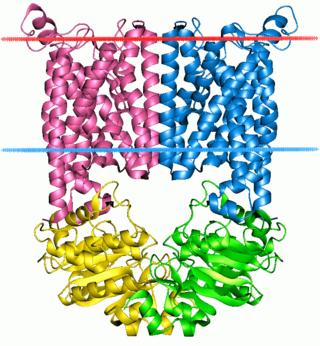
The ABC transporters, ATP synthase (ATP)-binding cassette transporters are a transport system superfamily that is one of the largest and possibly one of the oldest gene families. It is represented in all extant phyla, from prokaryotes to humans. ABC transporters belong to translocases.
Lipofuscin is the name given to fine yellow-brown pigment granules composed of lipid-containing residues of lysosomal digestion. It is considered to be one of the aging or "wear-and-tear" pigments, found in the liver, kidney, heart muscle, retina, adrenals, nerve cells, and ganglion cells.
Vitelliform macular dystrophy is an irregular autosomal dominant eye disorder which can cause progressive vision loss. This disorder affects the retina, specifically cells in a small area near the center of the retina called the macula. The macula is responsible for sharp central vision, which is needed for detailed tasks such as reading, driving, and recognizing faces. The condition is characterized by yellow, slightly elevated, round structures similar to the yolk of an egg.
Stargardt disease is the most common inherited single-gene retinal disease. In terms of the first description of the disease, it follows an autosomal recessive inheritance pattern, which has been later linked to bi-allelic ABCA4 gene variants (STGD1). However, there are Stargardt-like diseases with mimicking phenotypes that are referred to as STGD3 and STGD4, and have a autosomal dominant inheritance due to defects with ELOVL4 or PROM1 genes, respectively. It is characterized by macular degeneration that begins in childhood, adolescence or adulthood, resulting in progressive loss of vision.
ATP-binding cassette super-family G member 2 is a protein that in humans is encoded by the ABCG2 gene. ABCG2 has also been designated as CDw338. ABCG2 is a translocation protein used to actively pump drugs and other compounds against their concentration gradient using the bonding and hydrolysis of ATP as the energy source.
Peripherin-2 is a protein, that in humans is encoded by the PRPH2 gene. Peripherin-2 is found in the rod and cone cells of the retina of the eye. Defects in this protein result in one form of retinitis pigmentosa, an incurable blindness.
Bestrophin-1 (Best1) is a protein that, in humans, is encoded by the BEST1 gene.
Cone-rod homeobox protein is a protein that in humans is encoded by the CRX gene.
Retinaldehyde-binding protein 1 (RLBP1) also known as cellular retinaldehyde-binding protein (CRALBP) is a 36-kD water-soluble protein that in humans is encoded by the RLBP1 gene.
Rod cGMP-specific 3',5'-cyclic phosphodiesterase subunit beta is the beta subunit of the protein complex PDE6 that is encoded by the PDE6B gene. PDE6 is crucial in transmission and amplification of visual signal. The existence of this beta subunit is essential for normal PDE6 functioning. Mutations in this subunit are responsible for retinal degeneration such as retinitis pigmentosa or congenital stationary night blindness.
Elongation of very long chain fatty acids protein 4 is a protein that in humans is encoded by the ELOVL4 gene.
Cyclic nucleotide gated channel beta 3, also known as CNGB3, is a human gene encoding an ion channel protein.
Hemicentin-1 is a protein that in humans is encoded by the HMCN1 gene.
Putative ATP-binding cassette transporter sub-family C member 13 is a protein that is not present in humans. In humans, ABCC13 is a pseudogene.
Gene therapy using lentiviral vectors was being explored in early stage trials as of 2009.
Retinal gene therapy holds a promise in treating different forms of non-inherited and inherited blindness.
ATP-binding cassette, sub-family A (ABC1), member 5 is a protein that in humans is encoded by the ABCA5 gene.
Occult macular dystrophy (OMD) is a rare inherited degradation of the retina, characterized by progressive loss of function in the most sensitive part of the central retina (macula), the location of the highest concentration of light-sensitive cells (photoreceptors) but presenting no visible abnormality. "Occult" refers to the degradation in the fundus being difficult to discern. The disorder is called "dystrophy" instead of "degradation" to distinguish its genetic origin from other causes, such as age. OMD was first reported by Y. Miyake et al. in 1989.
This article incorporates text from the United States National Library of Medicine, which is in the public domain.