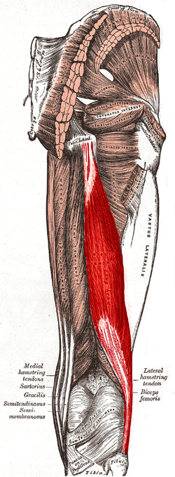
Biceps femoris tendon rupture | |
---|---|
Biceps femoris muscle |
Biceps femoris tendon rupture can occur when the biceps femoris is injured in sports that require explosive bending of the knee as seen in sprinting. If the athlete is fatigued or has not warmed up properly he/she may suffer a hamstring strain/rupture, which is the tearing of the hamstring muscle. Avulsion of the biceps femoris tendon is the complete pulling away of the tendon from the bone. This most commonly occurs where the long head attaches to the ischial tuberosity. Injuries to biceps femoris are more common than to other hamstring muscles.
One theory for this is the fact that each of the two heads are innervated by different branches of the sciatic nerve. In states of fatigue or when the muscle is not fully warmed up, uncoordinated firing of the nerves may cause the muscle to contract inappropriately during movement, leading to injury. Biceps femoris tendon avulsion may also be associated with an avulsion fracture which occurs when a piece of the bone is pulled away with the tendon, during forceful contraction.
Isolated avulsion is rare. [1]
The R.I.C.E. technique should be administered, however, a sports injury specialist is recommended for the possibility of surgery.
This section is empty. You can help by adding to it. (December 2017) |
The leg is the entire lower limb of the human body, including the foot, thigh or sometimes even the hip or buttock region. The major bones of the leg are the femur, tibia, and adjacent fibula.
The biceps or biceps brachii is a large muscle that lies on the front of the upper arm between the shoulder and the elbow. Both heads of the muscle arise on the scapula and join to form a single muscle belly which is attached to the upper forearm. While the biceps crosses both the shoulder and elbow joints, its main function is at the elbow where it flexes and supinates the forearm. Both these movements are used when opening a bottle with a corkscrew: first biceps screws in the cork (supination), then it pulls the cork out (flexion).
In human anatomy, a hamstring is any one of the three posterior thigh muscles between the hip and the knee.
The fibula or calf bone is a leg bone on the lateral side of the tibia, to which it is connected above and below. It is the smaller of the two bones and, in proportion to its length, the most slender of all the long bones. Its upper extremity is small, placed toward the back of the head of the tibia, below the knee joint and excluded from the formation of this joint. Its lower extremity inclines a little forward, so as to be on a plane anterior to that of the upper end; it projects below the tibia and forms the lateral part of the ankle joint.
A soft tissue injury is the damage of muscles, ligaments and tendons throughout the body. Common soft tissue injuries usually occur from a sprain, strain, a one-off blow resulting in a contusion or overuse of a particular part of the body. Soft tissue injuries can result in pain, swelling, bruising and loss of function.
Osgood–Schlatter disease (OSD) is inflammation of the patellar ligament at the tibial tuberosity (apophysitis) usually affecting adolescents during growth spurts. It is characterized by a painful bump just below the knee that is worse with activity and better with rest. Episodes of pain typically last a few weeks to months. One or both knees may be affected and flares may recur.
The biceps femoris is a muscle of the thigh located to the posterior, or back. As its name implies, it consists of two heads; the long head is considered part of the hamstring muscle group, while the short head is sometimes excluded from this characterization, as it only causes knee flexion and is activated by a separate nerve.
The semimembranosus muscle is the most medial of the three hamstring muscles in the thigh. It is so named because it has a flat tendon of origin. It lies posteromedially in the thigh, deep to the semitendinosus muscle. It extends the hip joint and flexes the knee joint.
The semitendinosus is a long superficial muscle in the back of the thigh. It is so named because it has a very long tendon of insertion. It lies posteromedially in the thigh, superficial to the semimembranosus.
The rectus femoris muscle is one of the four quadriceps muscles of the human body. The others are the vastus medialis, the vastus intermedius, and the vastus lateralis. All four parts of the quadriceps muscle attach to the patella by the quadriceps tendon.
The popliteus muscle in the leg is used for unlocking the knees when walking, by laterally rotating the femur on the tibia during the closed chain portion of the gait cycle. In open chain movements, the popliteus muscle medially rotates the tibia on the femur. It is also used when sitting down and standing up. It is the only muscle in the posterior (back) compartment of the lower leg that acts just on the knee and not on the ankle. The gastrocnemius muscle acts on both joints.
The knee examination, in medicine and physiotherapy, is performed as part of a physical examination, or when a patient presents with knee pain or a history that suggests a pathology of the knee joint.
The lateral collateral ligament is an extrinsic ligament of the knee located on the lateral side of the knee. Its superior attachment is at the lateral epicondyle of the femur ; its inferior attachment is at the lateral aspect of the head of fibula. The LCL is not fused with the joint capsule. Inferiorly, the LCL splits the tendon of insertion of the biceps femoris muscle.
The posterior compartment of the thigh is one of the fascial compartments that contains the knee flexors and hip extensors known as the hamstring muscles, as well as vascular and nervous elements, particularly the sciatic nerve.
Straining of the hamstring, also known as a pulled hamstring, is defined as an excessive stretch or tear of muscle fibers and related tissues. Hamstring injuries are common in athletes participating in many sports. Track and field athletes are particularly at risk, as hamstring injuries have been estimated to make up 29% of all injuries in sprinters. Hamstring injuries can also come with a hip injury from sprinting. Symptoms for a hip injury are pain, aching and discomfort while running or any physical exercise.
A patellar dislocation is a knee injury in which the patella (kneecap) slips out of its normal position. Often the knee is partly bent, painful and swollen. The patella is also often felt and seen out of place. Complications may include a patella fracture or arthritis.
Jersey finger, also known as rugby finger, is a finger-related tendon injury that is common in sport and can result in permanent loss of flexion of the end of the finger if not surgically repaired. The injury is common when one player grabs another's jersey with the tips of one or more fingers while that player is pulling or running away. It is the most common closed flexor tendon injury and occurs in the ring finger in 75% of cases.
Posterolateral corner injuries of the knee are injuries to a complex area formed by the interaction of multiple structures. Injuries to the posterolateral corner can be debilitating to the person and require recognition and treatment to avoid long term consequences. Injuries to the PLC often occur in combination with other ligamentous injuries to the knee; most commonly the anterior cruciate ligament (ACL) and posterior cruciate ligament (PCL). As with any injury, an understanding of the anatomy and functional interactions of the posterolateral corner is important to diagnosing and treating the injury.
Anatomical terminology is used to uniquely describe aspects of skeletal muscle, cardiac muscle, and smooth muscle such as their actions, structure, size, and location.
A biceps tendon rupture or bicep tear is a complete or partial rupture of a tendon of the biceps brachii muscle. It can affect any of the three biceps brachii tendons - the proximal tendon of the short head of the muscle belly, the proximal tendon of the long head of the muscle belly, or the distal tendon. The characteristic finding of a biceps tendon rupture is the Popeye sign. Patients often report an audible pop at the time of injury as well as pain, bruising, and swelling. Provocative physical exam maneuvers to assess for a rupture include Ludington's test, Hook test, and the Ruland biceps squeeze test. Treatment and prognosis are highly dependent on the site of the injury described in further detail below.